Limbal Stem Cell: An Evidence-Based Patient Guide

Limbal stem cells maintain the transparent corneal epithelium and support clear vision. Limbal stem cell deficiency may follow chemical injury, severe infection, inflammatory eye disease, contact lens overuse or multiple eye surgeries.
Key Takeaways
- Limbal stem cells maintain the transparent corneal epithelium and support clear vision.
- Limbal stem cell deficiency may follow chemical injury, severe infection, inflammatory eye disease, contact lens overuse or multiple eye surgeries.
- Diagnosis requires a detailed eye-surface examination and may include specialized imaging or laboratory testing.
- Medical treatment can protect the eye surface, while surgery may be considered for persistent or severe disease.
- Cell-based procedures require long-term follow-up because inflammation, dry eye and rejection can affect outcomes.
Limbal stem cells are specialized cells at the edge of the cornea that continually renew its clear outer surface. When they are lost or damaged, limbal stem cell deficiency can cause pain, light sensitivity and reduced vision; treatment ranges from surface protection and inflammation control to carefully selected cell-based surgery.
Overview: What Is a Limbal Stem Cell?
A limbal stem cell is a specialized regenerative cell located in the limbus, the narrow transition zone between the clear cornea and the white of the eye. These cells continually replace the corneal epithelium, the smooth protective layer covering the front of the eye. A healthy epithelium helps keep the cornea clear, comfortable and able to focus light properly.
When too few functioning cells remain, the condition is called limbal stem cell deficiency (LSCD). The cornea may become covered by blood-vessel-containing conjunctival tissue, become irregular or develop repeated surface breakdown. This can lead to discomfort and visual changes, but the outlook varies greatly depending on the cause, severity and health of the rest of the eye.
Limbal stem cell treatment is individualized. Some people improve with treatment of dry eye, inflammation and eyelid problems, while others may be candidates for limbal stem cell deficiency surgery or other corneal surface reconstruction. Assessment by an ophthalmologist with expertise in cornea and ocular surface disease is important before any procedure is considered.
Symptoms and the Pathology of Limbal Stem Cell Deficiency
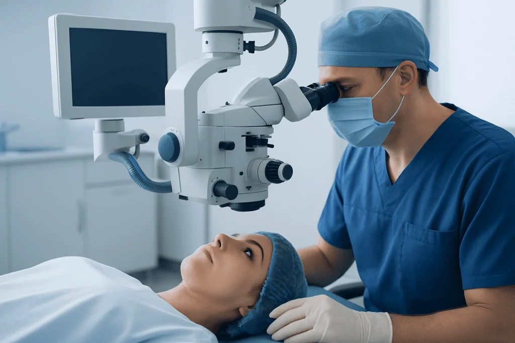
Limbal stem cell deficiency pathology involves failure of normal corneal epithelial renewal. Without sufficient limbal cells, the eye may not maintain a stable, transparent corneal surface. Instead, conjunctival cells can migrate over the cornea, a process sometimes called conjunctivalization. Surface inflammation, scarring and abnormal blood vessel growth may also occur.
Symptoms may affect one or both eyes and can range from mild to severe. They include blurred or fluctuating vision, redness, foreign-body sensation, burning, tearing, pain and sensitivity to light. Recurrent corneal erosions can cause episodes of more intense discomfort, particularly on waking.
Symptoms alone do not confirm LSCD. Dry eye disease, allergy, infection, eyelid disorders and other corneal conditions can produce similar complaints. A careful assessment is therefore needed to identify the underlying problem and select the safest management plan.
- Blurred, hazy or variable vision
- Persistent redness or irritation
- Light sensitivity and excessive tearing
- Repeated corneal surface defects or slow healing
- Visible changes to the corneal surface in more advanced cases
Causes and Risk Factors

LSCD can be acquired or, less commonly, inherited. A chemical or thermal burn is an important cause because it can directly damage the limbus and surrounding eye structures. Severe eye trauma, certain infections and repeated ocular surgeries may also affect limbal cell function.
Long-standing inflammation can contribute to disease. Examples include severe dry eye, ocular cicatricial pemphigoid, Stevens-Johnson syndrome and other disorders that affect the conjunctiva and eyelids. Chronic use of preserved topical eye medicines may irritate the surface in susceptible individuals, and prolonged or poorly tolerated contact lens wear can contribute to damage in some cases.
Risk is higher when both the limbal area and the supporting ocular environment are affected. Tear-film instability, eyelid malposition, incomplete blinking, autoimmune inflammation and corneal nerve problems can all make healing more difficult. Treating these contributing factors is often essential, whether or not surgery is planned.
How Doctors Diagnose and Assess Candidacy
Diagnosis begins with a history of symptoms, injuries, contact lens use, medications, autoimmune disease and previous eye procedures. During a slit-lamp examination, the ophthalmologist evaluates the cornea, limbus, conjunctiva, eyelids and tear film. Fluorescein staining can reveal unstable or damaged corneal epithelium.
Specialized tests may help clarify the extent of disease. These can include corneal topography, anterior-segment optical coherence tomography, in vivo confocal microscopy and impression cytology, in which surface cells are sampled for laboratory assessment. Not every patient needs every test; investigations are selected according to the clinical situation.
Candidacy for limbal stem cell therapy depends on more than the amount of limbal damage. The care team also assesses whether active inflammation is controlled, whether eyelids and tear production can support healing, whether the cornea has deep scarring, and whether vision could improve if the surface is restored. In one-sided disease, a patient’s healthy eye may sometimes provide cells; in both-eye disease, donor-based or laboratory-expanded approaches may be discussed.
Other causes of reduced vision must also be considered. Cataract, glaucoma, retinal disease and optic nerve damage may limit the visual benefit of ocular surface reconstruction even when the cornea becomes healthier.
Limbal Stem Cell Treatment and How Procedures Work
Initial limbal stem cell treatment commonly focuses on stabilizing the ocular surface. Depending on the cause, this may include preservative-free lubricants, treatment for eyelid or tear-film disorders, anti-inflammatory medicines prescribed by an ophthalmologist, protective contact lenses or measures to reduce exposure and irritation. Management of the underlying systemic disease is particularly important when autoimmune inflammation is present.
When surface disease remains significant despite appropriate medical care, limbal stem cell deficiency surgery may be considered. The goal is to re-establish a stable corneal epithelium by supplying viable limbal cells and creating an environment where they can survive. Approaches include autologous limbal grafting from the patient’s unaffected eye, small tissue graft techniques, cultivated limbal epithelial cell transplantation, or donor tissue transplantation when both eyes are involved.
In an autologous procedure, a carefully limited amount of limbal tissue may be taken from the healthier eye and placed on the affected cornea. This avoids immune rejection of the transplanted cells, but protecting the donor eye is a central priority. Donor-derived tissue may be necessary when both eyes have extensive disease; it generally requires systemic immune-suppressing treatment and close medical monitoring.
Some patients also need treatment for deeper corneal scarring after the surface has stabilized. A corneal transplant performed before control of LSCD may be less likely to remain clear, so the sequence of care matters. Corneal transplant evaluation may be discussed when scarring continues to limit vision after ocular surface reconstruction.
Step by Step, Recovery and Expected Benefits
Before surgery, the team aims to control inflammation, infection risk, dry eye and eyelid abnormalities. The exact procedure is selected according to whether disease affects one or both eyes, the availability of healthy limbal tissue and the condition of the cornea. Surgery is typically performed under local anesthesia with sedation or general anesthesia, depending on the procedure and individual needs.
During transplantation, abnormal surface tissue may be gently removed, and limbal tissue or cultivated cells are placed on the prepared eye surface. An amniotic membrane, sutures, tissue adhesive or a protective contact lens may be used to support healing. The patient receives detailed instructions about eye drops, shielding the eye and activity restrictions.
Early recovery often includes blurred vision, tearing, light sensitivity and discomfort that gradually improve as the epithelium heals. Follow-up visits are frequent at first because the surgeon needs to monitor epithelial closure, pressure within the eye, inflammation and infection. Visual recovery may take weeks to months, and it can be longer when scarring, inflammation or other eye disease is present.
The potential benefit is a more stable, comfortable ocular surface and, for suitable patients, improved corneal clarity and vision. Results cannot be predicted for every individual. Ongoing lubrication, management of dry eye and regular review may remain necessary after successful treatment.
Risks, Limits and the Role of Research
All eye procedures have risks. For limbal cell transplantation, these can include infection, persistent epithelial defects, inflammation, scarring, increased eye pressure, recurrence of LSCD and limited visual improvement. Donor-derived grafts can be rejected, and medicines used to reduce rejection can have important side effects that require monitoring by experienced clinicians.
Autologous transplantation avoids donor rejection but can carry a small risk to the eye that provides tissue. Surgeons minimize this risk by taking only an appropriate amount of tissue and confirming that the donor eye is healthy. Patients should discuss the possible effect on both eyes, likely alternatives and realistic goals before giving consent.
Limbal stem cell research is advancing in areas such as laboratory expansion of a patient’s own cells, tissue engineering and improved ways to support the eye surface. These developments are promising, but not every experimental approach is established, widely available or appropriate for routine care. A trustworthy treatment plan should explain the evidence supporting the proposed method, expected follow-up and any uncertainties.
When to Seek Medical Care
Prompt ophthalmic assessment is important after a chemical splash, thermal injury or significant eye trauma. The eye should be rinsed immediately with clean water or saline if a chemical exposure occurs, and urgent emergency care should be sought. Do not attempt to neutralize a chemical with another substance or delay care while looking for eye drops.
People should arrange an eye examination for persistent redness, pain, light sensitivity, recurrent surface irritation or reduced vision, especially after contact lens problems, eye surgery or inflammatory illness. Sudden vision loss, severe pain, a marked increase in redness, discharge or injury requires urgent medical attention.
For complex ocular surface disease, care may involve cornea specialists, rheumatology, dermatology or other teams depending on the underlying cause. Acibadem International’s multidisciplinary specialists and JCI-accredited hospitals diagnose and treat complex corneal and ocular surface conditions for international patients.
Frequently asked questions
What do limbal stem cells do?
Limbal stem cells renew the corneal epithelium, the clear outer layer of the eye. This renewal helps maintain a smooth, transparent corneal surface that protects the eye and supports clear vision.
Can limbal stem cell deficiency heal without surgery?
Mild or early disease may improve when the underlying cause is treated and the eye surface is protected. However, more extensive cell loss or persistent surface instability may require surgical assessment.
Is limbal stem cell therapy the same as a corneal transplant?
No. Limbal stem cell therapy aims to restore the cells that maintain the corneal surface, while a corneal transplant replaces damaged corneal tissue. Some people need one treatment, while others may require both in a carefully planned sequence.
Who may be eligible for limbal stem cell deficiency surgery?
Eligibility depends on the cause and severity of disease, whether one or both eyes are affected, and the condition of the eyelids, tears and deeper cornea. Active inflammation and infection generally need to be controlled before surgery is considered.
How long does recovery take after limbal stem cell transplantation?
The eye surface may begin healing in the first days to weeks, but stable vision can take several weeks or months. Recovery varies with the procedure used, the health of the eye surface and whether scarring or inflammation is present.
Can contact lenses cause limbal stem cell deficiency?
Long-term or poorly tolerated contact lens wear can contribute to limbal damage in some people, particularly if it causes chronic irritation, low oxygen exposure or inflammation. An eye specialist can advise whether contact lens use should be paused or modified.
References
- American Academy of Ophthalmology
- National Eye Institute
- European Society of Cataract and Refractive Surgeons
- International Society for Stem Cell Research
This article is for general information only and is not a substitute for professional medical advice. Please consult a qualified doctor about your individual situation.
Explore treatments in Turkey — costs, top hospitals & a free quote
JCI-accredited · board-certified surgeons · reply within 24h
Add us as a Preferred Source to see more of our trusted health content across Google Search, AI Overviews and Discover.