Lung Consolidation: A Complete Medical Overview

Lung consolidation describes dense lung tissue seen on examination or imaging when the air sacs are filled instead of air-filled. Common causes include pneumonia, aspiration, pulmonary edema, bleeding into the lung, and some inflammatory conditions.
Key Takeaways
- Lung consolidation describes dense lung tissue seen on examination or imaging when the air sacs are filled instead of air-filled.
- Common causes include pneumonia, aspiration, pulmonary edema, bleeding into the lung, and some inflammatory conditions.
- Symptoms may include cough, fever, chest discomfort, shortness of breath, or fatigue, but severity varies by cause.
- Diagnosis usually relies on a medical exam and chest imaging, often supported by blood tests or sputum testing.
- Treatment depends on the underlying cause and may include antibiotics, oxygen, fluids management, or other targeted care.
- Prompt medical review is important if breathing becomes difficult, oxygen levels fall, or symptoms worsen quickly.
Lung consolidation is a finding in which normally air-filled lung tissue becomes denser because the air spaces are filled with fluid, pus, blood, or inflammatory cells. It is not a disease itself, but a sign of an underlying problem that doctors evaluate with symptoms, imaging, and sometimes laboratory tests.
Overview: What lung consolidation means
Lung consolidation means that part of the lung has become solid-looking and denser than normal because the tiny air sacs, called alveoli, are no longer filled mainly with air. Instead, they contain material such as fluid, pus, blood, protein-rich fluid, or inflammatory cells. Doctors use the term as a clinical and imaging description rather than as a final diagnosis.
This finding is most commonly associated with infection, especially pneumonia, but it can also develop for other reasons. Aspiration of food or stomach contents, fluid buildup from heart-related causes, bleeding in the lung, and certain inflammatory or immune-related lung disorders can all produce a similar appearance. The meaning of lung consolidation depends on the person’s symptoms, medical history, and test results.
Consolidation may affect a small segment of one lung or involve a larger area. It can come on suddenly, as in a bacterial infection, or appear more gradually in chronic inflammatory conditions. Because the causes vary, the main goal is to identify why the lung tissue has become dense and to treat that underlying problem appropriately.
How lung consolidation affects breathing
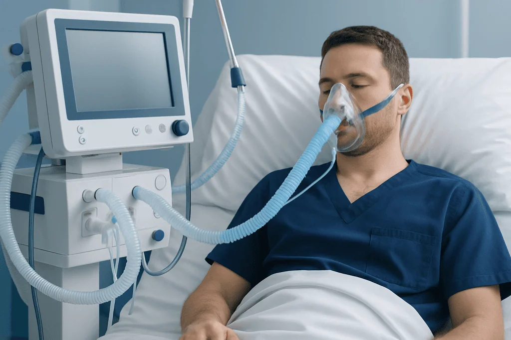
Healthy lungs work by moving oxygen through the alveoli into the bloodstream while removing carbon dioxide. When alveoli are filled with fluid or inflammatory material, gas exchange becomes less efficient. This can lead to shortness of breath, lower oxygen levels, and the sense that breathing requires more effort.
Not everyone experiences symptoms in the same way. A small area of consolidation may cause only mild cough or fatigue, while more extensive involvement can cause rapid breathing, chest discomfort, or marked breathlessness. People who are older, immunocompromised, or already have lung disease may become unwell more quickly.
During a physical exam, a clinician may hear crackles over the affected area or notice changes in how sound travels through the chest. These findings can suggest consolidation, but imaging is usually needed to confirm it. In many cases, a chest X-ray is the first test, and sometimes a thoracic CT scan helps define the pattern more clearly.
Symptoms and common patterns

The symptoms of lung consolidation depend on the cause, how much lung is involved, and how quickly it developed. Infection-related consolidation often causes fever, chills, cough, sputum production, chest pain that worsens with breathing, and shortness of breath. Other causes may produce a drier cough, wheezing, or symptoms linked to the underlying condition, such as leg swelling or trouble swallowing.
Some people notice reduced exercise tolerance, fatigue, or a general feeling of illness before more obvious breathing symptoms appear. In older adults, symptoms can sometimes be less typical and may include confusion, weakness, poor appetite, or worsening of an existing chronic illness. Children may breathe faster, appear less active, or have feeding difficulties.
- Cough, with or without mucus
- Shortness of breath or rapid breathing
- Fever or chills
- Chest discomfort, especially with deep breaths
- Fatigue or reduced stamina
- Low oxygen levels in more significant cases
Because these symptoms overlap with many chest conditions, consolidation is best understood as a clue rather than a diagnosis on its own. A person may have symptoms that resemble bronchitis, heart failure, aspiration, or lung cancer, so a careful assessment matters.
Causes and risk factors
The most common cause of lung consolidation is infection, particularly bacterial pneumonia. In pneumonia, the body’s immune response and the infection itself fill the alveoli with inflammatory fluid and cells. Viral infections can also cause dense areas in the lungs, though the imaging pattern may differ.
Another important cause is aspiration, when food, liquid, saliva, or stomach contents enter the airways. This may happen in people with swallowing problems, stroke, sedation, reflux, or reduced consciousness. Fluid overload or heart failure can also lead to dense lung areas, especially when pulmonary edema is present. Bleeding into the lungs, called alveolar hemorrhage, is less common but can also produce consolidation.
Some inflammatory and immune-mediated diseases can create persistent or recurrent consolidation. These include organizing pneumonia, eosinophilic lung disease, and certain autoimmune conditions. Rarely, a tumor can block an airway, leading to collapse and infection behind the blockage, which may appear as consolidation.
- Recent respiratory infection
- Smoking or chronic lung disease
- Older age or weakened immune system
- Swallowing difficulty or aspiration risk
- Heart disease or fluid retention states
- Recent hospitalization or immobility
Understanding risk factors helps guide the next steps in testing and treatment. For example, a person with sudden fever and cough may need evaluation for infection, while a person with recurrent right-sided consolidation and swallowing problems may need aspiration assessment.
How doctors diagnose lung consolidation
Diagnosis begins with a medical history and physical examination. Doctors ask about the onset of symptoms, fever, recent illness, smoking, reflux, travel, immune status, medication use, and any choking or swallowing difficulty. They also examine breathing rate, oxygen levels, chest sounds, and signs of infection or heart strain.
Imaging is central to diagnosis. A chest X-ray often shows the dense area and may suggest its location and extent. If the picture is unclear, symptoms are severe, or another cause is suspected, a CT scan provides more detail. Blood tests may look for inflammation, infection, anemia, or signs of organ stress. In some situations, sputum testing, blood cultures, or viral testing helps identify a specific infection.
When the cause remains uncertain or the consolidation does not clear as expected, doctors may expand the evaluation. This can include swallowing studies, heart evaluation, bronchoscopy, or targeted tests for autoimmune disease. If clinicians need a closer view of the airways or samples from the lungs, they may recommend bronchoscopy to help clarify the diagnosis.
Follow-up imaging is sometimes important, especially if symptoms persist, the person is at higher risk for an obstructing lesion, or the consolidation is unusually localized or recurrent. The purpose is not only to confirm improvement but also to make sure an underlying cause has not been missed.
Treatment options and recovery
Treatment for lung consolidation depends entirely on the cause. If the problem is bacterial pneumonia, antibiotics are often used along with rest, fluids, fever control, and close monitoring of breathing. If oxygen levels are low, oxygen therapy may be needed. Viral infections may be managed with supportive care unless a specific antiviral treatment is indicated.
When aspiration is involved, treatment may include antibiotics in selected cases, airway support, and addressing the reason aspiration occurred. This could mean swallowing assessment, diet adjustments, reflux management, or help with neurologic conditions. If fluid overload or heart failure is responsible, treatment focuses on removing excess fluid and improving heart function rather than treating infection.
More specialized causes need targeted therapy. Inflammatory lung disorders may require corticosteroids or other immune-modifying medicines under specialist supervision. If bleeding, airway obstruction, or an unusual mass is suspected, further procedures or referrals may be needed. In people with severe breathing difficulty, hospital-based supportive care can be essential while the cause is being treated.
Recovery time varies. Some people improve within days, while radiologic clearing can take longer, especially in older adults or those with chronic disease. Persistent symptoms should be reviewed rather than assumed to be part of normal healing. In advanced centers, multidisciplinary specialists, including at Acibadem International’s JCI-accredited hospitals, evaluate and treat respiratory conditions and may use tests such as pulmonary function testing during follow-up when appropriate.
Prevention, self-care, and when to seek medical care
Prevention depends on reducing the risk of the underlying causes. Staying up to date with recommended vaccinations, washing hands regularly, not smoking, and managing chronic illnesses can lower the risk of respiratory infections. People who have swallowing difficulties, reflux, or neurologic disease may benefit from strategies to reduce aspiration, such as posture changes during meals and specialist swallowing advice.
At home, supportive self-care can help recovery once a clinician has assessed the condition. Rest, good hydration, prescribed medicines taken exactly as directed, and monitoring symptoms are all important. However, self-care should not replace medical evaluation when consolidation is suspected, because some causes require prompt treatment and close observation.
Medical care should be sought promptly for shortness of breath, chest pain, bluish lips, confusion, high fever, coughing up blood, or symptoms that are rapidly worsening. People with weakened immune systems, chronic lung disease, heart disease, or older age should seek earlier review because they may become more unwell with fewer warning signs.
It is also sensible to contact a doctor if cough, fatigue, or fever does not improve, if symptoms return after partial recovery, or if follow-up imaging has been advised. Persistent or recurrent consolidation deserves reassessment so that uncommon causes are not overlooked.
Frequently asked questions
Is lung consolidation the same as pneumonia?
No. Lung consolidation is a descriptive finding that means part of the lung has become denser because the air spaces are filled with something other than air. Pneumonia is one common cause of consolidation, but not the only one.
Can lung consolidation go away on its own?
Sometimes mild causes improve as the underlying problem settles, but consolidation should not be assumed to be harmless. Because it may reflect infection, aspiration, fluid overload, bleeding, or inflammation, medical assessment is important to determine whether treatment is needed.
How is lung consolidation seen on imaging?
It often appears as a dense or white area on a chest X-ray or CT scan where air-filled lung should normally look darker. The exact pattern can give doctors clues about whether the cause is infection, fluid, inflammation, or another process.
Does lung consolidation always cause shortness of breath?
Not always. Small or localized areas may cause only cough, mild fatigue, or fever, and some people have few symptoms at first. Larger areas or disease in people with existing lung problems are more likely to cause breathlessness and low oxygen levels.
How long does it take for lung consolidation to clear?
Symptoms may improve in days to weeks, but the appearance on imaging can take longer to resolve. The timeline depends on the cause, the person’s age, overall health, and how much of the lung was affected.
When do doctors worry about persistent consolidation?
Doctors look more closely when consolidation does not improve as expected, keeps returning, or occurs with warning signs such as weight loss, coughing up blood, or repeated infections in the same area. In these situations, further imaging or procedures may be needed to rule out airway blockage, chronic inflammation, or other underlying conditions.
References
- American Thoracic Society
- Centers for Disease Control and Prevention
- National Heart, Lung, and Blood Institute
- Radiological Society of North America
- World Health Organization
This article is for general information only and is not a substitute for professional medical advice. Please consult a qualified doctor about your individual situation.
Explore treatments in Turkey — costs, top hospitals & a free quote
JCI-accredited · board-certified surgeons · reply within 24h
Add us as a Preferred Source to see more of our trusted health content across Google Search, AI Overviews and Discover.

More from the Health Library

Alirocumab — Explained by Medical Evidence, Not Myths

Buffalo Milk: An Evidence-Based Guide for Patients

Humectant — Explained by Medical Evidence, Not Myths

Indoor Rowing for Weight Loss: What Patients Need to Know

Healthy Fruits: An Evidence-Based Guide for Patients