Melanoma Stages and Treatment: How It Works, Results and What to Expect

Melanoma is staged from 0 to IV based on tumor thickness, ulceration and spread. Surgery is the main treatment for localized melanoma and may be curative when the cancer is caught early.
Key Takeaways
- Melanoma is staged from 0 to IV based on tumor thickness, ulceration and spread.
- Surgery is the main treatment for localized melanoma and may be curative when the cancer is caught early.
- A sentinel lymph node biopsy may help clarify the stage for selected thicker melanomas.
- Stage III and IV melanoma may be treated with immunotherapy, targeted therapy, surgery, radiation or clinical trials.
- A changing or new pigmented skin lesion should be assessed promptly by a qualified clinician.
- Follow-up after melanoma is important because new melanomas and recurrences can occur.
Melanoma stages and treatment are determined by how deeply the cancer has grown into the skin, whether it has ulcerated, and whether it has reached lymph nodes or distant organs. Many early melanomas can be treated successfully with surgery, while later-stage melanoma may require carefully planned combinations of surgery, immunotherapy, targeted therapy or radiation therapy.
Melanoma stages and treatment: an overview
Melanoma is a cancer that begins in melanocytes, the cells that make skin pigment. It can develop in an existing mole or as a new spot on the skin, and it can also occur in less common locations such as the eye, nail bed, mouth or genital area. Although melanoma is less common than several other skin cancers, it is more likely to spread if not diagnosed and treated early.
Melanoma stages and treatment are closely connected. Doctors use staging to describe the extent of disease and to guide discussions about treatment, follow-up and outlook. In general, stage 0 to stage II melanoma is confined to the skin, stage III involves nearby lymph nodes or nearby skin or tissue, and stage IV has spread to distant organs or distant areas of skin.
The stage is not based on appearance alone. Pathology results after a biopsy or surgery provide essential details, particularly the Breslow thickness, which measures how deeply the melanoma has grown. Ulceration, lymph node findings and imaging results may also affect the final stage and treatment plan.
How melanoma is staged
Staging usually follows the TNM system. The “T” category describes the primary tumor, including its thickness and whether ulceration is present. Ulceration means the skin over the melanoma has broken down when examined under the microscope; it can indicate a higher risk of spread than a melanoma of the same thickness without ulceration.
The “N” category records whether melanoma cells are found in regional lymph nodes or in small deposits in nearby skin or lymphatic channels. The “M” category describes spread to distant parts of the body, such as distant skin, lymph nodes, lungs, liver, brain, bone or other organs. Blood test findings may also contribute to assessment in advanced disease.
Stage 0, also called melanoma in situ, is limited to the top layer of skin. Stage I melanoma is thin and has not spread. Stage II melanoma is thicker and/or has ulceration but remains localized. Stage III means regional spread, most often to nearby lymph nodes. Stage IV indicates distant spread. These broad groups are useful, but each person’s situation requires individual interpretation by an experienced melanoma team.
- Stage 0: abnormal melanoma cells remain within the epidermis.
- Stages I-II: invasive melanoma is present in the skin but no regional or distant spread is identified.
- Stage III: melanoma has spread to regional lymph nodes or nearby tissue.
- Stage IV: melanoma has spread to distant sites.
How treatment works: choosing the right approach
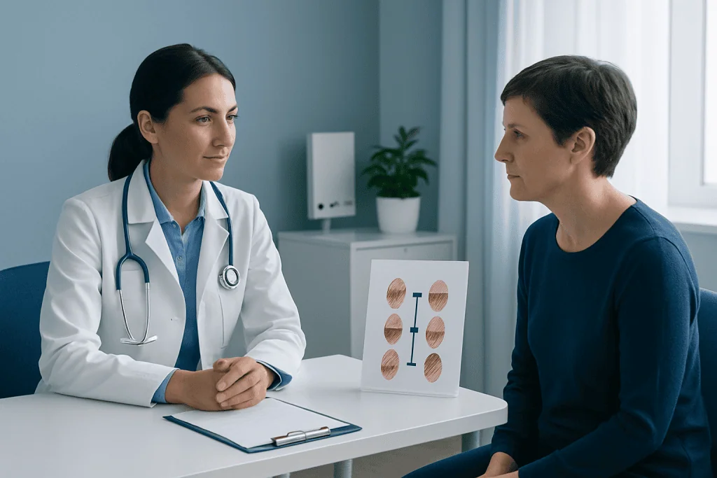
Treatment is tailored to the stage, tumor location, pathology findings, overall health and personal preferences. A dermatologist, surgical oncologist, medical oncologist, pathologist, radiologist and other specialists may work together to recommend a plan. The aim may be to remove all known cancer, reduce the risk of recurrence, control melanoma that has spread, relieve symptoms or combine these goals.
For localized melanoma, wide local excision is the usual first treatment. During this operation, the surgeon removes the melanoma site plus a measured margin of normal-looking skin around it. The margin size depends mainly on the thickness of the melanoma. The removed tissue is then examined to confirm that the edges are clear of cancer.
For some melanomas, particularly those with greater thickness or other higher-risk features, the team may recommend sentinel lymph node biopsy. This procedure identifies and removes the first lymph node or nodes likely to receive drainage from the tumor area. It can reveal microscopic spread that cannot be felt during an examination or seen on imaging.
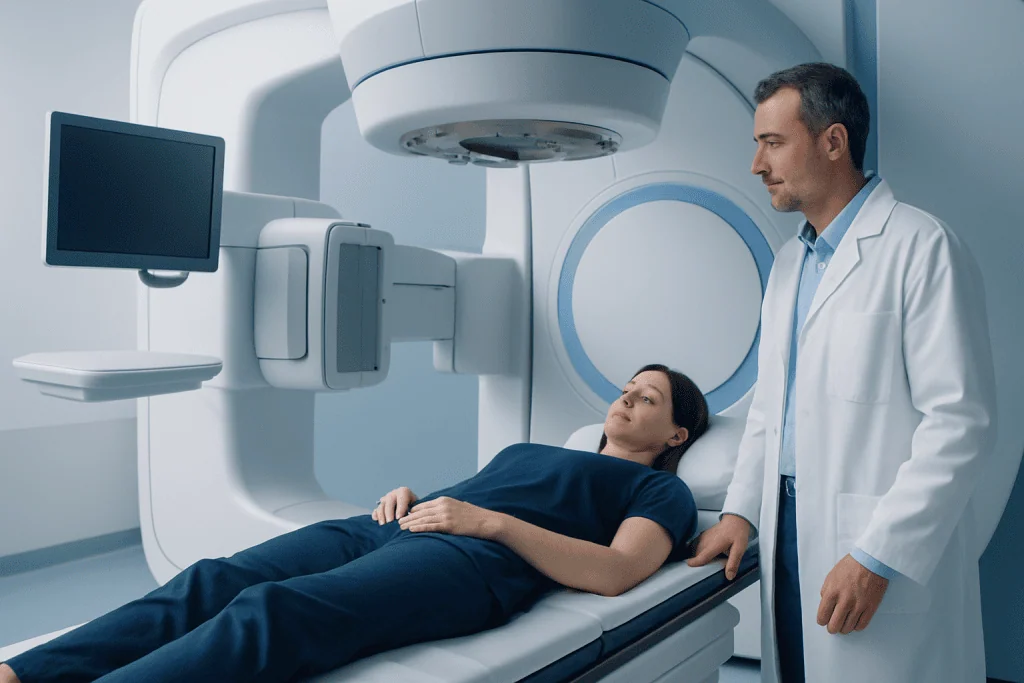
When melanoma has reached lymph nodes or distant sites, systemic treatments may be appropriate. Immunotherapy helps the immune system recognize and attack cancer cells. Targeted therapy may be used when tumor testing identifies particular genetic changes, commonly involving the BRAF gene. Radiation therapy can be useful in selected situations, such as treatment of a specific area at risk of recurrence or symptom control. Surgery may still have a role when a limited number of deposits can be safely removed.
Candidacy and the treatment process step by step
Anyone with a biopsy-confirmed melanoma should have a stage-appropriate assessment and treatment discussion. Before treatment, clinicians review the pathology report, examine the skin and lymph nodes, ask about personal and family history, and consider whether further procedures or scans are needed. Molecular testing is generally most relevant for advanced melanoma, where its results can help guide systemic treatment choices.
For surgery, the procedure commonly begins with marking the planned excision margin around the biopsy scar or remaining lesion. Local anesthetic is often sufficient for smaller excisions, while sedation or general anesthesia may be considered for larger operations, difficult locations or sentinel lymph node biopsy. The surgeon removes the tissue and closes the wound directly when possible; some areas require a skin graft or flap reconstruction.
A sentinel lymph node biopsy is performed using a tracer injected near the original melanoma site. The tracer helps the surgical team locate the sentinel node or nodes, which are removed through a small incision and reviewed by a pathologist. A negative result can be reassuring, although it does not eliminate the need for ongoing skin and lymph node checks.
Systemic treatment is usually delivered in repeated cycles under oncology supervision. Before and during treatment, blood tests, symptom reviews and imaging may be used to monitor response and side effects. Treatment decisions should be revisited over time, because melanoma biology, treatment response and a person’s priorities can change.
Recovery, benefits and possible risks
Recovery after a simple excision is often measured in days to a few weeks, depending on the wound size and location. Tenderness, bruising, swelling and a scar are expected. The care team provides individualized instructions about dressing changes, bathing, activity, pain relief and when stitches should be removed. Areas over joints, on the face, hands, feet or lower legs may need additional attention during healing.
The main benefit of surgery for early melanoma is that it can remove the cancer completely. Sentinel lymph node biopsy can provide more accurate staging and help identify people who may benefit from additional treatment or closer follow-up. However, it also has possible risks, including bleeding, infection, delayed healing, numbness, fluid collection and, less commonly, swelling of a limb caused by lymphatic disruption.
Immunotherapy and targeted therapy have improved options for many people with higher-risk or advanced melanoma, but they can cause side effects. Immunotherapy can lead to inflammation in organs such as the skin, bowel, lungs, liver, thyroid or other hormone-producing glands. Targeted therapies can cause fever, rash, fatigue, joint symptoms and other effects. Prompt reporting of new symptoms allows the oncology team to assess and manage side effects early.
Results vary substantially by stage and individual tumor features. No treatment can guarantee that melanoma will not return, but regular follow-up supports early detection of recurrence, treatment effects and new primary skin cancers. A personalized surveillance schedule is an important part of care.
How long does it take for melanoma to go from stage 1 to 4?
There is no reliable fixed timeline for melanoma to progress from stage I to stage IV. Some melanomas grow slowly over years, while others may grow or spread more quickly. Growth rate depends on the melanoma subtype, depth, genetic features, immune response and other biological factors that cannot be predicted precisely from a skin lesion alone.
Stage I does not automatically progress to stage IV. Many stage I melanomas are removed before they spread, and appropriate treatment and follow-up can substantially reduce risk. Because it is not possible to know how an individual untreated melanoma will behave, a suspicious changing lesion should be examined without delay rather than watched for a set period.
People previously treated for melanoma should attend their scheduled reviews and perform regular skin self-checks as advised. New symptoms, a changing mole, a firm new lump or unexplained persistent symptoms should be discussed with a healthcare professional.
What is the 2 week rule for melanoma?
The “2 week rule” is not a formal medical rule that can diagnose melanoma or determine whether a lesion is safe. It is often used informally to encourage prompt assessment when a new or changing skin mark does not settle, especially if it changes over a short period. A suspicious lesion should not be monitored at home for two weeks if it is clearly evolving, bleeding or otherwise concerning.
A practical approach is to seek medical review promptly for a spot that is new and changing, differs noticeably from other moles, or shows ABCDE warning signs: asymmetry, irregular border, varied color, increasing diameter or evolving appearance. Melanoma may also be pink, red, skin-colored or dark, so color alone is not enough to rule it out.
Photographing a lesion with the date can help document change, but photographs should not replace a clinical examination. A dermatologist may examine the lesion with dermoscopy and recommend a biopsy if melanoma is possible.
When to seek medical care
Medical assessment is recommended promptly for a mole or spot that is changing in size, shape, color, height or sensation. Other reasons to arrange review include persistent itching, bleeding, crusting, a sore that does not heal, a new dark streak under a nail, or a spot that looks unlike a person’s other marks. Most skin changes are not melanoma, but assessment is the safest way to identify lesions that need treatment.
People with a previous melanoma, many moles, a strong family history of melanoma, fair skin that burns easily, substantial ultraviolet exposure or a weakened immune system may need more regular professional skin examinations. Sun protection remains valuable for everyone: seek shade when practical, wear protective clothing and broad-spectrum sunscreen, and avoid tanning beds.
For people diagnosed with melanoma, urgent contact with the treating team is appropriate for concerning treatment side effects, such as severe or persistent diarrhea, shortness of breath, chest pain, high fever, confusion, severe headache, jaundice or rapidly worsening weakness. The care team can advise whether same-day assessment is needed.
Acibadem International’s multidisciplinary specialists and JCI-accredited hospitals diagnose and treat melanoma for international patients, with care plans based on pathology, staging and individual needs.
Frequently asked questions
Is stage 1 melanoma a big deal?
Stage I melanoma is an invasive skin cancer, so it should always be taken seriously and treated appropriately. The encouraging point is that it is usually localized and can often be treated with surgery. Ongoing follow-up is still important because a person can develop another melanoma or, less commonly, experience recurrence.
What is the average life expectancy after being diagnosed with melanoma?
There is no single average life expectancy that applies to everyone with melanoma. Outlook depends mainly on the stage at diagnosis, tumor thickness, ulceration, lymph node involvement, response to treatment, general health and other individual factors. Early melanoma generally has a much more favorable outlook than melanoma that has spread, and a treating specialist can provide the most meaningful personalized information.
Can melanoma be cured with surgery?
Surgery can be curative for many melanomas that are confined to the skin. The melanoma and a margin of surrounding skin are removed, and pathology helps confirm whether the excision was complete. Some people with higher-risk features need additional staging, treatment or closer surveillance after surgery.
Why might a sentinel lymph node biopsy be recommended?
A sentinel lymph node biopsy may be recommended when the melanoma has features associated with a greater possibility of microscopic lymph node spread. It identifies the first draining lymph node or nodes and allows them to be tested for melanoma cells. The result can refine staging and help guide discussions about follow-up and additional treatment.
Can melanoma return after treatment?
Yes, melanoma can recur after treatment, although recurrence risk varies widely between individuals. It may recur near the original site, in regional lymph nodes or elsewhere, and a new unrelated melanoma can also develop. Regular skin checks, follow-up visits and attention to new or changing lesions are therefore important.
Does every melanoma need immunotherapy?
No. Early-stage melanoma is commonly treated with surgery alone. Immunotherapy is more often considered for some stage III melanomas, stage IV melanoma, or selected higher-risk situations, depending on the pathology findings and the person’s overall circumstances. An oncology team weighs expected benefit against possible side effects before recommending it.
References
- American Cancer Society
- National Cancer Institute
- American Academy of Dermatology
- European Society for Medical Oncology
- National Comprehensive Cancer Network
This article is for general information only and is not a substitute for professional medical advice. Please consult a qualified doctor about your individual situation.
Melanoma in Turkey — costs, top hospitals & a free quote
JCI-accredited · board-certified surgeons · reply within 24h
Add us as a Preferred Source to see more of our trusted health content across Google Search, AI Overviews and Discover.

Medical Oncology Department
Medical treatment of cancer with chemotherapy, immunotherapy and targeted therapies under a multidisciplinary tumor board.
60 specialists in this unit