MR Angiography vs CT Angiography: Which Brain Vessel Scan Is Right?
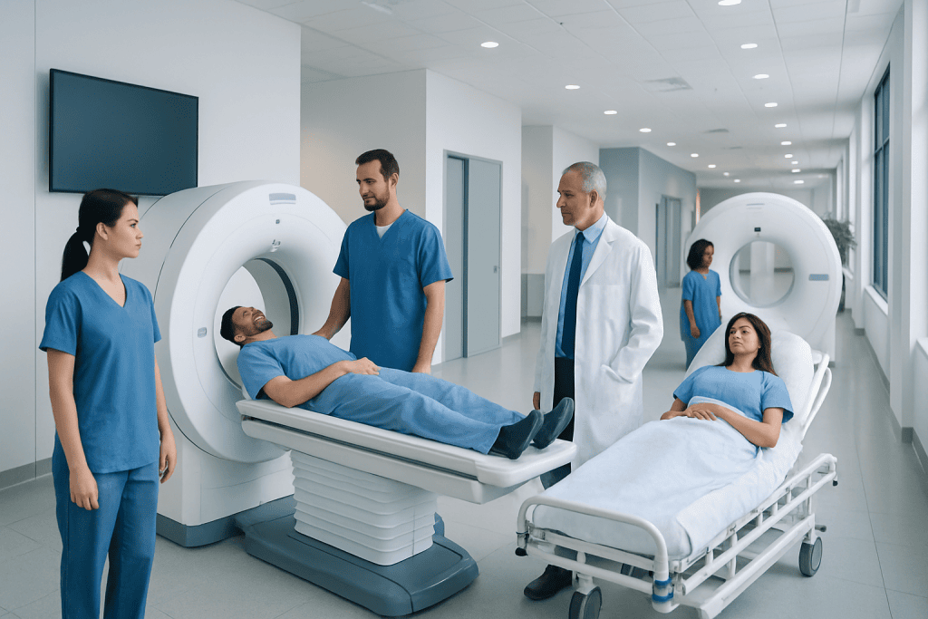
MRA and CTA both help doctors evaluate arteries and veins in and around the brain. CTA is usually faster and widely used in emergencies such as suspected stroke or bleeding.
Key Takeaways
- MRA and CTA both help doctors evaluate arteries and veins in and around the brain.
- CTA is usually faster and widely used in emergencies such as suspected stroke or bleeding.
- MRA avoids ionizing radiation and may be preferred when repeated imaging is needed.
- The best test depends on symptoms, medical history, contrast safety, kidney function, and the detail needed.
- A radiologist and treating doctor choose the most suitable scan based on the individual situation.
MR angiography (MRA) and CT angiography (CTA) are common imaging tests used to look at the brain’s blood vessels. Each has advantages, and the right choice depends on the clinical question, urgency, kidney function, contrast safety, and whether radiation should be avoided.
Overview: What MRA and CTA Show
MR angiography, often called MRA, and CT angiography, called CTA, are imaging methods that create detailed pictures of blood vessels. When used for the brain, they help doctors assess the arteries and veins supplying the brain and surrounding structures. These scans may be requested to investigate symptoms such as sudden severe headache, stroke-like symptoms, dizziness, visual changes, or concerns about an aneurysm or narrowing of an artery.
Although both tests aim to answer similar questions, they work in different ways. CTA uses computed tomography together with an iodine-based contrast dye to outline blood vessels. MRA uses magnetic resonance imaging and may be performed either without contrast or with a gadolinium-based contrast agent, depending on what needs to be seen.
In simple terms, CTA is often valued for speed and broad availability, especially in urgent care. MRA is often valued because it does not use ionizing radiation and can sometimes provide vessel images without any contrast injection at all. Neither test is universally “better”; each is useful in the right clinical setting.
How the Two Scans Differ
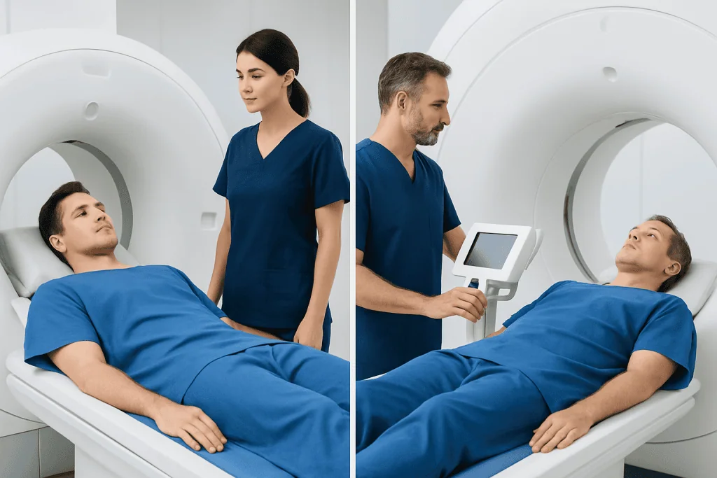
The most important difference is the technology. CTA uses X-rays, so it involves a small amount of ionizing radiation. It is usually completed very quickly, often within minutes, which is one reason it is commonly used in emergency departments. MRA uses a strong magnet and radio waves instead of radiation, but it generally takes longer and requires the patient to stay still in the scanner for a longer period.
Another key difference is contrast material. CTA almost always requires an iodine-based contrast injection through a vein. MRA may be done without contrast for many vessel studies, though contrast-enhanced MRA can improve detail in selected cases. This matters for people with prior contrast reactions, kidney disease, pregnancy-related concerns, or a need for repeated imaging over time.
The experience of the scan also differs. CT scanners are usually more open and may feel easier for people who are uncomfortable in enclosed spaces. MRI scanners are noisier and can feel more confining, which can be challenging for people with claustrophobia, severe anxiety, or difficulty lying still. Metal implants, certain medical devices, and fragments of metal in the body may also affect whether MRI-based testing is safe or practical.
When Doctors May Prefer CTA

CTA is frequently chosen when speed is essential. In a suspected stroke, transient ischemic attack, or sudden bleeding in or around the brain, doctors often need vessel imaging right away to guide urgent treatment decisions. CTA can quickly show major artery blockages, vessel narrowing, aneurysms, dissections, and some vascular malformations. It is commonly paired with a standard head CT in emergency care.
CTA can also be very helpful when detailed views of the vessel wall and nearby bone structures are important. For example, it may assist in evaluating trauma, skull base anatomy, or calcified atherosclerosis. In some cases, CTA gives especially crisp pictures of smaller vessels and is useful in pre-procedure planning.
Doctors may recommend CTA if MRI is not possible. This can happen when a person has an incompatible implanted device, cannot tolerate the MRI environment, or needs a scan that is faster and easier to complete. CTA is also widely available, which can make access simpler in urgent settings and smaller hospitals.
Even so, CTA is not the ideal option for everyone. Because it uses radiation and iodine contrast, doctors weigh the benefits carefully in people who are pregnant, have significantly reduced kidney function, or have had a previous serious contrast reaction.
When Doctors May Prefer MRA
MRA may be preferred when avoiding radiation is important. This can be especially relevant for younger patients, people who need repeated follow-up imaging, and some pregnant patients when imaging cannot be delayed and MRI is considered appropriate. Because some MRA techniques do not require contrast, MRA can also be useful when doctors want to reduce contrast exposure.
MRA is often used for planned, non-emergency evaluation of aneurysms, vessel narrowing, congenital vascular differences, and follow-up after treatment. It may also be part of a broader MRI brain examination when doctors want to assess both brain tissue and blood vessels in one session. In some situations, MRA can be combined with MRI to give a more complete picture of neurological symptoms.
For people with a history of iodine contrast allergy, MRA can sometimes offer an alternative, although gadolinium-based contrast may still be used in selected studies. If contrast is needed, kidney function and overall safety still matter. A medical team will decide whether non-contrast MRA, contrast-enhanced MRA, CTA, or another test is most suitable.
MRA is not always the first choice in emergencies because it usually takes longer and may be less practical for unstable patients. Still, in carefully selected cases, it provides excellent information without exposing the patient to radiation.
What Conditions These Scans Can Help Diagnose
Both MRA and CTA can help detect a range of cerebrovascular problems. Common examples include aneurysms, arterial narrowing due to atherosclerosis, vessel occlusion, arterial dissection, and some vascular malformations. They may also be used when doctors suspect reduced blood flow to part of the brain or want to investigate symptoms such as repeated transient neurological episodes.
In suspected stroke, CTA is often used early because it can rapidly identify large vessel blockages that may need urgent treatment. It may be part of a pathway that leads to interventional neuroradiology treatment in selected patients. MRA can also evaluate stroke-related vessel changes, particularly in a more stable or follow-up setting.
For aneurysm assessment, both tests can be valuable. CTA may be favored when very quick answers are needed or when there is concern for bleeding. MRA is often useful for surveillance over time, especially when doctors want to limit radiation exposure. If a vascular condition is confirmed, imaging findings may be discussed together with related conditions such as brain aneurysm.
These scans may also support evaluation of disorders affecting blood flow outside the brain itself, including arteries in the neck. In some cases, doctors compare MRA or CTA results with ultrasound, standard MRI, or conventional catheter angiography for the clearest diagnosis.
How Doctors Decide Which Scan Is Right
The choice between MRA and CTA is based on the clinical question first. If the main issue is urgent diagnosis in a time-sensitive situation, CTA is often favored because of its speed. If the concern is long-term monitoring or reducing radiation exposure, MRA may be more attractive. The exact area of interest, the size of vessels being assessed, and whether nearby brain tissue also needs evaluation all influence the decision.
Medical history is equally important. Doctors consider kidney function, pregnancy, known allergies to contrast agents, implanted devices, and whether the patient can lie flat and still. They also review prior scans and any previous vascular procedures. A person who cannot safely undergo MRI may be directed toward CTA, while someone who should avoid radiation or iodine contrast may be steered toward MRA if feasible.
Image quality can depend on the patient as well as the technology. Movement during the scan can reduce clarity, especially during MRI. Fast or irregular blood flow patterns may affect how vessels appear on MRA, while dense calcium or metal artifacts may influence CTA interpretation. Because of these trade-offs, the “best” scan is the one most likely to answer the doctor’s question safely and clearly for that individual.
Sometimes the answer is not one or the other. A patient may start with CTA in the emergency setting and later have MRA for follow-up. In more complex cases, specialists may recommend neuroradiology evaluation or even catheter angiography if noninvasive imaging does not fully explain the problem.
Preparing for the Scan and What to Expect
Preparation is usually simple. Patients may be asked about kidney disease, diabetes medications, allergies, pregnancy, and implanted devices. Before MRI-based testing, the team will ask detailed questions about pacemakers, clips, hearing implants, metal fragments, and other hardware. Jewelry and metallic items generally need to be removed before MRI.
For CTA, an intravenous line is typically placed so iodine contrast can be injected during the scan. The scan itself is brief, and some people notice a warm sensation or metallic taste for a short time after contrast injection. For MRA, the test may take longer, and patients hear loud tapping or thumping sounds from the machine. Ear protection is usually provided.
It is helpful to stay as still as possible for either study, because movement can blur the images. People who feel anxious in enclosed spaces should tell the care team in advance. Children and some adults may need additional support or tailored planning to complete the test comfortably.
After the scan, most people can return to normal activities unless their doctor gives different instructions. Results are reviewed by a radiologist or neuroradiologist and then discussed with the treating doctor, who explains what the images mean and whether any further testing or treatment is needed.
Risks, Limitations, and When to Seek Medical Advice
Both MRA and CTA are considered routine and generally safe when appropriately selected, but each has limitations. CTA exposes the patient to radiation and uses iodine contrast, which can cause allergic reactions in some people and may not be suitable for certain patients with reduced kidney function. MRA avoids radiation, but MRI may not be possible for everyone and can be difficult for those with claustrophobia or incompatible devices.
Neither test replaces a medical assessment. A normal scan does not always rule out every cause of symptoms, and sometimes findings need confirmation with another imaging study. In certain complex vascular disorders, catheter angiography may still be needed because it can provide very high-resolution images and may allow treatment during the same procedure.
People should seek prompt medical attention for sudden weakness, facial drooping, speech difficulty, loss of vision, sudden severe headache, confusion, seizure, or sudden loss of balance. These symptoms can signal a medical emergency, and urgent imaging may be necessary. For non-urgent questions about which scan is appropriate, a neurologist, neurosurgeon, or neuroradiology team can help guide the decision.
At the end of the diagnostic pathway, care is often multidisciplinary. Acibadem International’s specialists in neurology, neurosurgery, and neuroradiology at JCI-accredited hospitals evaluate and treat international patients who need advanced brain vessel imaging and follow-up care.
Frequently asked questions
Which is better for brain blood vessels, MRA or CTA?
Neither test is always better in every situation. CTA is often preferred when speed is critical, while MRA may be preferred when avoiding radiation or repeated contrast exposure is important. The best choice depends on the reason for the scan and the patient’s medical history.
Does MRA use radiation?
No. MRA uses a magnetic field and radio waves rather than X-rays, so it does not expose the patient to ionizing radiation. This is one reason it may be chosen for follow-up imaging or in selected patients who should minimize radiation exposure.
Why is CTA often used in emergencies?
CTA is very fast and widely available, which makes it useful in urgent situations such as suspected stroke or sudden bleeding. It can quickly show major blockages, narrowed arteries, aneurysms, and other important vessel problems that may need immediate treatment decisions.
Can MRA be done without contrast dye?
Yes. Many MRA studies can be performed without contrast, depending on which blood vessels need to be evaluated and how much detail is required. In some cases, however, contrast-enhanced MRA provides clearer or more complete information.
Is CTA safe for people with kidney problems?
CTA uses iodine-based contrast, so doctors review kidney function before the scan. Some people with reduced kidney function may still be able to have CTA, but the decision is individualized. In certain cases, MRA without contrast or another imaging test may be considered instead.
What if a patient is claustrophobic?
CTA is often easier for people with claustrophobia because the scanner is quicker and usually feels less enclosed. MRI-based scans can be more challenging, but the care team can often offer support, planning, and strategies to make the test more comfortable.
References
- American College of Radiology
- Radiological Society of North America
- National Institute of Neurological Disorders and Stroke
- European Society of Radiology
- World Health Organization
This article is for general information only and is not a substitute for professional medical advice. Please consult a qualified doctor about your individual situation.
Explore treatments in Turkey — costs, top hospitals & a free quote
JCI-accredited · board-certified surgeons · reply within 24h
Add us as a Preferred Source to see more of our trusted health content across Google Search, AI Overviews and Discover.