MRI Advantages Over CT: An Evidence-Based Guide for Patients

MRI usually provides more detailed images of the brain, spinal cord, joints, ligaments, and many internal organs. MRI does not use ionizing radiation, which can matter when repeat imaging is needed.
Key Takeaways
- MRI usually provides more detailed images of the brain, spinal cord, joints, ligaments, and many internal organs.
- MRI does not use ionizing radiation, which can matter when repeat imaging is needed.
- CT is often faster, more available, and especially useful for trauma, bone injury, and some emergencies.
- The best scan is the one that answers the medical question safely and quickly.
- People with certain implants, severe claustrophobia, or kidney concerns may need special planning before MRI or contrast imaging.
MRI advantages over CT mainly involve clearer views of soft tissues and no exposure to ionizing radiation. However, CT can still be the better choice in some urgent situations, so the right test depends on the body area, the clinical question, and a doctor’s judgment.
Overview: Is MRI Better Than CT?
When patients ask about mri advantages over ct, the short answer is that MRI is often better for seeing soft tissues in detail and it does not use ionizing radiation. CT, however, is usually faster and may be more helpful in emergencies, especially when doctors need quick answers about bleeding, fractures, or certain chest and abdominal problems.
Rather than thinking of one scan as universally superior, it is more accurate to see MRI and CT as different tools. Each creates images in a different way and each has strengths, limits, and safety considerations. A doctor chooses between them based on symptoms, the part of the body being examined, how urgent the situation is, and whether contrast material is needed.
MRI uses a strong magnetic field and radio waves to create detailed images. CT uses X-rays and computer processing to produce cross-sectional images of the body. Because they work differently, they are not interchangeable in every case.
For many non-emergency problems involving the brain, spine, muscles, ligaments, and some organs, MRI may provide the most useful information. For major injuries, suspected internal bleeding, and many urgent evaluations, CT may remain the first choice because of speed and practicality.
How MRI and CT Differ in Practice
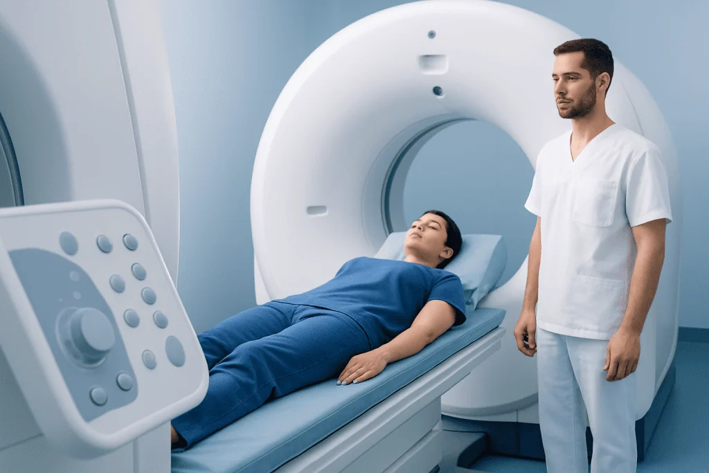
The main practical difference is image type and acquisition speed. CT scans are typically completed within minutes and are widely used in emergency departments because they quickly show major injuries, bleeding, lung problems, kidney stones, and many bone conditions. MRI scans usually take longer, and the patient must remain still in a magnet during the study.
MRI tends to separate soft tissues more clearly than CT. This means structures such as the brain, spinal cord, discs, tendons, cartilage, pelvic organs, liver tissue in selected cases, and some tumors can be assessed with greater contrast and definition. CT, by contrast, is excellent for bone detail and is often preferred when calcifications, fractures, or acute trauma are suspected.
Another major difference is radiation. CT uses ionizing radiation, while MRI does not. This does not mean CT is unsafe when appropriately used, but it is one reason MRI may be preferred when repeat imaging is expected or when avoiding radiation is especially desirable.
- MRI strengths: soft-tissue detail, multiplanar imaging, no ionizing radiation
- CT strengths: speed, broad availability, excellent bone imaging, emergency use
- Shared role: both may use contrast agents to improve diagnostic accuracy in selected situations
Key MRI Advantages Over CT

The most important MRI advantage is superior soft-tissue contrast. In simple terms, MRI can often distinguish between nearby soft tissues more clearly than CT. This helps doctors evaluate problems such as disc disease, ligament tears, brain abnormalities, some liver lesions, joint injuries, and suspected nerve compression with more confidence.
A second major advantage is the lack of ionizing radiation. This can be especially relevant for younger patients, for those who may need several scans over time, and for conditions that require ongoing follow-up. Avoiding radiation does not automatically make MRI the right test, but it can be a meaningful factor in choosing the most suitable imaging method.
MRI also allows excellent imaging in multiple planes without repositioning the patient in the same way CT may require. This can improve assessment of complex anatomy such as the spine, pelvis, and joints. In conditions involving the brain and nervous system, MRI is often central to evaluating concerns such as Parkinson’s disease, multiple sclerosis, stroke patterns after initial emergency care, or unexplained neurological symptoms.
Another advantage is tissue characterization. MRI can help distinguish inflammation, scarring, edema, dead tissue, and some tumor features more effectively than CT. For patients undergoing advanced evaluation, doctors may recommend MRI imaging as the most informative next step when the main question involves soft tissue rather than bone or urgent trauma.
When CT May Still Be the Better Choice
CT is often preferred when time matters most. In trauma, suspected internal bleeding, severe chest pain, shortness of breath, or concern for a life-threatening emergency, CT can frequently deliver fast, reliable information that helps doctors make immediate decisions. In these situations, speed can be as important as image detail.
CT is also highly effective for evaluating bone injuries, lung disease, sinus disease, kidney stones, and many abdominal emergencies. A patient with a suspected fracture may benefit more from CT than MRI, especially if the goal is to define bone anatomy quickly and clearly. CT angiography can also be very useful in assessing blood vessels in urgent settings.
Some patients are not ideal candidates for MRI. Certain implanted devices, metallic fragments in the body, or severe claustrophobia may limit MRI use, although modern MRI safety screening and newer compatible devices have expanded access. CT may be the more practical choice when MRI is unsafe, not available promptly, or unlikely to change treatment decisions.
In some care pathways, the two tests complement one another rather than compete. For example, a CT scan may first identify an urgent problem, and MRI may then add detail about tissue injury, inflammation, or recovery planning. This is common in neurological and musculoskeletal medicine, including situations where brain surgery or neurosurgical treatment is being considered and precise preoperative imaging is needed.
Conditions Where MRI Often Has an Edge
MRI is commonly favored for problems involving the brain and spine. It is often more sensitive than CT for many tumors, demyelinating disease, spinal cord compression, disc disease, ligament injury, and subtle brain changes. In a patient with persistent headaches, numbness, seizures, or chronic back pain, MRI may answer questions that CT cannot fully resolve.
Joints and sports injuries are another area where MRI often stands out. It can show cartilage, tendons, ligaments, menisci, marrow edema, and muscle injury in ways that CT generally cannot match. Patients being assessed for chronic knee pain, shoulder instability, or suspected soft-tissue tears may therefore be directed toward MRI rather than CT.
In abdominal and pelvic imaging, the choice is more nuanced. CT remains essential for many acute conditions, but MRI may offer better characterization of liver lesions, pelvic organs, rectal disease, and some inflammatory conditions. It can also be useful when doctors want more clarity after an earlier CT or ultrasound result.
MRI can also support treatment planning and follow-up in cancer care, complex neurological disorders, and selected cardiovascular conditions. In some situations, specialists may use MRI after initial evaluation of stroke to better define the affected tissue and guide recovery planning, though emergency stroke imaging often begins with CT.
Safety, Preparation, and Contrast Considerations
Both MRI and CT are generally safe when used appropriately, but each has important precautions. For MRI, the magnetic field means patients must tell the imaging team about pacemakers, aneurysm clips, cochlear implants, metal fragments, insulin pumps, or prior surgeries. Many implants are now MRI-conditional, but formal safety screening is still essential.
CT involves radiation exposure, so doctors weigh the expected benefit against the need for imaging. In modern practice, imaging teams aim to use the lowest dose needed for diagnostic quality. MRI avoids radiation, but some patients find the enclosed space uncomfortable, and scan times are usually longer.
Both tests may involve contrast material. CT contrast is iodine-based, while MRI contrast is often gadolinium-based. Contrast can improve the visibility of tumors, inflammation, blood vessels, and organ detail, but it may not be suitable for everyone. Kidney function, allergy history, and the exact clinical question all influence whether contrast is used.
Practical preparation varies by scan type. Patients may be asked to avoid food for a short time, remove metal items, change into a gown, or arrive early for screening. If anxiety or claustrophobia is a concern, it is helpful to mention this in advance so the care team can discuss supportive options.
How Doctors Decide Which Scan to Order
The most useful question is not simply “Which is better?” but “Which test best answers this medical question?” Doctors consider symptoms, examination findings, urgency, age, prior imaging, medical history, implants, pregnancy status, and whether surgery or another procedure may follow. Imaging is most effective when tailored to a specific concern.
For example, sudden major trauma may call for CT because rapid detection of bleeding or fractures is critical. Progressive weakness, chronic joint pain, or suspected spinal cord problems may point toward MRI because soft-tissue and nerve detail matter most. If one test is inconclusive, the other may be recommended as a second step.
Radiologists and referring specialists often make these decisions together. Their aim is to avoid unnecessary scans while choosing the imaging method most likely to provide a clear diagnosis. This is why two people with similar pain in different clinical contexts may be advised to have different tests.
Near the end of a patient’s care pathway, imaging may also guide treatment planning. At Acibadem International, multidisciplinary specialists in JCI-accredited hospitals evaluate international patients and use advanced imaging to help diagnose and treat complex conditions in a coordinated way.
When to Seek Medical Care
Medical attention is important when symptoms are sudden, severe, or worsening. Patients should seek urgent care for signs such as sudden weakness, trouble speaking, severe head injury, chest pain, severe shortness of breath, seizures, or severe abdominal pain. In these cases, the choice of imaging is often part of emergency assessment and should not be delayed.
Non-urgent medical review is also appropriate when symptoms persist without a clear explanation. Ongoing headaches, back pain with numbness, joint instability, unexplained neurological symptoms, or recurrent abdominal complaints may all need evaluation. A clinician can decide whether MRI, CT, ultrasound, blood tests, or another approach is most appropriate.
It is also wise to contact a doctor before imaging if there is pregnancy, kidney disease, an implanted device, or a previous reaction to contrast material. These issues do not always prevent scanning, but they may change the safest and most informative test.
Patients should avoid self-ordering assumptions based on general online comparisons. MRI advantages over CT are real, but the best imaging decision is individualized and should be made with a qualified healthcare professional.
Frequently asked questions
What are the main MRI advantages over CT?
MRI’s main advantages are better soft-tissue detail and the fact that it does not use ionizing radiation. This often makes it especially useful for the brain, spine, joints, muscles, ligaments, and some internal organs.
Is MRI always safer than CT?
Not always in every situation. MRI avoids radiation, but it may not be suitable for some people with certain implants, metal fragments, or severe claustrophobia, and contrast use may require extra caution in some patients.
Why would a doctor order a CT instead of an MRI?
CT is often faster and is widely used in emergencies. It is especially helpful for trauma, suspected bleeding, many lung and abdominal emergencies, fractures, and kidney stones.
Does MRI show more than CT for back and joint problems?
In many cases, yes. MRI usually gives more detailed information about discs, nerves, ligaments, cartilage, tendons, and muscle injuries, which is why it is commonly preferred for chronic spine and joint evaluations.
Do MRI and CT both use contrast dye?
Both can use contrast agents, but they are different types. CT generally uses iodine-based contrast, while MRI often uses gadolinium-based contrast, and the decision depends on the clinical need and the patient’s medical history.
Which scan is better for cancer detection?
Neither test is best for every cancer-related question. MRI may better characterize some soft-tissue tumors and help with treatment planning, while CT is often used for rapid assessment, staging in certain cancers, and evaluating the chest or abdomen.
This article is for general information only and is not a substitute for professional medical advice. Please consult a qualified doctor about your individual situation.
Explore treatments in Turkey — costs, top hospitals & a free quote
JCI-accredited · board-certified surgeons · reply within 24h
Add us as a Preferred Source to see more of our trusted health content across Google Search, AI Overviews and Discover.

More from the Health Library

Lingam Massage: A Complete Medical Overview

Omphalocele — Explained by Medical Evidence, Not Myths

Esophageal Perforation: An Evidence-Based Guide for Patients

Tummy Rumbling After Eating: An Evidence-Based Guide for Patients

Blood Alcohol Level: What Patients Need to Know