MRI brain with and without contrast cpt: Symptoms, Causes, and Treatment — Complete Patient Guide
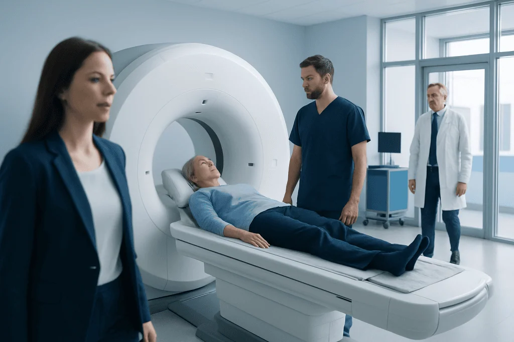
A brain MRI with and without contrast includes images taken before and after contrast is injected. The CPT wording relates to billing and documentation, not to a different patient experience.
Key Takeaways
- A brain MRI with and without contrast includes images taken before and after contrast is injected.
- The CPT wording relates to billing and documentation, not to a different patient experience.
- Doctors may order this study to evaluate tumors, inflammation, infection, stroke, multiple sclerosis, or unexplained neurological symptoms.
- MRI does not use radiation, but contrast is not needed for every brain scan.
- Patients should tell the care team about kidney disease, allergies, implants, claustrophobia, or pregnancy before the scan.
MRI brain with and without contrast CPT refers to the medical coding used for a brain MRI performed both before and after contrast material is given. For patients, the more important point is that this type of scan helps doctors see brain structures in greater detail when evaluating symptoms such as headaches, seizures, weakness, or suspected neurological disease.
Overview: what “MRI brain with and without contrast CPT” means
MRI brain with and without contrast CPT is a phrase patients often see on an order, insurance document, or radiology report request. “MRI brain” means magnetic resonance imaging of the brain. “With and without contrast” means images are taken first without contrast material and then again after a contrast agent is given through a vein, so the radiologist can compare both sets of images.
The term “CPT” stands for Current Procedural Terminology, a coding system used for medical billing and documentation. It helps hospitals, clinics, and insurers identify which imaging study was performed. For most patients, the key question is not the code itself, but why the scan was ordered and what it may help doctors detect.
A brain MRI gives highly detailed images of the brain, surrounding tissues, and blood vessel-related structures. Adding contrast can make certain abnormalities easier to see, including inflammation, tumors, infection, or active disease changes. In some situations, a doctor may order a scan without contrast only; in others, both parts are important to answer a specific clinical question.
Why a doctor may order this type of brain MRI
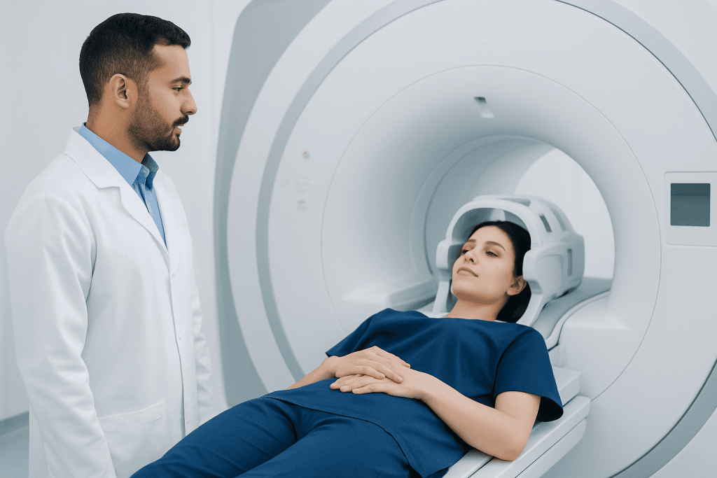
Doctors use a brain MRI with and without contrast when they need more detailed information than a physical exam alone can provide. It may be ordered for ongoing headaches with concerning features, seizures, weakness on one side of the body, changes in speech, memory problems, balance issues, or unexplained visual symptoms. It is also commonly used when there is concern for infection, inflammation, bleeding, or a mass.
Contrast helps highlight areas where the blood-brain barrier is disrupted or where there is unusual blood flow. This can be useful when evaluating conditions such as brain tumors, abscesses, inflammatory disorders, and some vascular problems. It can also help in follow-up imaging when a known condition needs to be monitored over time.
In clinical practice, this scan may be part of the evaluation of suspected brain tumor, demyelinating disease such as multiple sclerosis, or complications related to stroke. The exact reason for using contrast depends on the person’s symptoms, medical history, and what the doctor needs to confirm or rule out.
Symptoms and situations that may lead to the test
A brain MRI with and without contrast is not a disease itself, so it does not cause symptoms. Instead, it is ordered because a person has symptoms or signs that need evaluation. Common reasons include persistent or worsening headaches, new seizures, numbness, weakness, dizziness, fainting, personality or memory changes, and difficulty with coordination or walking.
It may also be recommended after head injury when symptoms do not fully improve, or when symptoms raise concern for a structural brain problem. In some people, MRI is used after an abnormal CT scan, or when CT findings need clarification. It can also be used to monitor a known neurological condition, check whether treatment is working, or look for recurrence of a previously treated lesion.
Doctors consider not only the symptom itself but also the pattern. For example, headaches combined with vomiting, vision changes, weakness, fever, cancer history, or immune system problems may require more urgent imaging. A seizure occurring for the first time in adulthood is another common reason to investigate the brain in detail.
How the scan works and what to expect
MRI uses a powerful magnet and radio waves to create detailed images of the brain. Unlike X-rays or CT scans, MRI does not use ionizing radiation. During a brain MRI, the patient lies on a table that moves into the scanner. The machine makes loud tapping or knocking sounds, so ear protection is usually provided.
For a scan ordered “with and without contrast,” the first part of the exam is performed without contrast. Then a contrast agent, usually gadolinium-based, is injected through an IV line, and additional images are taken. The contrast helps certain tissues or abnormalities appear more clearly. The entire appointment may take longer than a non-contrast study because both image sets are needed.
Patients are usually asked to remove metal objects and tell the imaging team about pacemakers, aneurysm clips, cochlear implants, shrapnel, medication pumps, or other implanted devices. Some implants are MRI-safe and some are not, so this screening is important. If a person has severe claustrophobia, the care team may discuss ways to make the test more comfortable. In some centers, advanced imaging evaluation may be coordinated with related services such as neuroradiology or neurology depending on the symptoms being investigated.
Possible causes and risk factors behind abnormal results
Abnormal findings on a brain MRI can have many causes, ranging from relatively minor issues to conditions that need specialist care. MRI may show signs of stroke, prior injury, inflammation, infection, fluid buildup, structural differences, bleeding, or a tumor. In some cases, the scan may reveal changes that explain symptoms clearly; in others, it may show nonspecific findings that need clinical correlation.
Risk factors for having an abnormal brain MRI depend on the suspected condition. For example, age, high blood pressure, diabetes, smoking, and irregular heart rhythm may increase stroke risk. A weakened immune system can raise the risk of certain infections. A personal history of cancer may increase concern that symptoms could be related to spread to the brain. Autoimmune conditions may make inflammatory causes more likely.
It is also important to know that an abnormal MRI does not always mean a serious disease, and a normal MRI does not explain every symptom. Some headache disorders, dizziness syndromes, or seizure-related conditions may require additional testing even when MRI results are reassuring. The scan is one part of a broader medical assessment.
Diagnosis, interpretation, and related tests
A radiologist interprets the images and sends a report to the doctor who ordered the test. The report typically describes whether the brain tissue looks normal, whether there are areas of swelling, bleeding, abnormal enhancement, or other structural changes, and whether any follow-up is recommended. The ordering doctor then explains what the findings mean in the context of the patient’s symptoms and exam.
Sometimes the MRI provides a clear answer right away. In other situations, it may lead to more testing, such as blood work, EEG, lumbar puncture, vascular imaging, or specialist consultation. For example, a patient with seizures may need a neurological assessment, while someone with findings suggestive of a mass may be referred for further treatment planning.
If the MRI detects a lesion that needs more evaluation, doctors may involve specialists in neurosurgery or radiation oncology depending on the diagnosis. At the diagnostic stage, the main goal is accuracy: understanding whether an imaging finding is active disease, an old change, or an incidental finding that may not be causing symptoms.
Treatment options after the scan
Treatment depends entirely on what the MRI shows and on the patient’s symptoms. A normal scan may support conservative care, observation, or treatment focused on symptom management. For example, a person with a normal MRI but chronic headaches may still need a structured headache evaluation, lifestyle advice, or medication review.
If MRI identifies a specific problem, treatment is directed at the underlying cause. This may include medicines for infection or inflammation, therapies to reduce seizure risk, rehabilitation after stroke, or targeted treatment for tumors or vascular abnormalities. Some findings require urgent care, while others can be followed over time with repeat imaging.
For international patients who need coordinated evaluation, Acibadem International offers diagnosis and treatment through multidisciplinary specialists and JCI-accredited hospitals. When MRI findings point to a neurological condition, treatment planning may involve neurologists, neuroradiologists, neurosurgeons, and rehabilitation experts working together.
Safety, self-care, and when to seek medical care
Brain MRI is generally considered safe, and many people complete it without any difficulty. Still, patients should tell the care team about kidney disease, prior reactions to contrast material, implanted devices, pregnancy, or anxiety in enclosed spaces. Following preparation instructions and arriving with a complete medication and medical history can help the scan go smoothly.
After contrast MRI, most people can return to normal activities unless their doctor advises otherwise. Drinking fluids may be recommended as part of routine self-care, although individual instructions vary. If a sedative was used for claustrophobia, the patient may need someone else to drive them home and should follow the imaging center’s guidance carefully.
Medical care should be sought promptly for symptoms such as sudden weakness, trouble speaking, a seizure, severe or abrupt “worst-ever” headache, confusion, fainting, high fever with neurological symptoms, or new vision loss. Even if an MRI has been scheduled, urgent symptoms should not wait for a routine appointment. If symptoms are mild but persistent, arranging a timely review with a qualified doctor is the safest next step.
Frequently asked questions
What does MRI brain with and without contrast CPT mean?
It refers to a brain MRI that is performed in two parts: first without contrast and then again after contrast is injected. The CPT portion is the coding term used for medical documentation and billing, not a different kind of scan from the patient’s point of view.
Why would a doctor order a brain MRI with and without contrast instead of without contrast only?
Contrast can make some abnormalities easier to detect or characterize. Doctors may choose this approach when they need to look more closely for inflammation, tumors, infection, active multiple sclerosis lesions, or certain vascular and postoperative changes.
Is contrast for a brain MRI safe?
For many patients, gadolinium-based contrast is used safely and without major problems. However, the imaging team should know about kidney disease, past contrast reactions, pregnancy, or other important health issues before the scan.
How long does a brain MRI with and without contrast take?
It often takes longer than a non-contrast MRI because images are taken both before and after the injection. The exact time varies by center and by the type of images needed, but patients should expect the process to include screening, positioning, scanning, and IV contrast administration.
Will the MRI itself tell exactly what is causing symptoms?
Sometimes it does, but not always. MRI is a very useful tool, yet doctors still interpret the images alongside symptoms, examination findings, medical history, and sometimes other tests.
Can I eat or take my usual medicines before the scan?
Many patients can continue normal routines, but instructions vary depending on the facility, whether sedation is planned, and the person’s medical conditions. It is best to follow the specific preparation guidance given by the imaging center or ordering doctor.
References
- American College of Radiology
- Radiological Society of North America
- National Institute of Neurological Disorders and Stroke
- National Institute of Biomedical Imaging and Bioengineering
- Mayo Clinic
This article is for general information only and is not a substitute for professional medical advice. Please consult a qualified doctor about your individual situation.
Explore treatments in Turkey — costs, top hospitals & a free quote
JCI-accredited · board-certified surgeons · reply within 24h
Add us as a Preferred Source to see more of our trusted health content across Google Search, AI Overviews and Discover.