Neoplasm of Uncertain Behavior: What Patients Need to Know

A neoplasm of uncertain behavior is a descriptive medical term, not a final diagnosis. It means doctors need more information to predict how a growth may behave over time.
Key Takeaways
- A neoplasm of uncertain behavior is a descriptive medical term, not a final diagnosis.
- It means doctors need more information to predict how a growth may behave over time.
- Further evaluation may include repeat imaging, biopsy review, laboratory tests, or surgical removal.
- Many such growths are ultimately found to be noncancerous, but some require cancer-style treatment.
- Follow-up timing matters because changes in size, shape, or cell features can clarify the diagnosis.
A neoplasm of uncertain behavior is an abnormal growth that cannot be confidently classified as benign or malignant based on the information available so far. It does not automatically mean cancer, but it does mean the growth needs careful evaluation, follow-up, and sometimes removal to reach a clearer diagnosis.
Overview: what a neoplasm of uncertain behavior means
A neoplasm of uncertain behavior is a growth of abnormal cells that cannot yet be clearly labeled as benign (noncancerous) or malignant (cancerous). In practice, this term is often used when a biopsy, scan, or examination shows changes that are real but not definitive enough to predict exactly how the growth will behave. It is a medical description of uncertainty, not a conclusion that a person has cancer.
Doctors may use this term for growths found in many parts of the body, including the skin, thyroid, breast, digestive tract, soft tissues, and reproductive organs. Sometimes the uncertainty comes from limited tissue in a biopsy sample. In other cases, the cells have some unusual features but do not meet the full criteria for a specific benign or malignant diagnosis.
For patients, the most important point is that this result usually leads to a next step rather than an immediate final answer. That next step may be watchful follow-up, another biopsy, review by a specialist pathologist, or removal of the growth. The goal is to understand the lesion more clearly and choose treatment that fits the actual level of risk.
Why this diagnosis can happen
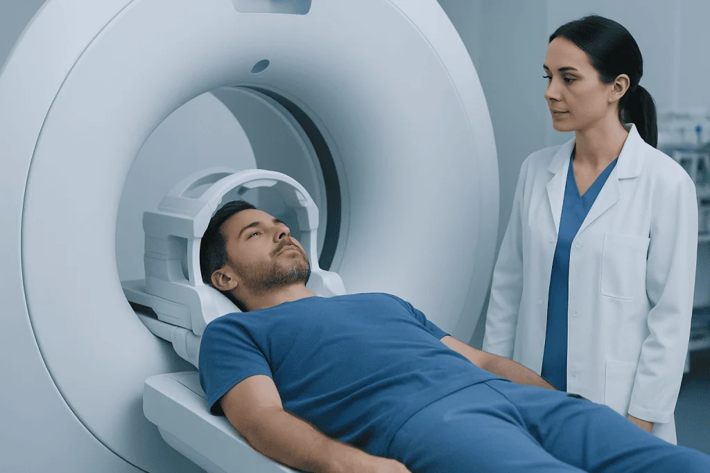
Pathology and imaging are highly useful, but they do not always provide a complete picture at the first evaluation. A pathologist studies the shape, arrangement, and activity of cells under a microscope. If the cells show mixed or borderline features, the report may use cautious language such as uncertain behavior, indeterminate, atypical, or suspicious but not diagnostic.
Uncertainty can also happen because some growths change over time. A lesion may begin with mild abnormalities and later show clearer signs of benign stability or malignant potential. This is one reason follow-up imaging or repeat tissue sampling may be recommended rather than making assumptions based on a single snapshot in time.
In some situations, the location of the growth makes interpretation more complex. For example, a lump in a gland, a small deep tissue lesion, or an abnormal area in an organ may require combined input from radiology, pathology, surgery, and oncology. When specialists compare the scan findings with the microscopic findings, the diagnosis often becomes clearer.
Patients may also see similar but different terms in reports. A cancer diagnosis is made when features clearly support malignancy, while a lesion with uncertain behavior sits in a gray zone that requires careful judgment. That distinction is important because it helps avoid both under-treatment and over-treatment.
Possible symptoms and how these growths are found

Many neoplasms of uncertain behavior cause no symptoms at all and are found incidentally during imaging, screening tests, skin checks, or surgery for another reason. Others are discovered because a person notices a lump, a new skin lesion, localized swelling, bleeding, pain, or changes in bowel, urinary, or hormonal function. Symptoms depend much more on the location of the growth than on the term uncertain behavior itself.
Examples may include a skin spot that changes appearance, a thyroid nodule found on ultrasound, a breast lesion seen on mammography, or a polyp detected during endoscopy. Some people only learn about the issue after a pathology report from a removed mole, cyst, or polyp. In these cases, the report may recommend clinical correlation or further excision.
It is understandable to feel worried when symptoms are vague or when a report seems noncommittal. However, uncertainty in medicine often reflects careful, responsible communication. Rather than forcing a premature label, the care team uses the information available and recommends the safest way to reach a more reliable answer.
Causes and risk factors
A neoplasm of uncertain behavior is not one single disease, so there is no single cause. In general, neoplasms arise when cell growth becomes abnormal. This may relate to aging, inherited factors, chronic irritation, inflammation, hormonal influences, immune changes, environmental exposures, or random DNA changes that occur over time. In many patients, no specific cause can be identified.
Risk factors vary by body site. For example, sun exposure may matter for some skin lesions, while smoking, certain infections, family history, radiation exposure, or hormonal factors may be relevant in other organs. A doctor will usually interpret the pathology result together with personal history, symptoms, imaging findings, and family history to estimate the likelihood that a lesion is harmless, precancerous, or malignant.
Having a growth with uncertain behavior does not mean a person did something wrong. It also does not guarantee that cancer will develop. What matters most is the biological behavior of the specific lesion and whether it shows change over time, invasion into nearby tissue, or cellular features associated with a higher chance of progression.
If there is concern that the lesion may represent or evolve into a more serious condition, referral to specialists in oncology care or surgery may be appropriate. Risk assessment is individualized, and the same pathology phrase can lead to different recommendations in different patients.
How doctors confirm the diagnosis
Evaluation usually begins with a medical history, physical examination, and review of the original imaging or pathology report. Doctors often ask when the lesion was first noticed, whether it has changed, and whether there are related symptoms such as pain, bleeding, weight loss, fever, or fatigue. Family history and previous biopsy results can also be important.
Tests depend on the location of the growth. They may include ultrasound, mammography, CT, MRI, endoscopy, blood tests, dermoscopy, or repeat tissue sampling. Sometimes the best next step is a second opinion from a subspecialty pathologist, especially when the microscopic features are borderline. In selected cases, molecular testing or special stains can help refine the diagnosis.
A biopsy can be very informative, but it may sample only part of a lesion. If the findings do not match the imaging appearance or clinical exam, doctors may recommend a larger biopsy or complete removal. This is common when a small sample shows atypical cells but cannot rule out a more significant process elsewhere in the tissue.
Imaging can also help define size, depth, nearby structures, and whether there are signs that suggest a benign pattern or something more concerning. Depending on the organ involved, doctors may use MRI imaging or PET-CT imaging as part of the broader work-up, although not every patient needs advanced scans.
Treatment options and follow-up planning
Treatment depends on where the neoplasm is located, how suspicious it appears, whether it is causing symptoms, and what the pathology report shows. In some situations, watchful waiting is reasonable. This means scheduled checkups and repeat imaging or exams to look for growth, change in shape, or new symptoms. Observation is more likely when the lesion is small, stable, and low risk.
In other cases, doctors recommend removing the lesion completely. Surgical excision may serve both diagnostic and therapeutic purposes because the entire specimen can be examined in more detail than a small biopsy. If the final pathology shows a benign lesion, no further treatment may be needed beyond routine follow-up. If it shows malignant features, treatment can then be planned more precisely.
Additional treatments depend on the final diagnosis and body site. Some patients may need endoscopic removal, localized procedures, medication, or cancer-directed care such as surgery, radiation therapy, or systemic treatment. The aim is to match treatment intensity to the actual risk, avoiding unnecessary procedures when possible while not delaying needed care.
Multidisciplinary review is often helpful for difficult cases. Near the end of the care pathway, patients may benefit from centers where radiologists, pathologists, surgeons, and oncologists discuss findings together. Acibadem International’s multidisciplinary specialists in JCI-accredited hospitals diagnose and treat complex neoplastic conditions for international patients when coordinated expert evaluation is needed.
Self-care, monitoring, and questions to ask
While waiting for more information, patients can take practical steps to support their care. Keeping copies of pathology reports, imaging results, and procedure notes can make second opinions and follow-up visits more efficient. It is also helpful to note any changes in symptoms, lesion size, bleeding, pain, skin color, or general health.
Healthy routines do not replace medical treatment, but they can support overall well-being. These include not smoking, protecting the skin from excessive sun exposure, following recommended screening tests, eating a balanced diet, and keeping chronic conditions such as diabetes under good control. Patients should avoid self-diagnosing based on online images because many growths look similar but behave differently.
Useful questions for a doctor may include:
- What organ or tissue is involved, and how certain is the diagnosis?
- Does the report suggest low, intermediate, or higher concern?
- Do I need repeat imaging, a second pathology review, or complete removal?
- What symptoms should prompt earlier review?
- How often should follow-up be scheduled?
For some people, the uncertainty itself is the hardest part. Clear communication can reduce anxiety. Asking for the result in plain language and requesting a written plan for next steps can make the situation easier to manage.
When to seek medical care
Medical care should be sought promptly if a known lesion grows quickly, becomes painful, starts bleeding, ulcerates, changes color or shape, or causes new pressure-related symptoms. The same is true if there is unexplained weight loss, persistent fatigue, new difficulty swallowing, changes in bowel habits, or other organ-specific symptoms that are new or worsening.
Even when there are no urgent symptoms, follow-up appointments should not be skipped. A neoplasm of uncertain behavior often becomes clearer only with time, repeat testing, or complete tissue examination. Delays can make monitoring less reliable and may postpone treatment if the lesion proves to be more significant than first thought.
Urgent assessment is especially important if there are signs of infection after a biopsy or procedure, such as fever, spreading redness, drainage, or severe pain. Anyone who feels unsure about a pathology report should ask the treating doctor to explain the result directly and to outline the next recommended step.
Frequently asked questions
Is a neoplasm of uncertain behavior cancer?
Not necessarily. The term means the available information does not yet clearly show whether the growth is benign or malignant. Some lesions are later confirmed to be noncancerous, while others need additional treatment after more testing.
Why would a pathology report use uncertain language?
Pathologists use cautious language when the cell features are not definitive or when the tissue sample is limited. This helps avoid giving an inaccurate final label too early. It usually means more clinical correlation, follow-up, or tissue evaluation is needed.
Will I always need surgery?
No. Some lesions can be safely monitored with repeat exams or imaging if they appear low risk and are not causing symptoms. Surgery is more likely if the lesion is growing, symptomatic, hard to classify, or located in a place where complete removal is the best way to reach a diagnosis.
Can a biopsy miss part of the problem?
Yes, that can happen in some cases because a biopsy samples only part of a lesion. If the biopsy result does not match the scan or physical findings, doctors may recommend a repeat biopsy or complete removal. This is one reason follow-up plans are important.
How often should follow-up be done?
There is no single schedule for everyone. Timing depends on the organ involved, the size and appearance of the lesion, the pathology wording, and personal risk factors. The treating doctor will usually recommend a timetable for repeat imaging, examination, or further procedures.
Should I get a second opinion?
A second opinion can be very helpful if the pathology is complex, the lesion is in a sensitive area, or treatment decisions are difficult. Review by a subspecialty pathologist or multidisciplinary team may clarify whether the lesion needs observation, removal, or more extensive care.
References
- National Cancer Institute
- American Cancer Society
- World Health Organization
- College of American Pathologists
- Mayo Clinic
This article is for general information only and is not a substitute for professional medical advice. Please consult a qualified doctor about your individual situation.
Oncology care in Turkey — second opinion and treatment plan
JCI-accredited · board-certified surgeons · reply within 24h
Add us as a Preferred Source to see more of our trusted health content across Google Search, AI Overviews and Discover.

More from the Health Library



Related Specialists
Dr. Hamdi Tüfekçi
Anesthesiology
Assoc. Prof. Dr. Fatih Aygün
Pediatric Intensive Care Unit
Prof. Dr. Arel Gereli
Orthopedic Surgery & Traumatology
Dr. Can Turan (m)
Intensive Care