Neurointerventional Radiology vs Open Brain Surgery: How Doctors Choose

Neurointerventional radiology treats many brain and blood vessel conditions through tiny tubes inserted into blood vessels rather than a large surgical opening. Open brain surgery may be preferred when direct access, tissue removal, or pressure relief is needed.
Key Takeaways
- Neurointerventional radiology treats many brain and blood vessel conditions through tiny tubes inserted into blood vessels rather than a large surgical opening.
- Open brain surgery may be preferred when direct access, tissue removal, or pressure relief is needed.
- The best choice depends on the condition, anatomy, urgency, and the patient’s general health.
- Many decisions are made by a multidisciplinary team that includes neurologists, neurosurgeons, and interventional specialists.
- In some cases, both techniques may be used at different stages of treatment.
Neurointerventional radiology and open brain surgery are two different ways to treat brain and blood vessel conditions. Doctors choose between them by looking at the diagnosis, the exact location of the problem, the patient’s overall health, and which approach offers the best balance of safety and effectiveness.
Overview
When doctors compare neurointerventional radiology with open brain surgery, they are deciding between two very different treatment paths for conditions affecting the brain, spinal cord, or blood vessels of the nervous system. Neurointerventional radiology, also called interventional neuroradiology or endovascular neurosurgery in some settings, uses thin catheters guided through blood vessels to reach a problem area from inside the body. Open brain surgery involves making a surgical opening to directly see and treat the brain or nearby structures.
Neither method is automatically better in every situation. Each has strengths, limitations, and specific uses. Neurointerventional procedures are often less invasive and may shorten recovery for selected patients. Open surgery may be the safer or more complete option when a surgeon needs direct access to remove a tumor, repair a structural problem, stop bleeding, or relieve dangerous pressure.
Doctors do not choose based on technique alone. They look at what is being treated, how urgent the problem is, where it is located, what the imaging shows, and whether the planned treatment goal is to close a blood vessel abnormality, restore blood flow, remove tissue, or decompress the brain. The final decision is often made by a team rather than one doctor working alone.
What Neurointerventional Radiology Is
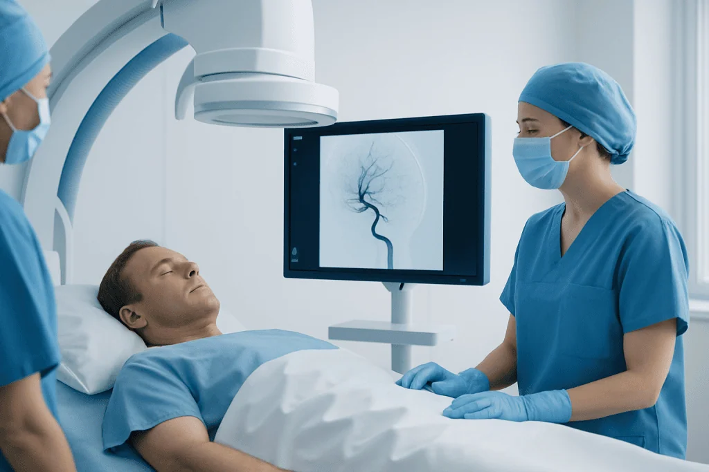
Neurointerventional radiology uses imaging guidance to diagnose and treat certain neurological and cerebrovascular conditions from within the blood vessels. A specialist inserts a small catheter, usually through an artery in the wrist or groin, and carefully guides it toward the brain or spine while watching live imaging. Through that catheter, the doctor can deliver tiny devices, coils, stents, or medication.
This approach is commonly used for problems such as some brain aneurysms, acute ischemic stroke caused by a blocked artery, arteriovenous malformations in selected cases, dural arteriovenous fistulas, and narrowing of certain blood vessels. It may also help control bleeding or improve blood flow. Procedures such as interventional radiology can sometimes treat the underlying issue without opening the skull.
The main advantage is that treatment can often be performed through a very small access point. This may mean less disruption to surrounding tissues and, in many cases, a faster recovery. However, not every condition can be reached safely from inside a blood vessel, and some lesions are shaped or positioned in ways that make endovascular treatment incomplete or unsuitable.
What Open Brain Surgery Is

Open brain surgery, often called craniotomy when part of the skull is temporarily opened, gives the surgical team direct access to the brain and surrounding structures. This allows the surgeon to see the area, remove abnormal tissue, repair certain problems directly, and control bleeding under direct vision. In neurosurgery, this approach remains essential for many serious and complex conditions.
Doctors may recommend open surgery when a lesion must be physically removed, when swelling or pressure needs urgent relief, or when a problem cannot be treated safely through the blood vessels. Examples include many brain tumors, some hemorrhages, infections that need drainage, certain vascular malformations, skull base abnormalities, and traumatic injuries. A patient with a mass lesion, for example, may need brain tumor surgery rather than a catheter-based procedure because the treatment goal is tissue removal.
Although open surgery is more invasive than endovascular treatment, it may offer the most definitive solution in the right case. It can also provide a tissue sample for diagnosis, which is important when doctors need to identify the exact nature of a tumor or another abnormality. Recovery may be longer, but direct surgery can be life-saving and sometimes cannot be replaced by a minimally invasive technique.
How Doctors Decide Between the Two
The first and most important factor is the diagnosis. If the problem is inside a blood vessel, such as a blocked artery in an acute stroke or certain aneurysms, neurointerventional treatment may be strongly considered. If the problem is a mass, widespread bleeding, severe swelling, or something that needs direct removal or decompression, open surgery may be more appropriate. The treatment goal matters just as much as the diagnosis itself.
Location and anatomy also guide the decision. Some aneurysms have a shape, neck size, or branch pattern that is more suitable for endovascular coiling or stent-assisted treatment, while others are better treated with surgical clipping. Some deep or hard-to-reach areas may favor a catheter-based approach because it avoids passing through sensitive brain tissue. In other situations, nearby structures, vessel tortuosity, or access difficulties may make open surgery safer.
Urgency is another key point. In acute ischemic stroke caused by a large artery blockage, time-sensitive stroke and cerebrovascular treatment may involve mechanical thrombectomy through a catheter to restore blood flow quickly. On the other hand, a patient with a large brain bleed and dangerous pressure may need emergency open surgery to remove blood and reduce compression. Sometimes one method stabilizes the patient first, and another is used later for definitive treatment.
Doctors also consider age, overall health, current medications, bleeding risk, kidney function, previous surgeries, and the ability to tolerate anesthesia. A frail patient may benefit from the least invasive effective option, but a minimally invasive route is not chosen if it reduces the chance of complete treatment. The aim is always the best overall outcome, not simply the smallest procedure.
Conditions Commonly Considered for Either Approach
One of the best known examples is a brain aneurysm. Some aneurysms can be treated through the blood vessels with coils, flow-diverting devices, or stents, while others are better managed with surgical clipping. The decision depends on aneurysm size, shape, location, whether it has ruptured, and the patient’s age and health status. Both methods are established, and each can be appropriate in different circumstances.
Stroke care also shows how choice depends on the type of problem. A blocked artery causing an ischemic stroke may respond to clot retrieval through a catheter, while a hemorrhagic stroke may require neurosurgical management if there is significant bleeding or pressure. Conditions such as stroke are therefore not treated by one single method; treatment changes according to the mechanism of injury seen on imaging.
Arteriovenous malformations, fistulas, and certain vascular tumors may be treated with endovascular embolization, open surgery, radiosurgery, or a combination. In some cases, embolization is used before surgery to reduce blood flow and make later removal safer. For tumors, open surgery is often necessary when diagnosis, decompression, or removal is the main goal, though catheter-based techniques may support treatment planning or bleeding control.
- Vessel problems often favor endovascular treatment when anatomy is suitable.
- Mass lesions often favor open surgery when tissue removal is needed.
- Some conditions are best treated with a staged or combined plan.
- Imaging findings are central to choosing the safest route.
Benefits, Risks, and Recovery Differences
Neurointerventional radiology is often appealing because it avoids a large incision and does not usually require opening the skull. This can reduce hospital stay for selected patients and may allow faster return to daily activities. There may be less postoperative discomfort and less visible scarring. For some vascular conditions, it offers excellent results with a lower burden on the body.
At the same time, catheter-based procedures have their own risks. These can include bleeding at the access site, blood vessel injury, stroke, contrast-related complications, and the possibility that the lesion is not fully treated and needs follow-up procedures. Some patients require long-term imaging surveillance after endovascular treatment, especially when there is a risk of recurrence or device-related follow-up needs.
Open brain surgery carries risks related to the operation itself, such as infection, bleeding, seizures, swelling, or neurological deficits depending on the area being treated. Recovery can take longer, and rehabilitation may be needed in some cases. However, surgery may provide immediate decompression, complete removal of a lesion, or direct repair that cannot be achieved through a catheter.
Because each option has different trade-offs, doctors explain not only the procedure but also the expected recovery, follow-up imaging, potential need for additional treatment, and the possible effects on speech, movement, memory, or independence. Shared decision-making helps patients and families understand why one path is being recommended.
The Role of Imaging and Team-Based Planning
Modern imaging is central to choosing the right treatment. CT, MRI, angiography, and sometimes perfusion studies help doctors see the exact size, location, and behavior of a lesion. Imaging may show whether a vessel is blocked, whether bleeding is active, how much brain tissue is at risk, or whether surrounding anatomy makes one procedure safer than another. In many cases, the decision cannot be made accurately without detailed imaging.
These decisions are often reviewed by a multidisciplinary team. Depending on the condition, this may include a neurologist, interventional neuroradiologist, neurosurgeon, anesthesiologist, intensive care specialist, and rehabilitation experts. Team discussion is especially valuable when more than one good option exists or when a combined strategy may offer the best result.
Patients and families may hear that a case is being discussed in terms of feasibility, durability, and risk profile. Feasibility means whether the technique can actually reach and treat the problem. Durability means how likely the treatment is to remain effective over time. Risk profile means the immediate and long-term safety considerations. In experienced centers, this balanced planning helps match the treatment to the individual rather than the diagnosis alone.
What Patients Should Ask and When to Seek Care
Patients can feel more confident when they understand why one treatment is recommended over another. Helpful questions include: What is the exact diagnosis? Is the goal to remove, repair, block, or restore blood flow? Why is a catheter-based approach or open surgery more suitable in this case? What are the main benefits and risks? Will more than one procedure be needed? These questions can support clear, informed discussions with the care team.
Urgent symptoms should never be ignored. Sudden weakness, facial drooping, difficulty speaking, severe new headache, seizures, loss of consciousness, sudden vision changes, or confusion need prompt medical attention. These symptoms may be related to stroke, bleeding, or other neurological emergencies where fast evaluation can make a major difference.
For non-emergency findings such as a known aneurysm, vascular malformation, or newly discovered brain lesion, evaluation by specialists is still important even when symptoms are mild. In advanced centers, including Acibadem International, multidisciplinary specialists in JCI-accredited hospitals evaluate these conditions and plan individualized care for international patients, including options such as neurosurgery when direct surgical treatment is needed.
In many cases, the choice is not a contest between two competing procedures. It is a careful medical judgment about which method offers the safest and most effective treatment for that particular patient at that particular time. A qualified doctor can explain the reasoning and help guide the next step.
Frequently asked questions
Is neurointerventional radiology safer than open brain surgery?
It is often less invasive, but it is not automatically safer in every case. Safety depends on the condition being treated, the anatomy, the urgency, and the experience of the treating team. For some problems, open surgery is actually the safer and more effective choice.
Which conditions are most often treated with neurointerventional radiology?
It is commonly used for certain brain aneurysms, some types of stroke caused by blocked arteries, and selected vascular malformations or fistulas. The key factor is whether the problem can be reached and treated through the blood vessels. Detailed imaging helps determine this.
Why would a doctor recommend open brain surgery instead of a minimally invasive procedure?
Open surgery may be needed when tissue must be removed, pressure in the brain must be relieved, or direct access offers the best chance of complete treatment. This is often the case with many brain tumors, some hemorrhages, and structural abnormalities. The recommendation is based on what the patient needs most, not just on the size of the incision.
Can both treatments be used for the same patient?
Yes. Some patients benefit from a combined approach, such as embolization before surgery to reduce bleeding risk, or emergency endovascular treatment followed by later surgery. Treatment planning is often staged to improve safety and effectiveness.
How do doctors know which option is best?
They use neurological examination, imaging studies such as CT, MRI, and angiography, and an assessment of the patient’s general health. The exact diagnosis, anatomy, and treatment goal are all important. In many hospitals, the decision is made by a multidisciplinary team.
Is recovery always faster after neurointerventional treatment?
Recovery is often shorter because the procedure is less invasive, but not always. Some endovascular treatments still require close monitoring, follow-up imaging, or additional procedures. Recovery also depends on the underlying condition, especially if there has been a stroke or bleeding.
References
- World Stroke Organization
- American Association of Neurological Surgeons
- Society of NeuroInterventional Surgery
- National Institute of Neurological Disorders and Stroke
- Radiological Society of North America
This article is for general information only and is not a substitute for professional medical advice. Please consult a qualified doctor about your individual situation.
Joint and spine care in Turkey — expert assessment & treatment
JCI-accredited · board-certified surgeons · reply within 24h
Add us as a Preferred Source to see more of our trusted health content across Google Search, AI Overviews and Discover.