Neurointerventional Radiology: What Happens Before, During, and After Treatment

Neurointerventional radiology uses small catheters and live imaging instead of large surgical openings. It may be used for conditions such as stroke, aneurysms, arteriovenous malformations, and narrowed blood vessels.
Key Takeaways
- Neurointerventional radiology uses small catheters and live imaging instead of large surgical openings.
- It may be used for conditions such as stroke, aneurysms, arteriovenous malformations, and narrowed blood vessels.
- Preparation often includes scans, blood tests, medication review, and fasting instructions.
- Recovery depends on the procedure, but monitoring after treatment is an important part of safe care.
- Follow-up visits and imaging help confirm healing and guide long-term management.
Neurointerventional radiology is a minimally invasive specialty that diagnoses and treats certain conditions of the brain, spine, head, and neck blood vessels using imaging guidance and tiny catheters. Understanding what happens before, during, and after treatment can help patients and families feel more prepared and informed.
Overview
Neurointerventional radiology is a highly specialized field that treats disorders affecting blood vessels in and around the brain, spinal cord, head, and neck. It is also called endovascular neurosurgery or interventional neuroradiology. Instead of making a large incision, the specialist usually guides a very thin tube called a catheter through a blood vessel, often starting from the wrist or groin, and navigates it to the area that needs treatment.
Using real-time imaging, the doctor can diagnose a problem and, in many cases, treat it during the same session. Treatments may include removing a clot, closing off an aneurysm, opening a narrowed artery, or reducing abnormal blood flow in a malformation. Because these procedures are minimally invasive, they may reduce recovery time compared with some open surgical approaches, although they still require expert planning and careful monitoring.
Neurointerventional radiology may be used in emergencies, such as stroke, or in planned care for conditions found on scans or after symptoms develop. The exact treatment depends on the diagnosis, the patient’s overall health, and whether the goal is urgent treatment, symptom relief, or prevention of future complications.
Which conditions can neurointerventional radiology treat?
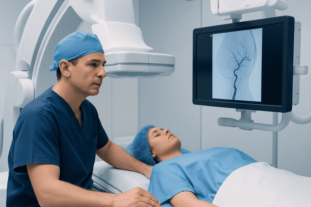
This specialty is often used for conditions involving abnormal blood flow, bleeding risk, or blocked vessels. One of the best-known examples is brain aneurysm, where a weak area in a blood vessel wall may need treatment to lower the chance of rupture. Another example is ischemic stroke caused by a clot blocking blood flow to part of the brain.
It may also be used for arteriovenous malformations, dural arteriovenous fistulas, carotid or intracranial artery narrowing, certain tumors with a rich blood supply, and some causes of spinal vascular symptoms. In selected cases, it can support treatment planning by providing highly detailed imaging of blood vessels through cerebral angiography.
- Acute ischemic stroke, sometimes with stroke treatment to remove a clot
- Brain aneurysms treated with techniques such as coiling or flow diversion
- Arteriovenous malformations and fistulas that may need embolization
- Narrowed carotid or brain arteries that may require stenting or angioplasty
- Bleeding or vascular complications after trauma or surgery
Not every patient with these conditions is treated in the same way. Some people are better served by medication, conventional surgery, observation, or a combination of approaches decided by a multidisciplinary team.
What happens before treatment?

Before treatment, the care team first confirms the diagnosis and reviews whether a neurointerventional procedure is the safest and most effective option. This usually includes a medical history, physical and neurological examination, and a review of current medicines, especially blood thinners, diabetes medicines, and any history of allergy to contrast dye. Patients should mention pregnancy, kidney problems, previous bleeding issues, and implanted devices.
Imaging tests are central to planning. Depending on the situation, these may include CT, MRI, CT angiography, MR angiography, ultrasound, or a diagnostic angiogram. Blood tests may also be done to check kidney function, blood counts, and clotting. In urgent cases such as stroke, this evaluation happens very quickly so treatment is not delayed.
Patients are commonly asked not to eat or drink for a period before the procedure, although instructions vary. The team explains which regular medicines should be taken, paused, or adjusted. Consent is obtained after discussing the reason for treatment, possible benefits, alternatives, and known risks such as bleeding, vessel injury, contrast reaction, or stroke-related complications.
It is also helpful to prepare for the practical side of treatment. Patients may need someone to accompany them, especially if sedation or anesthesia is planned. Many procedures require a short hospital stay, while some complex or urgent treatments need intensive monitoring afterward.
What happens during the procedure?
On the day of treatment, the patient changes into a hospital gown and an intravenous line is placed for fluids and medicines. Depending on the procedure, care may be provided under local anesthesia with sedation or under general anesthesia. The skin at the catheter entry site, usually the wrist or groin, is cleaned and numbed.
The specialist inserts a catheter into the artery and carefully guides it using X-ray imaging to the target area. Contrast dye is often injected so blood vessels can be seen clearly. The patient does not feel the catheter moving inside the blood vessels, but may notice pressure at the entry site or a brief warm sensation when contrast is injected.
The treatment itself depends on the condition. A stroke procedure may use a device to remove a clot. An aneurysm may be treated with tiny coils or a flow-diverting stent, as part of brain aneurysm treatment. A vascular malformation or fistula may be embolized by delivering materials that block abnormal vessels. Narrowed arteries may sometimes be opened with angioplasty or stenting.
Throughout the procedure, the team closely monitors heart rate, blood pressure, breathing, and neurological status. When treatment is complete, the catheter is removed and pressure or a closure device is used to reduce bleeding at the puncture site. The procedure may take less than an hour or several hours, depending on the diagnosis and complexity.
What happens after treatment?
After the procedure, the patient is moved to a recovery area, stroke unit, intensive care unit, or hospital room depending on what was done and how they are feeling. Nurses and doctors monitor blood pressure, pulse, neurological signs, and the catheter insertion site. If the groin was used, the patient may need to lie flat for a period. If the wrist was used, movement restrictions are usually different.
It is normal to feel tired after sedation or anesthesia, and some people have mild soreness or bruising where the catheter was inserted. The team also watches for more important issues such as bleeding, worsening headache, weakness, speech changes, confusion, allergic reaction, or changes in kidney function. Reporting new symptoms promptly helps the team respond early if needed.
The length of stay varies. Some planned diagnostic angiograms or simpler treatments may allow discharge the same day or the next day, while emergency stroke care and more complex procedures often require longer observation. Before leaving, patients receive advice about activity, bathing, driving, medications, hydration, and follow-up appointments.
Follow-up imaging is often an important part of care, especially after aneurysm treatment, stent placement, or embolization. These checks help confirm that the treated area remains stable and that no further intervention is needed.
Benefits, risks, and recovery
The main benefit of neurointerventional radiology is that it can treat certain serious conditions without the larger incisions used in open surgery. For many patients, this means smaller wounds, less blood loss, shorter hospital stays, and a faster return to daily activities. In urgent conditions such as stroke, rapid endovascular treatment can be a key part of protecting brain function.
At the same time, these are complex procedures performed in delicate blood vessels, so risks must be weighed carefully. Possible risks include bleeding at the puncture site, infection, blood vessel damage, stroke, clot formation, contrast allergy, and kidney strain from contrast dye. Device-specific risks, such as stent-related clotting or the need for blood-thinning medicines, may also apply in some cases.
Recovery is different for every patient. It depends on the underlying condition, whether the situation was elective or an emergency, the type of treatment performed, and the person’s general health. Some people recover quickly from the procedure itself but still need rehabilitation if the original condition, such as stroke, caused symptoms.
A balanced discussion with the care team is important. Doctors consider whether a minimally invasive approach is the best choice on its own or whether it should be combined with neurosurgery or medical therapy for the safest long-term result.
Self-care, follow-up, and when to seek medical help
Good aftercare supports recovery and lowers the chance of complications. Patients should take medicines exactly as prescribed, especially antiplatelet or blood-thinning drugs if a stent or flow diverter was used. They should also follow instructions about lifting, exercise, hydration, and wound care at the catheter site. Missing follow-up visits can delay the detection of problems that may not cause symptoms right away.
Long-term prevention depends on the underlying condition. This may include controlling blood pressure, cholesterol, and diabetes; stopping smoking; staying physically active within the doctor’s advice; and maintaining regular medical checkups. For some vascular conditions, repeat imaging is part of standard care even when the patient feels well.
Patients should seek urgent medical attention if they develop sudden weakness, facial drooping, trouble speaking, severe or unusual headache, loss of vision, chest pain, shortness of breath, heavy bleeding, a rapidly enlarging lump at the puncture site, or significant swelling, numbness, or color change in the arm or leg used for access. Fever, increasing redness, or drainage from the puncture site should also be reported.
Care is often coordinated by neurologists, neurosurgeons, interventional neuroradiologists, anesthesiologists, rehabilitation specialists, and intensive care teams. For international patients, Acibadem International’s multidisciplinary specialists and JCI-accredited hospitals diagnose and treat neurovascular conditions with coordinated evaluation, including options such as interventional radiology when appropriate.
Frequently asked questions
Is neurointerventional radiology the same as brain surgery?
Not exactly. Neurointerventional radiology is a minimally invasive approach that uses catheters and imaging guidance rather than a large opening in the skull. In some situations it may replace open surgery, while in others it complements it.
Will the patient be awake during the procedure?
Some procedures are done with local anesthesia and sedation, while others require general anesthesia. The choice depends on the type of treatment, the patient’s condition, and how still the patient needs to remain. The care team explains the plan in advance whenever possible.
How long does recovery take?
Recovery varies widely based on the condition being treated and whether the procedure was planned or done in an emergency. Some patients go home the same day or the next day, while others need longer monitoring or rehabilitation. The underlying illness often affects recovery more than the catheter procedure itself.
Is neurointerventional radiology only used for stroke?
No. It is also used for aneurysms, arteriovenous malformations, fistulas, narrowed arteries, and other vascular problems involving the brain and spine. In some cases it is also used for detailed diagnosis through angiography.
Are there risks from the contrast dye?
Contrast dye is commonly used to help doctors see blood vessels clearly. Most people tolerate it well, but some may have an allergic reaction or temporary kidney strain, especially if kidney function is already reduced. That is why the team checks medical history and blood tests beforehand.
Why are follow-up scans needed if the treatment was successful?
Follow-up imaging helps confirm that the treated vessel remains stable and that blood flow is as expected. Some conditions, such as aneurysms or vascular malformations, can change over time. Regular checks help doctors decide whether further treatment or continued observation is needed.
References
- World Health Organization
- American Stroke Association
- Society of NeuroInterventional Surgery
- Radiological Society of North America
- National Institute of Neurological Disorders and Stroke
This article is for general information only and is not a substitute for professional medical advice. Please consult a qualified doctor about your individual situation.
Joint and spine care in Turkey — expert assessment & treatment
JCI-accredited · board-certified surgeons · reply within 24h
Add us as a Preferred Source to see more of our trusted health content across Google Search, AI Overviews and Discover.