Orbital Tumors: Symptoms, Imaging, and When Orbit Surgery Is Needed

Orbital tumors may be benign or malignant and can arise from different tissues in the eye socket. Common symptoms include eye bulging, double vision, pain, eyelid swelling, and changes in vision.
Key Takeaways
- Orbital tumors may be benign or malignant and can arise from different tissues in the eye socket.
- Common symptoms include eye bulging, double vision, pain, eyelid swelling, and changes in vision.
- Imaging tests such as MRI and CT scans are central to diagnosis and treatment planning.
- Not every orbital tumor needs immediate surgery; some are monitored closely over time.
- Surgery may be needed to confirm the diagnosis, relieve pressure, remove the tumor, or protect vision.
- Evaluation often involves ophthalmology, radiology, pathology, and sometimes oncology or neurosurgery specialists.
Orbital tumors are growths that develop in or around the eye socket and can affect vision, eye movement, or the appearance of the eye. Care usually begins with a detailed eye examination and imaging, while orbit surgery is considered when a tumor threatens vision, causes symptoms, or needs tissue diagnosis.
Overview of orbital tumors
Orbital tumors are abnormal growths located in the orbit, the bony space that contains the eye, eye muscles, nerves, blood vessels, and connective tissue. These tumors may begin in the orbit itself, spread from nearby structures such as the eyelids or sinuses, or rarely reach the orbit from another part of the body. Some orbital tumors are benign, meaning they do not spread to distant organs, while others are malignant and require more urgent treatment.
The term “orbital tumor” describes a wide range of conditions rather than a single disease. Examples include vascular lesions, inflammatory masses, tumors of the lacrimal gland, nerve sheath tumors, lymphoid tumors, and metastatic disease. Because the orbit is a small and delicate space, even a noncancerous growth can cause important symptoms if it presses on the eye, optic nerve, or eye muscles.
Care is tailored to the individual. Doctors consider the patient’s age, symptoms, imaging findings, rate of growth, and the suspected type of tumor. Some lesions can be observed safely with regular follow-up, while others require biopsy, medication, radiation therapy, or surgery. A careful evaluation by eye specialists, often including neuro-ophthalmology and orbital surgery teams, helps guide the next steps.
Symptoms and signs to watch for
Symptoms of orbital tumors can develop gradually or, less commonly, over a short period of time. One of the best-known signs is proptosis, which means the eye appears to bulge or shift forward. Some people notice that one eye looks more prominent than the other in photographs or in the mirror. Others first become aware of swelling around the eyelids or a feeling of fullness behind the eye.
Vision-related symptoms are also important. These may include blurred vision, double vision, reduced side vision, trouble moving the eye, or pain with eye movement. If a tumor presses on the optic nerve, vision may become dimmer or colors may look less vivid. Some people develop headaches, tearing, or a pressure sensation around the eye.
Symptoms vary depending on the type and location of the lesion. For example, a mass near the eye muscles may mainly cause double vision, while one near the optic nerve may affect sight more directly. In children, warning signs can include a rapidly developing bulging eye, eyelid droop, or an eye that seems out of alignment. Any persistent or unexplained change around the eye deserves prompt medical assessment.
- Bulging or displacement of one eye
- Double vision or limited eye movement
- Blurred vision or decreased visual clarity
- Pain, pressure, or headaches around the eye
- Eyelid swelling or a visible lump
- Color vision changes or visual field loss
Causes and risk factors

Orbital tumors can arise from many different tissues in the orbit. Some develop from blood vessels, nerves, fat, muscles, connective tissue, or the lacrimal gland that produces tears. Others are related to inflammatory disorders that can mimic a tumor on imaging and examination. In adults, certain orbital masses are more common than others, but the exact cause of many cases is not fully understood.
Age can influence which diagnoses are more likely. In children, doctors may think about developmental cysts, vascular lesions, or rare childhood cancers. In adults, possibilities may include cavernous venous malformations, lacrimal gland tumors, meningioma involving the optic nerve sheath, lymphoma, or spread from nearby sinus disease. Sometimes the orbit is affected by cancers that started elsewhere in the body.
Risk factors depend on the specific condition. A personal history of cancer can raise concern for metastasis. A history of chronic inflammation, immune system disorders, or previous radiation exposure may also be relevant in some patients. However, many people diagnosed with an orbital tumor have no clear risk factor. That is why symptoms, examination findings, and imaging are so important in making an accurate diagnosis.
How orbital tumors are diagnosed
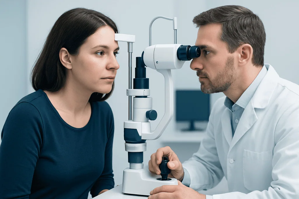
Diagnosis begins with a detailed medical history and eye examination. The doctor will ask when symptoms started, whether they are getting worse, and whether there is pain, double vision, or vision loss. The examination may include checking visual acuity, color vision, pupil reactions, eye alignment, eye movements, visual fields, and the appearance of the optic nerve and retina. A comprehensive eye examination is usually the first step.
Imaging plays a central role in evaluating orbital tumors. MRI is often preferred because it gives excellent detail about the soft tissues, optic nerve, muscles, and the relationship of the mass to surrounding structures. CT scanning is also valuable, especially when doctors need to assess the bony walls of the orbit or detect calcification. In some situations, ultrasound or additional body imaging may be useful.
Blood tests are not always needed, but they may help when inflammation, infection, thyroid eye disease, or systemic cancer is part of the differential diagnosis. When imaging alone cannot provide a confident diagnosis, a biopsy may be recommended. This means taking a small tissue sample so a pathologist can examine the cells under a microscope. Tissue diagnosis is especially important when lymphoma, lacrimal gland tumors, or metastatic disease is suspected.
Because orbital disorders can affect both vision and the nervous system, some patients benefit from assessment through general ophthalmology and subspecialty teams. The goal is not simply to identify a mass, but to understand exactly what it is and how likely it is to affect vision or overall health.
What imaging can show
Imaging helps doctors answer several key questions: where the mass is located, what tissue it may arise from, whether it is well-defined or infiltrative, and whether nearby structures are compressed or invaded. These details help narrow the diagnosis and determine whether observation, biopsy, or treatment is most appropriate. Imaging is also important for surgical planning if an operation is being considered.
On MRI, doctors can evaluate the shape of the lesion, how it behaves with contrast dye, and whether it involves the optic nerve, extraocular muscles, or the orbital apex, the deep part of the eye socket. CT scans are useful when bone erosion, sinus involvement, or calcified lesions are suspected. Imaging can also show whether a tumor extends beyond the orbit toward the brain, sinuses, or face.
Although imaging often strongly suggests a diagnosis, scans do not always give a final answer. Some benign and malignant conditions can look similar. For that reason, doctors interpret imaging together with the clinical examination and, when necessary, biopsy results. Follow-up imaging may also be used over time to check whether a known lesion is stable or changing.
Treatment options and when orbit surgery is needed
Treatment depends on the tumor type, its location, the severity of symptoms, and whether there is a risk to vision or overall health. Not all orbital tumors need immediate intervention. A small, slow-growing, clearly benign-appearing lesion with no symptoms may be monitored with regular examinations and repeat imaging. Observation is often appropriate when the benefits of surgery do not clearly outweigh the risks.
Orbit surgery is generally considered when a tumor is causing vision changes, compressing the optic nerve, producing significant eye bulging, restricting eye movement, causing pain, growing over time, or when tissue is needed to establish a diagnosis. In some cases, surgery aims to remove the entire tumor. In others, the goal is to obtain a biopsy, reduce pressure within the orbit, or remove as much tumor as is safely possible while protecting important structures.
The surgical approach varies with the size and position of the lesion. Some tumors can be reached through small incisions hidden in the eyelid crease or conjunctiva, while deeper or more complex cases may require a more extensive approach. Surgeons work carefully around the eye muscles, optic nerve, and blood vessels. Depending on the diagnosis, additional treatment such as steroids, radiation therapy, chemotherapy, or targeted therapy may be recommended after surgery or instead of surgery.
Patients are often cared for by teams that include orbital and oculoplastic surgery specialists, radiologists, pathologists, and oncologists when needed. In selected cases, broader eye surgery services and multidisciplinary planning help tailor treatment to the individual lesion and the patient’s visual needs.
Recovery, follow-up, and self-care
Recovery after treatment depends on the type of orbital tumor and the therapy used. After surgery, temporary swelling, bruising, and mild discomfort around the eye are common. Doctors usually give instructions about cold compresses, eye protection, wound care, and activity limits. Follow-up visits are important to monitor healing, vision, eye movements, and the pathology results if tissue was removed.
Even when a tumor is benign, follow-up matters because some lesions can recur or slowly change over time. Repeat examinations and imaging may be scheduled at intervals based on the diagnosis. People treated for malignant tumors may need longer-term monitoring and care from oncology teams as well. Keeping all appointments helps doctors detect any new problems early.
Self-care does not replace medical treatment, but it can support recovery. Patients should take medicines exactly as directed, avoid rubbing the eye, and report worsening pain, increased swelling, new double vision, fever, or any drop in vision. If the diagnosis affects appearance as well as function, emotional support can also be valuable. Near the end of the care journey, some international patients may choose centers such as Acibadem International, where multidisciplinary specialists at JCI-accredited hospitals evaluate and treat orbital conditions.
When to see a doctor
Any persistent eye bulging, unexplained swelling around one eye, new double vision, or changes in eyesight should be evaluated by a qualified doctor. These symptoms do not always mean a tumor is present, but they should not be ignored. Early assessment can help distinguish orbital tumors from infections, inflammation, thyroid-related eye disease, or other eye conditions.
Urgent medical attention is especially important if symptoms progress quickly, vision suddenly worsens, severe pain develops, or the eye becomes difficult to move. Children with a newly bulging eye or rapidly changing eye appearance should be seen promptly. A timely diagnosis can protect vision and allow treatment to begin when needed.
Many orbital problems are first noticed by general practitioners, ophthalmologists, or emergency doctors. Referral to an orbital specialist may follow if imaging or examination suggests a mass in the eye socket. The safest approach is to seek professional advice rather than trying to guess the cause based on symptoms alone.
Frequently asked questions
Are orbital tumors always cancer?
No. Many orbital tumors are benign and do not spread to other parts of the body. However, even benign tumors may still need treatment if they press on the eye, optic nerve, or eye muscles.
What is the difference between an orbital tumor and an eye tumor?
An orbital tumor develops in the eye socket around the eye, not necessarily inside the eyeball itself. An eye tumor usually refers to a growth in structures of the eye, such as the retina, choroid, or other intraocular tissues.
Which scan is better for orbital tumors, MRI or CT?
Both can be useful, and doctors often choose based on the clinical question. MRI usually gives better detail of soft tissues, while CT is especially helpful for assessing bone and certain calcified lesions.
Do all orbital tumors need surgery?
No. Some tumors can be monitored safely with regular examinations and imaging if they appear stable and are not causing symptoms. Surgery is usually considered when vision is at risk, symptoms are significant, the mass is growing, or a biopsy is needed.
Can an orbital tumor cause double vision?
Yes. Double vision can happen when a tumor affects the eye muscles or the nerves that control eye movement. Pressure within the orbit can also change how the eyes line up and move together.
Is biopsy always necessary?
Not always. If imaging and examination strongly suggest a benign lesion and the plan is careful monitoring, biopsy may not be needed right away. When the diagnosis is uncertain or cancer is a concern, tissue sampling is often important.
References
- American Academy of Ophthalmology
- National Eye Institute
- National Cancer Institute
- American Association for Pediatric Ophthalmology and Strabismus
- Radiological Society of North America
This article is for general information only and is not a substitute for professional medical advice. Please consult a qualified doctor about your individual situation.
Orbit Surgery in Turkey — costs, top hospitals & a free quote
JCI-accredited · board-certified surgeons · reply within 24h
Add us as a Preferred Source to see more of our trusted health content across Google Search, AI Overviews and Discover.