Osteochondritis Dissecans Knee Surgery: Procedure, Recovery and Results
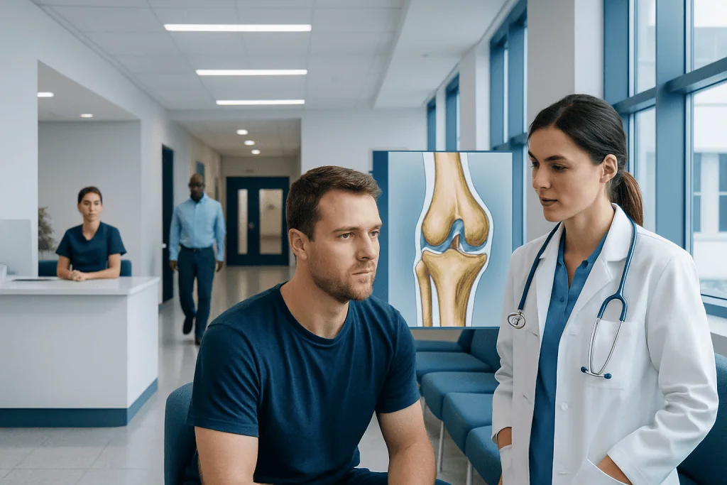
Surgery is not needed for every OCD lesion; stable lesions, especially in growing children, may heal without an operation. Arthroscopy helps the surgeon assess cartilage and bone stability and may allow treatment through small incisions.
Key Takeaways
- Surgery is not needed for every OCD lesion; stable lesions, especially in growing children, may heal without an operation.
- Arthroscopy helps the surgeon assess cartilage and bone stability and may allow treatment through small incisions.
- Common surgical approaches include drilling, fixation of a fragment, and cartilage-restoration procedures for irreparable defects.
- Recovery can take several months, while a return to impact sports often takes longer and depends on healing and rehabilitation progress.
- Early assessment is important when knee pain is persistent or accompanied by swelling, catching, locking, or loss of motion.
Osteochondritis dissecans knee surgery may be recommended when a damaged area of cartilage and underlying bone is unstable, detached, or has not healed with activity changes and rehabilitation. The procedure is tailored to the lesion’s size, location, stability, and the patient’s age, with recovery commonly requiring protected weight-bearing and structured physiotherapy.
Overview: What Is Osteochondritis Dissecans Knee Surgery?
Osteochondritis dissecans knee surgery is a group of procedures used to treat a focal area of injured bone beneath the knee cartilage. In osteochondritis dissecans (OCD), blood supply to a small part of the bone may be disrupted. The overlying cartilage and bone fragment can become unstable and, in some cases, partially or completely separate from the joint surface.
Surgery aims to preserve the person’s own cartilage whenever possible, restore a smooth joint surface, and reduce the risk of ongoing symptoms or later joint damage. It is usually considered when a lesion is unstable, displaced, large, painful despite appropriate non-surgical treatment, or found in an adult with limited healing potential.
The knee is the most commonly affected joint, often involving the inner portion of the thigh bone (medial femoral condyle). Treatment decisions are individualized after imaging, examination, and discussion of activity goals. OCD should not be confused with generalized arthritis: it is a localized cartilage-and-bone problem, although an untreated unstable lesion can contribute to earlier wear of the joint.
How Serious Is Osteochondritis Dissecans?
Osteochondritis dissecans ranges from a stable lesion that can heal to an unstable lesion that may damage the joint surface. Its seriousness depends on whether the cartilage remains intact, whether the bone-and-cartilage fragment is securely attached, the size and location of the lesion, and whether the growth plates are still open.
In children and adolescents who are still growing, stable lesions often have a better capacity to heal with reduced impact activity, monitoring, and rehabilitation. In skeletally mature adolescents and adults, lesions are less likely to heal without treatment and may require closer assessment. A loose fragment inside the knee can cause sudden catching or locking and should be evaluated promptly.
Even when symptoms are manageable, persistent OCD should be followed by an orthopedic specialist. Timely care can help protect the cartilage and guide a safe return to school, work, exercise, or sport.
Candidacy, Assessment and Diagnosis

Before recommending osteochondritis dissecans knee surgery, an orthopedic surgeon considers symptoms, physical examination findings, skeletal maturity, previous treatment, and imaging. Common symptoms include activity-related knee pain, recurrent swelling, tenderness, reduced range of motion, a feeling that the knee gives way, catching, or locking.
Plain X-rays can show the location and appearance of the affected bone. Magnetic resonance imaging (MRI) is especially useful for evaluating the cartilage, surrounding bone, fluid behind the lesion, and features that may suggest instability. In some situations, the final assessment of stability is made during arthroscopy, when the surgeon views and gently tests the area directly.
Non-surgical management may be appropriate for a stable lesion, particularly in younger patients. It can include temporarily stopping running, jumping, pivoting sports, or other painful activities; guided physiotherapy; and scheduled imaging follow-up. Surgery becomes more likely if symptoms continue, healing does not occur, or imaging indicates an unstable or detached fragment.
- Potential surgical candidates: people with unstable, displaced, or persistent lesions.
- Important planning factors: age, growth status, lesion size, cartilage condition, alignment, and desired activity level.
- Shared decision-making: the surgeon explains realistic goals, rehabilitation demands, and alternatives before treatment.
How the Procedure Works: Step-by-Step
Most OCD knee operations are performed using arthroscopy, a minimally invasive technique in which a small camera and specialized instruments are introduced through small incisions around the knee. The patient receives anesthesia, and the surgical team examines the joint, including the OCD lesion, cartilage surfaces, menisci, and ligaments. The exact operation depends on what is found.
For a stable lesion with intact cartilage, the surgeon may perform drilling. Small channels are made in the bone to encourage blood flow and healing at the lesion. Drilling can be performed from outside the joint or through the cartilage surface, depending on the lesion and the surgeon’s approach.
If the cartilage-and-bone fragment is viable but unstable, it may be repositioned and secured with implants such as screws, pins, or bioabsorbable devices. Damaged edges are cleaned as needed to create a suitable healing surface. If a fragment cannot be preserved or there is a larger cartilage-and-bone defect, cartilage restoration may be considered, such as marrow-stimulation techniques, osteochondral grafting, or other restorative procedures selected for the individual defect.
After the procedure, the knee is dressed and a brace may be used. Some patients go home the same day, while others may need a short hospital stay depending on the operation and their general health. A focused knee-cartilage evaluation and knee arthroscopy plan can help determine the most appropriate technique.
What Happens During Stage 4 of Osteochondritis Dissecans?
Staging systems vary slightly, particularly between MRI-based and arthroscopic classifications. In general, stage 4 osteochondritis dissecans describes a lesion in which the cartilage-and-bone fragment has become detached or is a loose body within the knee joint.
At this stage, symptoms may include more frequent swelling, pain, catching, locking, or a sudden inability to fully straighten the knee. Because the normal joint surface is disrupted, repeated mechanical symptoms may further irritate the cartilage. Stage 4 usually requires assessment by an orthopedic surgeon and often surgical treatment.
During surgery, the surgeon determines whether the fragment is healthy enough to be fixed back into place. If not, it may be removed and the remaining defect treated with a cartilage-repair or restoration technique. The aim is to relieve mechanical symptoms and preserve the best possible joint surface for long-term function.
How Long Does It Take to Recover From Osteochondritis Dissecans Surgery?
Recovery from osteochondritis dissecans knee surgery varies widely because the procedure ranges from drilling of a stable lesion to fixation or cartilage restoration for a larger defect. Many patients need crutches and limited or no weight-bearing for several weeks, particularly after fixation or grafting. The treating team provides specific instructions based on the procedure and healing needs.
Early rehabilitation usually focuses on controlling swelling, maintaining safe knee motion, and activating the thigh muscles. A brace may be used to protect the knee. As healing progresses, physiotherapy typically advances to gradual weight-bearing, strength training, balance work, and movement control. Follow-up appointments and imaging may be used to check bone healing and guide activity progression.
Daily activities may become easier over the first several weeks to months, but returning to running, jumping, pivoting, or competitive sport commonly takes many months. A return to sport should be based on healing, knee motion, strength, functional testing, and the surgeon’s advice rather than a calendar date alone. Trying to return too early can place the repair at risk.
- First weeks: wound care, swelling control, protected walking, and prescribed motion exercises.
- Following months: progressive strengthening and gradual restoration of normal walking and function.
- Later rehabilitation: sport-specific training only after adequate healing and functional recovery.
Benefits, Risks and Expected Results
The potential benefits of OCD knee surgery include stabilizing a threatened fragment, encouraging bone healing, removing a loose body, reducing pain and mechanical symptoms, and preserving the joint surface. The best opportunity for durable function is often when the patient’s own viable cartilage and bone can be retained and healed.
As with any operation, there are risks. These can include infection, bleeding, blood clots, stiffness, persistent swelling or pain, anesthesia-related complications, failure of bone healing, implant-related issues, and the need for additional surgery. Cartilage restoration procedures may also have procedure-specific limitations, and no surgery can completely eliminate the future possibility of cartilage degeneration.
What is the success rate of OCD knee surgery? There is no single rate that applies to every patient because studies evaluate different ages, lesion stages, operations, and definitions of success. In general, stable juvenile lesions and repairable fragments tend to have more favorable healing potential than large, chronic, detached lesions in adults. The orthopedic team can discuss expected outcomes based on the individual MRI findings, arthroscopic assessment, and proposed technique.
For international patients, Acibadem International’s multidisciplinary specialists and JCI-accredited hospitals provide evaluation and treatment planning for knee cartilage and orthopedic conditions. A structured rehabilitation program remains an important part of any surgical result.
When to Seek Medical Care
Medical assessment is appropriate for knee pain that persists beyond a short period of rest, repeatedly returns with sports or activity, or is associated with swelling. Children, adolescents, and active adults should not simply continue impact exercise through recurrent joint symptoms, especially when one knee is consistently affected.
More urgent evaluation is advisable if the knee locks, cannot fully straighten, suddenly becomes very swollen, feels unstable after an injury, or pain makes weight-bearing difficult. These symptoms do not always mean OCD, but they can indicate a problem that benefits from timely examination.
Until reviewed by a clinician, avoiding painful high-impact activity may help prevent further irritation. A qualified orthopedic specialist can clarify the diagnosis, explain whether surgery is needed, and coordinate imaging, anesthesia assessment, and rehabilitation when appropriate.
Frequently asked questions
Is osteochondritis dissecans knee surgery always necessary?
No. Stable OCD lesions, particularly in children and adolescents with open growth plates, may heal with activity modification, monitoring, and rehabilitation. Surgery is more often considered for unstable, detached, persistent, or symptomatic lesions that do not improve with non-surgical care.
How long does it take to recover from osteochondritis dissecans surgery?
Recovery depends on the surgical technique and the lesion’s size and stability. Protected weight-bearing may be needed for weeks, while full rehabilitation and return to impact sports can take many months. The surgeon and physiotherapist should guide progression based on healing and functional recovery.
What is the success rate of OCD knee surgery?
A single success rate is not reliable because outcomes differ by age, lesion stage, procedure, and the definition of success used in research. Repairable lesions in younger people generally have better healing potential than chronic, large, or detached lesions. An orthopedic surgeon can discuss prognosis using the person’s imaging and surgical findings.
How serious is osteochondritis dissecans?
OCD can be mild and stable or more serious when the cartilage-and-bone fragment becomes unstable or detached. Without appropriate assessment and management, an unstable lesion can lead to ongoing pain, locking, and damage to the joint surface. Early evaluation helps identify which lesions need protection, monitoring, or surgery.
What happens during stage 4 of osteochondritis dissecans?
Stage 4 generally refers to a detached fragment or loose body in the knee, although staging terminology can vary. It may cause catching, locking, swelling, and pain. Surgical evaluation is commonly needed to determine whether the fragment can be fixed or whether the defect requires cartilage restoration.
Can a person return to sports after OCD knee surgery?
Many people can return to sports, but the timing and level of return depend on healing, strength, movement quality, and the type of sport. High-impact and pivoting activities are usually reintroduced gradually after medical clearance. Completing rehabilitation is important for reducing the risk of reinjury or overload.
References
- American Academy of Orthopaedic Surgeons
- OrthoInfo by the American Academy of Orthopaedic Surgeons
- American Orthopaedic Society for Sports Medicine
- National Institute of Arthritis and Musculoskeletal and Skin Diseases
This article is for general information only and is not a substitute for professional medical advice. Please consult a qualified doctor about your individual situation.
Joint and spine care in Turkey — expert assessment & treatment
JCI-accredited · board-certified surgeons · reply within 24h
Add us as a Preferred Source to see more of our trusted health content across Google Search, AI Overviews and Discover.

More from the Health Library


Related Specialists

Dr. Hülya Yıldız Bayar
Neurology
Assoc. Prof. Dr. Tunç Özdemir
Pediatric Surgery
Dr. Mukhtar Shahgaldıyev
Physical Medicine and Rehabilitation
Asst. Prof. Dr. Ceyda Çaytemel
Dermatology