

Parkinson’s Brain MRI: Preparation, Procedure and Results
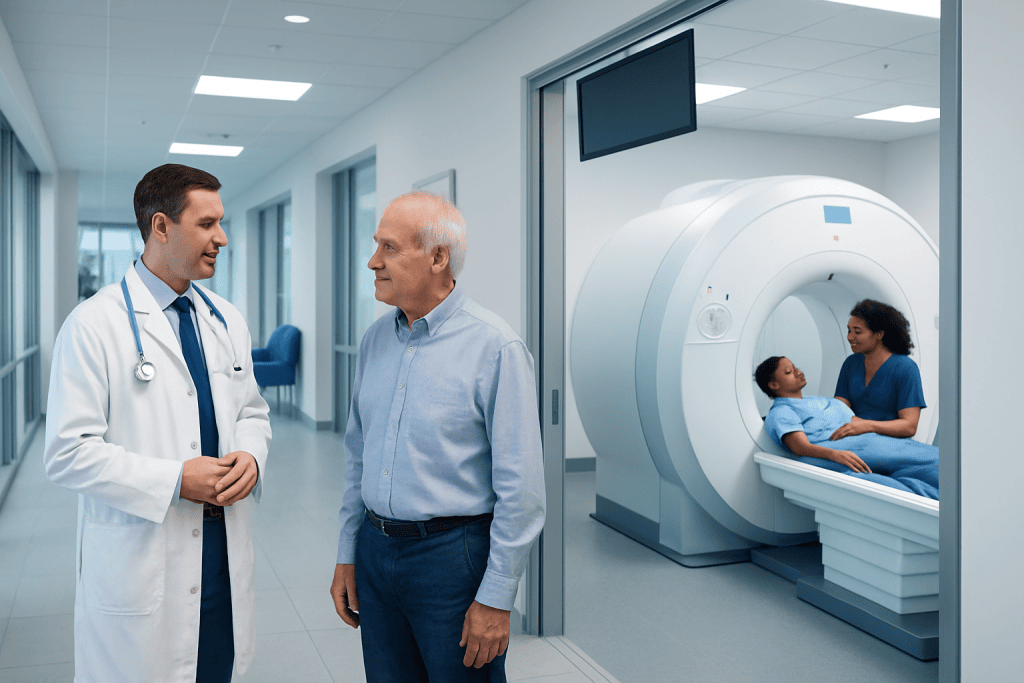
A routine brain MRI usually cannot diagnose Parkinson's disease by itself. MRI is valuable for ruling out structural or vascular causes of tremor, stiffness and slowed movement.
Key Takeaways
- A routine brain MRI usually cannot diagnose Parkinson's disease by itself.
- MRI is valuable for ruling out structural or vascular causes of tremor, stiffness and slowed movement.
- Preparation mainly involves MRI safety screening, including discussion of implants, metal fragments and kidney problems if contrast may be used.
- The scan commonly takes about 20 to 60 minutes, depending on the sequences needed and whether contrast is required.
- A neurologist diagnoses Parkinson's disease primarily through a detailed clinical assessment over time.
- New sudden neurological symptoms, such as weakness, speech difficulty or a severe sudden headache, need urgent medical assessment.
A Parkinson's brain MRI is usually used to look for other brain conditions that can resemble Parkinson's disease, rather than to confirm Parkinson's itself. The scan is painless, does not use radiation, and is interpreted alongside a neurological examination, symptom history and, when needed, other tests.
Parkinson's Brain MRI: What It Is and Why It Is Used
A Parkinson’s brain MRI is a magnetic resonance imaging scan of the brain performed when a person has symptoms such as tremor, stiffness, slowed movement, balance changes or difficulty walking. It uses a strong magnetic field and radio waves to create detailed images of brain structures. Unlike CT scanning and X-rays, MRI does not use ionising radiation.
In most people, a standard MRI does not show a specific image that confirms Parkinson’s disease. Instead, it helps a neurologist exclude other conditions that may cause similar symptoms, including previous small strokes, fluid-pressure disorders, brain tumours, inflammation, or patterns of brain change associated with atypical parkinsonism. This makes MRI an important supporting part of a careful assessment.
People being assessed for movement symptoms may also benefit from information about Parkinson's disease and related neurological conditions. The exact imaging plan is individual: some people need a routine non-contrast scan, while others need additional sequences or contrast dye to answer a specific clinical question.
How a Parkinson's Brain MRI Works and Who May Need One
MRI works by detecting signals from hydrogen atoms in the body’s tissues after they are placed in a magnetic field. A computer turns these signals into detailed cross-sectional images. Different MRI sequences highlight different features, such as fluid, blood vessels, areas of prior injury and subtle changes in brain tissue.
A neurologist may request a brain MRI when symptoms are unusual, begin at a younger age, progress quickly, affect one side in an unexpected way, or include early falls, marked cognitive changes, eye movement problems, weakness, numbness or other signs not typical of uncomplicated Parkinson’s disease. It may also be requested when the diagnosis remains uncertain after an examination.
MRI is suitable for many people, but the imaging team must check for safety before scheduling or scanning. Some implanted devices, metallic clips, cochlear implants, certain neurostimulators, retained metal fragments and older pacemakers may require special planning or may make MRI unsuitable. Modern MRI-conditional devices can often be scanned under specific protocols, so people should never assume that an implant automatically prevents an MRI.
- Tell the team about all implants, surgeries and possible metal exposure.
- Report pregnancy, kidney disease, allergies and severe claustrophobia.
- Bring prior scans or reports if they are available and relevant.
What Not to Do Before an MRI of the Brain?

Before a brain MRI, a person should not wear or bring metal objects into the scan room. This includes jewellery, watches, hearing aids, hairpins, removable dental appliances, belts, coins, keys and bank cards. Some cosmetics, tattoos and medicated skin patches may contain metallic substances, so it is helpful to ask the imaging centre for its instructions in advance.
People should not skip the MRI safety questionnaire or leave out information about implants, prior surgeries, shrapnel injuries, metal work or medication patches. Even a small metal fragment near the eyes can be important. Staff may arrange further checks, such as an X-ray, if there is concern about prior metal exposure.
For most non-contrast brain MRI scans, normal eating, drinking and regular medicines can continue unless the care team gives different instructions. If sedation is planned because of anxiety or claustrophobia, fasting and transport arrangements may be required. When contrast is being considered, the team may ask about kidney function and prior reactions to contrast agents.
It is also best not to self-medicate with a sedative before arriving unless it has been prescribed or approved by the clinician arranging the scan. Sedating medicines can affect safety, driving and the ability to complete screening. A person with Parkinson’s symptoms should ask whether usual movement-disorder medicines should be taken on schedule; in many cases, keeping to the usual regimen is appropriate.
What Happens During the Procedure?
On arrival, the patient checks in and completes a safety screening process. They change into clothing without metal if needed, remove personal items and may have an intravenous line placed if contrast is planned. A radiographer explains the scan, provides hearing protection and offers a call button to use if help is needed.
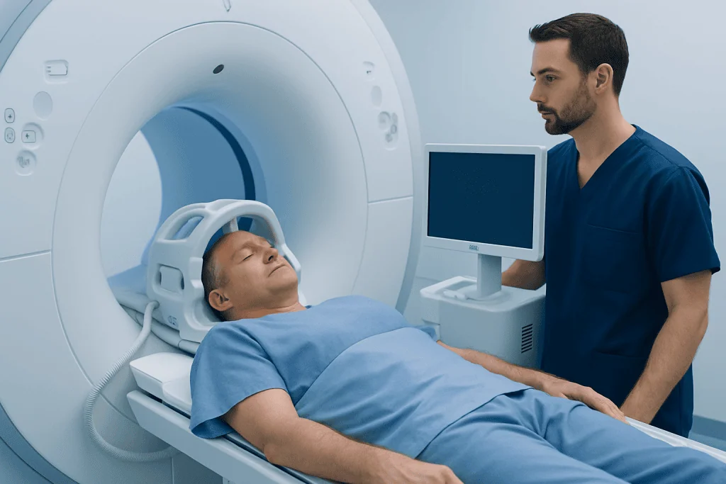
The patient lies on a padded table, usually on their back, with the head positioned in a special support called a head coil. The table then moves slowly into the MRI scanner. Keeping still is important because movement can blur the images. The scanner makes loud tapping and knocking sounds during image acquisition, but the test itself should not be painful.
A routine brain MRI commonly lasts around 20 to 60 minutes. Some centres provide music, a mirror or communication through an intercom. If contrast is used, gadolinium-based contrast material is injected through the IV partway through the examination to improve visibility of certain tissues or blood vessels.
Advanced imaging may occasionally be considered in specialist settings, but it is not a replacement for a clinical assessment. For patients who require a broader movement-disorder evaluation, neurology care can coordinate imaging, examination and follow-up planning.
Can You See Parkinson's Disease on a Brain MRI?
In typical Parkinson’s disease, a conventional brain MRI is often normal or shows only age-related changes. Therefore, a brain MRI cannot usually confirm or exclude Parkinson’s disease on its own. Parkinson’s is primarily a clinical diagnosis based on the pattern of symptoms and findings during a neurological examination.
However, MRI can show conditions that can mimic Parkinson’s disease or contribute to similar symptoms. These include strokes affecting movement pathways, normal-pressure hydrocephalus, brain masses, significant small-vessel disease and some structural changes associated with other neurodegenerative disorders. Identifying one of these findings can change the diagnosis and treatment plan.
Specialised MRI techniques may detect subtle changes in research or expert-centre settings, but their role varies and results must be interpreted carefully. Other tests may sometimes be useful when the diagnosis is unclear, including dopamine transporter imaging in selected cases. No scan result should be considered independently of the person’s symptoms, examination and medical history.
Imaging is one part of a wider process that may also include assessment of tremor, walking, sleep, cognition, autonomic symptoms and medication response. Depending on the findings, a clinician may discuss essential tremor or other possible causes of shaking and slowed movement.
How Does a Neurologist Know If You Have Parkinson's?
A neurologist diagnoses Parkinson’s disease by taking a detailed history and performing a focused neurological examination. The core movement feature is bradykinesia, meaning slowness and reduced amplitude of movement, together with tremor at rest and/or rigidity. The clinician also evaluates posture, balance, facial expression, handwriting, voice, arm swing and the pattern of walking.
The timing and progression of symptoms are important. Parkinson’s disease often begins gradually and may initially affect one side more than the other. A neurologist also asks about non-movement symptoms, such as reduced sense of smell, constipation, sleep behaviour changes, mood symptoms and urinary changes, while recognising that these symptoms have many possible causes.
The assessment includes looking for features that suggest an alternative diagnosis, such as very early severe falls, early marked blood-pressure instability, prominent eye movement abnormalities, rapid decline or signs of a stroke-related disorder. Blood tests, MRI and other investigations may be used to rule out other explanations when appropriate.
Follow-up matters because the diagnosis can become clearer over time. A person’s response to Parkinson’s medication may provide supporting information, but it is not the sole diagnostic test. Care may include movement-disorder specialists, physiotherapists, occupational therapists, speech and language therapists and other professionals, including physical therapy and rehabilitation when mobility or daily activities are affected.
Results, Recovery, Benefits and Possible Risks
After a non-sedated MRI, most people can return to normal activities immediately. If contrast was used, the IV is removed after the scan and normal hydration is generally encouraged unless a clinician advises otherwise. People who received sedation should follow the centre’s instructions, avoid driving and arrange for a responsible adult to accompany them home.
A radiologist reviews the images and sends a report to the clinician who ordered the scan. The neurologist then explains what the findings mean in the context of the person’s symptoms. MRI results may be reported within days, although timing differs between facilities and urgent findings are handled through established clinical communication processes.
The main benefit of MRI is its detailed view of the brain without radiation exposure. It can provide reassurance when no concerning structural cause is found and can identify findings that need further assessment. Still, a normal MRI does not mean symptoms should be ignored, and an abnormality on MRI does not automatically explain every symptom.
Risks are low but include discomfort from lying still, anxiety in enclosed spaces and noise exposure without proper hearing protection. Gadolinium contrast is generally well tolerated, although allergic-like reactions are uncommon and additional caution is needed for people with severe kidney impairment. The strongest safety concern is interaction between the MRI magnet and certain metal objects or devices, which is why complete screening is essential.
Do Doctors Typically Notify You Right Away If MRI Results Are Bad?
Not usually. In many settings, a radiologist reviews the images after the scan and sends a report to the referring doctor, who discusses the result at a follow-up appointment or by telephone. The time needed depends on the urgency of the request, local processes and whether comparison with earlier scans is required.
If the images show a finding that needs urgent action, the radiology team generally follows a process to alert the responsible clinician promptly. The patient may be asked to remain at the facility, contacted quickly, or directed to urgent care depending on the situation. This does not mean every unexpected finding is an emergency; many changes need thoughtful interpretation and planned follow-up.
Patients can ask before leaving how and when results will be communicated, who to contact if they have not heard back, and whether they can access a written report. It is reasonable to discuss questions with the doctor who ordered the MRI, because that clinician can relate the report to the person’s examination and medical history.
When to Seek Medical Care
A person should arrange medical assessment for a new or persistent tremor, increasing stiffness, slowed movement, changes in walking, recurrent falls, loss of dexterity or symptoms that interfere with work, self-care or quality of life. Early evaluation does not mean a person has Parkinson’s disease; these symptoms can have several causes, many of which can be assessed and managed.
Urgent medical care is needed for sudden weakness or numbness on one side, facial drooping, sudden speech or vision changes, a severe sudden headache, fainting, new seizures, sudden confusion or an abrupt major change in balance. These symptoms may indicate a time-sensitive neurological problem and should not wait for a routine MRI appointment.
For people travelling for care, Acibadem International’s multidisciplinary specialists and JCI-accredited hospitals can coordinate neurological assessment, imaging and treatment planning for international patients. A qualified clinician can help determine whether a Parkinson’s brain MRI is useful and explain the next appropriate steps.
Frequently asked questions
Does a brain MRI diagnose Parkinson's disease?
A standard brain MRI does not usually diagnose Parkinson's disease. It is mainly used to exclude other structural or vascular conditions that can cause similar symptoms. A neurologist makes the diagnosis primarily from the medical history and neurological examination.
Do I need contrast for a Parkinson's brain MRI?
Not always. Many brain MRI scans for Parkinson's-like symptoms are performed without contrast. Contrast may be added if the clinician needs to investigate inflammation, a tumour, blood-vessel changes or another specific concern.
Can I eat before a brain MRI?
Most people can eat and drink normally before a non-contrast brain MRI. Instructions may differ if sedation or contrast is planned, or if another test is being performed at the same visit. The imaging centre should provide individual preparation guidance.
What if I am claustrophobic during the MRI?
Claustrophobia is common and can often be managed with preparation, communication with the radiographer, music, a mirror or breaks when appropriate. In some cases, a clinician may recommend prescribed medication or an alternative scanner arrangement. It is best to mention this when the MRI is booked.
How long does it take to receive brain MRI results?
The reporting time varies by facility and clinical urgency. Many results are available within a few days, but the ordering clinician may need time to review the report and discuss its relevance. Urgent findings are generally communicated promptly through the healthcare team.
Is MRI safe if I have a pacemaker or other implant?
Some implants are MRI-conditional and can be scanned safely under defined protocols, while others may not be suitable. The MRI team needs the device type, manufacturer information and details of implantation before the appointment. A person should always disclose every implanted device and any history of metal injury.
References
- Parkinson's Foundation
- National Institute of Neurological Disorders and Stroke
- American College of Radiology
- National Health Service
- Radiological Society of North America
This article is for general information only and is not a substitute for professional medical advice. Please consult a qualified doctor about your individual situation.
Explore treatments in Turkey — costs, top hospitals & a free quote
JCI-accredited · board-certified surgeons · reply within 24h
Add us as a Preferred Source to see more of our trusted health content across Google Search, AI Overviews and Discover.

More from the Health Library



Related Specialists

Fzt. Ahsen Ercan
Physical Medicine & Rehabilitation
Prof. Dr. Gürhan Şişman
Gastroenterology
Assoc. Prof. Dr. Süleyman Şahin
Medical Oncology
Dr. Melda Koyuncu
Dermatology