

PET Scan of Whole Body: Preparation, Procedure and Results
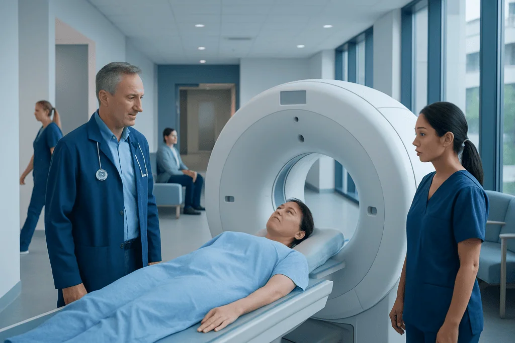
A whole-body PET scan usually combines PET and CT images to show both metabolic activity and body structures. Preparation often includes fasting for several hours, avoiding strenuous exercise, and following individualized instructions for diabetes medicines.
Key Takeaways
- A whole-body PET scan usually combines PET and CT images to show both metabolic activity and body structures.
- Preparation often includes fasting for several hours, avoiding strenuous exercise, and following individualized instructions for diabetes medicines.
- PET scans can identify suspicious areas but cannot diagnose cancer on their own; results must be interpreted with medical history, examination findings, and other tests.
- The scan itself is painless, although the appointment commonly takes two to three hours because time is needed for tracer uptake.
- Most people can return to usual activities soon afterward and should drink fluids unless their care team advises otherwise.
A PET scan of whole body is an imaging test that uses a small amount of radioactive tracer to show areas of increased or reduced metabolic activity throughout the body. It is commonly combined with CT imaging and may help doctors diagnose, stage, monitor, or assess treatment response for several conditions.
Overview: what a PET scan of whole body shows
A PET scan of whole body, also called a whole-body positron emission tomography scan, is a nuclear medicine imaging examination that shows how tissues are functioning. It is frequently performed together with computed tomography (CT), creating a PET-CT scan. PET highlights metabolic activity, while CT provides detailed information about anatomy, helping clinicians locate and interpret findings more accurately.
Most PET scans use a tracer called fluorodeoxyglucose (FDG), a glucose-like substance tagged with a very small amount of radioactive material. After injection, active cells absorb the tracer. Many cancers may use glucose more rapidly than surrounding tissue, but infection, inflammation, healing tissue, and normal organs can also show increased uptake. For this reason, a PET scan is one important part of an overall medical assessment rather than a stand-alone diagnosis.
Doctors may request whole-body PET imaging to investigate a concerning finding, determine the extent of a known cancer, assess response to treatment, look for recurrence, or evaluate selected inflammatory and infectious conditions. The exact area scanned and the tracer used depend on the clinical question.
How the scan works and who may need it
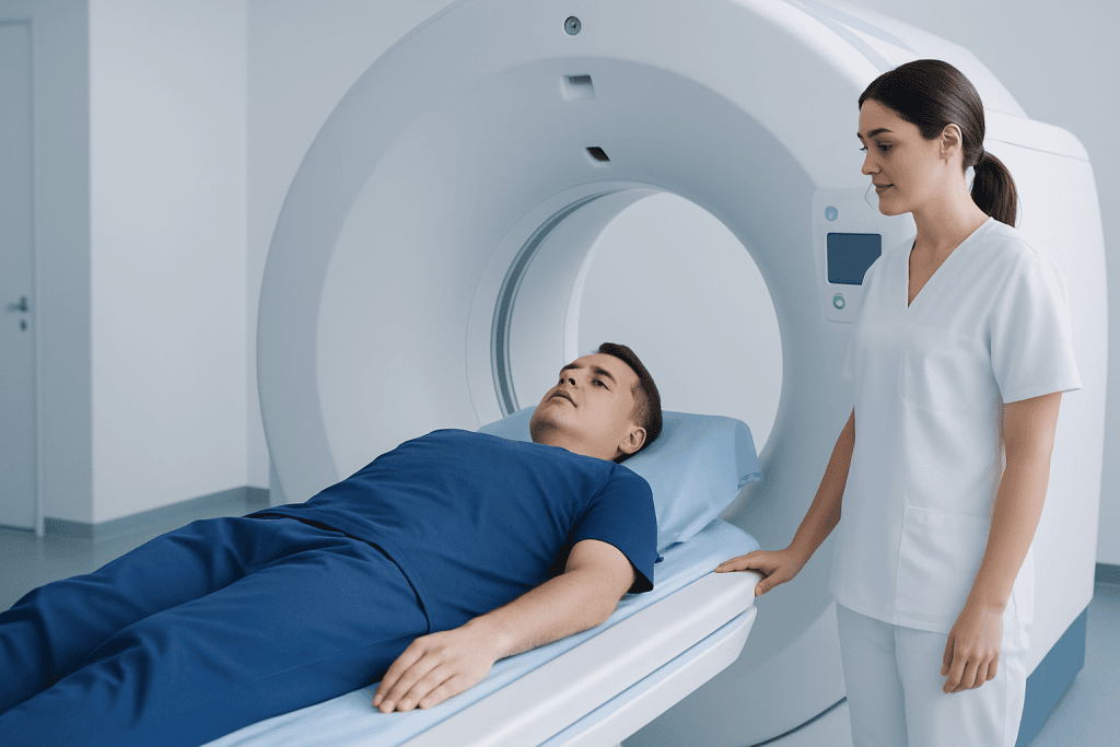
After the tracer enters the bloodstream, it circulates and is taken up by tissues. A PET scanner detects signals released as the tracer decays and uses them to create images of metabolic activity. The accompanying CT scan adds anatomical detail, allowing the radiologist or nuclear medicine physician to compare areas of tracer uptake with organs, lymph nodes, bones, and other structures.
Whole-body PET-CT is most often used in oncology. It can support the evaluation of conditions such as lung cancer and other cancers when a specialist needs to understand disease extent or treatment response. It may also be used in specific circumstances for unexplained inflammation, certain infections, or some heart and brain conditions, although these examinations may require different tracers or protocols.
Not everyone needs a PET scan. A doctor considers symptoms, prior imaging, laboratory results, the type of suspected or confirmed condition, and whether the answer is likely to change care. Pregnancy, breastfeeding, diabetes, kidney problems, recent surgery, infection, and recent treatments should be discussed before scheduling because they can affect safety planning or image interpretation.
Preparing for a whole-body PET scan
Preparation is essential because blood sugar, physical activity, and some medicines can influence how the tracer is distributed. The imaging center provides the most important instructions, which should always take priority. For an FDG PET scan, many people are asked to eat a low-carbohydrate diet the day before and to fast for several hours before the appointment; plain water is often allowed and encouraged.
What not to do 24 hours before a PET scan? Unless the imaging team gives different advice, people are usually asked to avoid strenuous exercise for at least 24 hours. Heavy exercise can increase glucose uptake in muscles and make images more difficult to interpret. They may also be asked to avoid sugar-containing drinks, candy, chewing gum, smoking, alcohol, and unnecessary caffeine, depending on the protocol. Keeping warm and avoiding shivering before the scan can also help prevent normal muscle or fat activity from affecting the images.
What are the best foods to eat the day before a PET scan? If a low-carbohydrate preparation is requested, suitable foods may include eggs, fish, poultry, meat, tofu, non-starchy vegetables, cheese, and healthy fats such as olive oil or avocado. Bread, rice, pasta, potatoes, cereals, sweets, fruit juice, and sweetened drinks are commonly limited. Requirements vary, especially for scans that use tracers other than FDG, so the patient should not make major dietary changes without checking the instructions from the imaging department.
People with diabetes need individualized guidance. Fasting and diabetes medicines must be coordinated safely to avoid low or high blood glucose, either of which may compromise the scan. The patient should tell the team about all medicines, insulin use, allergies, pregnancy or possible pregnancy, breastfeeding, and any difficulty lying flat or staying still.
Step by step: what happens during the procedure
On arrival, staff confirm the patient’s details, medical history, current medicines, and preparation. Blood glucose may be checked before FDG is administered. An intravenous line is placed, and the tracer is injected through a vein. The injection is generally brief and feels similar to a routine blood test or intravenous medication.
The patient then rests quietly for about 45 to 60 minutes while the tracer is absorbed. During this uptake period, they are usually asked to remain relaxed, limit talking and movement, and stay comfortably warm. This quiet waiting period is a key reason the overall appointment takes longer than the time spent in the scanner.
For the scan, the patient lies on a padded table that moves slowly through a ring-shaped scanner. The equipment is open at both ends and is typically less confining than a traditional MRI scanner. It is important to remain still; the imaging portion often takes 20 to 45 minutes, depending on the protocol and body area covered. Staff can communicate with the patient throughout the examination.
Some PET-CT studies use CT contrast material. If contrast is planned, the patient may feel temporary warmth or a metallic taste. The team should be told in advance about prior contrast reactions, asthma, kidney disease, or thyroid conditions. In appropriate clinical settings, PET-CT may be part of the assessment before or after chemotherapy treatment or other cancer therapies.
Benefits, limits, risks, and recovery timeline
A key benefit of whole-body PET-CT is that it can reveal changes in cellular activity before an abnormality causes clear structural changes on CT or other imaging. It can help clinicians identify sites that may need further evaluation, guide biopsy planning, determine disease stage, and assess whether treatment is having the expected effect. It may reduce uncertainty when used alongside other appropriate tests.
However, PET findings are not specific to cancer. Inflammation, infection, recent surgery, radiation treatment, vaccination, and normal activity in organs such as the brain, heart, kidneys, and bladder can cause tracer uptake. Conversely, some cancers have low FDG uptake or may be too small to detect. A suspicious result may therefore lead to follow-up imaging, repeat testing, or a biopsy rather than an immediate conclusion.
The radiation exposure from the tracer and CT component is carefully considered against the expected medical benefit. Allergic reactions to the PET tracer are very uncommon; if CT contrast is used, contrast-related reactions are possible but also uncommon. The intravenous injection may cause mild bruising or discomfort. Patients should discuss pregnancy or breastfeeding with their care team, as timing or feeding recommendations may need adjustment.
Recovery is usually immediate. Most people can eat, drink, drive, and return to usual activities after the scan unless they received sedation or were given other instructions. Drinking extra water for the rest of the day may help clear the tracer through urine. For several hours, the imaging department may advise limiting prolonged close contact with pregnant people and young children, based on the tracer and local protocol.
Understanding the report and timing of results
Do you get your PET scan results immediately? Usually, no. Although the images are available as soon as the scan is complete, a radiologist or nuclear medicine physician needs time to review them carefully, compare them with earlier scans, and prepare a formal report. The referring doctor then discusses what the findings mean in the context of the person’s health and other test results.
Timing varies by hospital, the complexity of the scan, and whether urgent review is needed. Some results are discussed within a few days, while others may take longer. A patient can ask the imaging center or referring clinician when to expect the report and how it will be communicated.
A report may describe areas of increased, decreased, or normal tracer uptake and may use terms such as “avid,” “hypermetabolic,” or “physiologic.” These descriptions are not diagnoses by themselves. The next step might be no action, clinical follow-up, additional imaging, laboratory testing, or tissue sampling. When cancer care is needed, PET findings can help inform an individualized plan that may include radiotherapy or surgery alongside systemic treatments.
When to seek medical care
Patients should contact the imaging department before the appointment if they are ill, have a fever, have recently had surgery or a vaccination, cannot follow the fasting instructions, or have blood glucose that is difficult to manage. These factors do not necessarily prevent a scan, but the team may need to adjust scheduling or preparation to obtain reliable images safely.
After the scan, urgent medical care is appropriate for signs of a severe allergic reaction, such as trouble breathing, swelling of the face or throat, widespread hives, or fainting. These reactions are rare. Mild tenderness or bruising at the injection site generally settles without treatment, but persistent redness, swelling, or worsening pain should be reported to a healthcare professional.
A PET scan is ordered to answer a specific clinical question and should be reviewed with the referring doctor rather than interpreted alone. Acibadem International’s multidisciplinary specialists and JCI-accredited hospitals diagnose and treat complex conditions for international patients, coordinating imaging findings with appropriate clinical care.
Frequently asked questions
Will a full body PET scan show any cancer?
A full-body PET scan can detect many areas with unusually high metabolic activity, which may be caused by cancer. However, it cannot show every cancer, and increased tracer uptake can also occur with infection, inflammation, healing, or normal body functions. A doctor may recommend additional imaging or a biopsy to confirm the cause of an abnormal finding.
How long does a PET scan of whole body take?
The scan appointment commonly lasts about two to three hours. Much of this time is the quiet uptake period after the tracer injection, often around 45 to 60 minutes. The actual imaging time is commonly 20 to 45 minutes, but it can vary with the type of examination.
Can a person drink water before a PET scan?
Plain water is often permitted and may be encouraged before an FDG PET scan, even during the fasting period. Drinks containing sugar, milk, juice, alcohol, or sweeteners may interfere with preparation. The patient should follow the specific instructions provided by the imaging center.
Is a whole-body PET scan painful?
The scan itself is painless. A person may feel a brief pinch when the intravenous line is placed and may need to lie still on the scanning table. Anyone with pain, anxiety, claustrophobia, or difficulty lying flat should tell the imaging team beforehand so they can help plan the examination.
Can I take my regular medicines before a PET scan?
Many routine medicines can be taken with plain water, but instructions differ depending on the scan and the medication. Diabetes medicines, insulin, steroid medicines, and drugs that affect blood sugar may require special timing. Patients should not stop prescribed medication unless their clinician or imaging team tells them to do so.
What happens if a PET scan result is abnormal?
An abnormal result means that an area showed tracer uptake or another feature that needs clinical interpretation; it does not automatically mean cancer. The referring doctor will consider the scan together with symptoms, examination findings, blood tests, prior imaging, and medical history. Further testing may be advised when it is needed to clarify the finding.
References
- National Cancer Institute
- Radiological Society of North America
- Society of Nuclear Medicine and Molecular Imaging
- American College of Radiology
- International Atomic Energy Agency
This article is for general information only and is not a substitute for professional medical advice. Please consult a qualified doctor about your individual situation.
Explore treatments in Turkey — costs, top hospitals & a free quote
JCI-accredited · board-certified surgeons · reply within 24h
Add us as a Preferred Source to see more of our trusted health content across Google Search, AI Overviews and Discover.

More from the Health Library


Related Specialists

Prof. Dr. Gül Esen İçten
Breast Clinic Clinical Service
Dr. Ecem Ertürk
Dermatology
Fzt. Busenur Sezer
Physical Medicine & Rehabilitation
Prof. Dr. Mehdi S. Öğüt
Ophthalmology