Pleural Effusion: Fluid Around the Lung, Causes, and Drainage Options
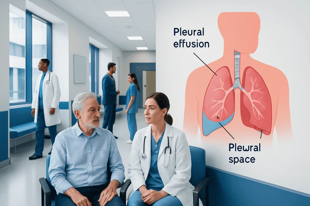
Pleural effusion means fluid has collected around the lung, which can make breathing uncomfortable or difficult. Common causes include heart failure, pneumonia, cancer, pulmonary embolism, liver disease, kidney disease, and inflammatory conditions.
Key Takeaways
- Pleural effusion means fluid has collected around the lung, which can make breathing uncomfortable or difficult.
- Common causes include heart failure, pneumonia, cancer, pulmonary embolism, liver disease, kidney disease, and inflammatory conditions.
- Diagnosis usually involves a physical examination, chest imaging, and sometimes thoracentesis to test the fluid.
- Drainage options include thoracentesis, chest tube drainage, pleural catheter placement, and procedures to prevent recurrence in selected cases.
- Anyone with new shortness of breath, chest pain, fever, or worsening symptoms should seek medical care promptly.
Pleural effusion is a buildup of fluid in the pleural space, the thin gap between the lung and the chest wall. It is a sign of an underlying condition, and treatment focuses on relieving symptoms, identifying the cause, and preventing the fluid from returning.
Overview
Pleural effusion is the medical term for a buildup of fluid around the lung. The lungs are covered by a thin membrane called the pleura, and the inside of the chest wall is lined by another pleural layer. Normally, a very small amount of lubricating fluid sits between these layers so the lungs can move smoothly during breathing. When extra fluid collects in this space, it can limit lung expansion and cause symptoms.
Pleural effusion is not a disease by itself. It is usually a sign that another condition is affecting the lungs, heart, blood vessels, liver, kidneys, immune system, or chest cavity. Some effusions are small and found incidentally on an X-ray or scan. Others are large enough to cause breathlessness, cough, or chest discomfort.
Doctors often describe pleural fluid as transudate or exudate. A transudative effusion is commonly related to pressure or fluid-balance problems, such as heart failure or low blood protein. An exudative effusion is usually linked to inflammation, infection, cancer, blood clots, or injury. This distinction helps guide testing and treatment.
Symptoms
The symptoms of pleural effusion depend on the amount of fluid, how quickly it develops, and the underlying cause. A small effusion may cause no symptoms at all. A larger or rapidly developing effusion can make breathing feel restricted because the lung cannot fully expand.
Common symptoms may include shortness of breath, especially with activity or when lying flat; chest discomfort; a dry cough; and a feeling of heaviness or pressure on one side of the chest. Some people describe sharp chest pain that worsens with deep breathing or coughing, which may suggest pleural irritation.
Symptoms related to the cause may also be present. For example, fever, chills, and sputum production can occur with pneumonia. Leg swelling and fatigue may be seen with heart failure. Unexplained weight loss, persistent cough, or coughing blood require careful medical assessment. The pattern of symptoms helps the healthcare team decide how urgently to investigate and whether drainage is needed.
Causes and Risk Factors

Many conditions can lead to fluid around the lung. One of the most common causes is heart failure, where pressure changes in the blood vessels can push fluid into the pleural space. Lung infections, including pneumonia, can cause an inflammatory effusion and sometimes pus in the pleural space, known as empyema. Pulmonary embolism, a blood clot in the lung, may also cause pleural fluid and chest pain.
Cancer can cause pleural effusion when it affects the pleura directly or blocks normal fluid drainage. Lung cancer, breast cancer, lymphoma, and cancers that spread from other organs are among the possible causes. Liver cirrhosis, kidney disease, low blood protein, pancreatitis, autoimmune diseases such as rheumatoid arthritis or lupus, and complications after surgery or chest injury can also contribute.
Risk factors depend on the underlying condition. Smoking, chronic lung disease, previous cancer, heart disease, recent surgery, immobility, blood clotting disorders, infections, and immune system problems may increase the chance of developing pleural effusion. Because the list of possible causes is broad, medical evaluation is important even when symptoms are mild.
- Transudative causes often involve fluid balance, pressure, or protein levels.
- Exudative causes often involve inflammation, infection, cancer, or blood clots.
- Recurrent effusions need careful follow-up to identify and control the cause.
Diagnosis
Diagnosis begins with a medical history and physical examination. A doctor may listen for reduced breath sounds over the affected area and may check for signs of heart, liver, kidney, infection, or clotting problems. The pattern of symptoms, medical history, medications, and recent illnesses helps narrow the possible causes.
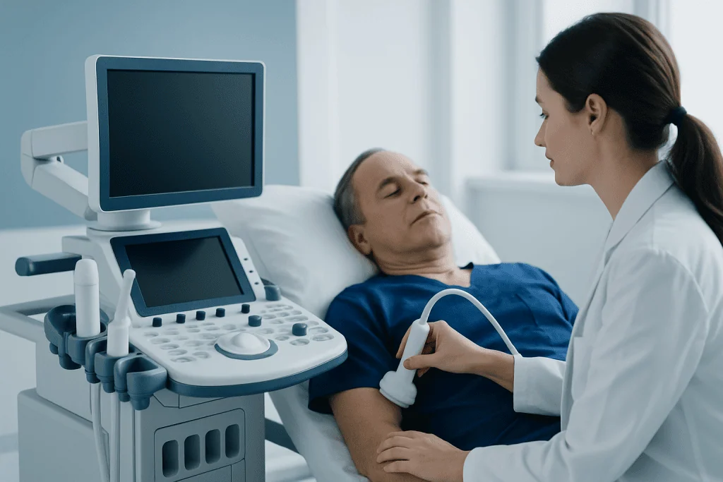
Imaging is central to confirming pleural effusion. A chest X-ray can show fluid, especially when the effusion is moderate or large. Ultrasound is very useful because it can detect smaller amounts of fluid, show whether the fluid is free-flowing or divided into pockets, and guide safe drainage. Computed tomography, or CT, may be recommended when doctors need more detail about the lungs, pleura, lymph nodes, or chest structures.
In many cases, a procedure called thoracentesis is performed to remove a sample of fluid for testing. The laboratory can measure protein, lactate dehydrogenase, glucose, pH, cell counts, and other markers. Tests may also look for bacteria, tuberculosis, malignant cells, or signs of inflammation. Blood tests, echocardiography, sputum studies, or additional imaging may be used depending on the suspected cause.
Treatment and Drainage Options
Treatment depends on the cause, the size of the effusion, how quickly it developed, and how much it affects breathing. If the effusion is small and the cause is clear, doctors may treat the underlying condition and monitor the fluid with follow-up examinations or imaging. For example, pleural effusion related to heart failure may improve with medicines that reduce fluid overload, while infection-related effusions require appropriate antimicrobial treatment and monitoring.
Thoracentesis is a common drainage procedure used for diagnosis and symptom relief. A doctor uses ultrasound guidance in many cases, numbs the skin, and places a needle or small catheter into the pleural space to remove fluid. Patients often notice easier breathing after a significant amount of fluid is removed. The healthcare team monitors comfort, oxygen levels, and any post-procedure symptoms.
Some effusions need more than a single thoracentesis. A chest tube may be placed when the fluid is infected, thick, bloody, or difficult to drain. In recurrent effusions, especially when fluid keeps returning despite treatment of the cause, options may include an indwelling pleural catheter for intermittent home or clinic drainage, or pleurodesis, a procedure that helps the pleural layers stick together to reduce reaccumulation. Surgery, such as video-assisted thoracoscopic surgery, may be considered in selected cases to break down pockets of fluid, take biopsies, or treat complex pleural disease.
Each drainage option has benefits and possible risks, such as pain, bleeding, infection, low blood pressure, or pneumothorax, which means air around the lung. These complications are not common when procedures are carefully planned, but they are the reason pleural drainage should be performed by trained healthcare professionals with appropriate monitoring.
Prevention and Self-care
Not all pleural effusions can be prevented, because they often result from medical conditions that require professional care. However, controlling known health problems can reduce the chance of recurrence. People with heart failure, kidney disease, liver disease, autoimmune disease, or cancer should follow their treatment plan and attend regular follow-up appointments.
Self-care focuses on supporting breathing and recognizing changes early. Patients should take prescribed medicines as directed, avoid smoking and secondhand smoke, stay current with recommended vaccinations, and report symptoms such as fever, increasing cough, swelling, or worsening shortness of breath. Gentle activity may be helpful when approved by a doctor, but overexertion should be avoided during active breathing symptoms.
After a drainage procedure, patients are usually given instructions about wound care, activity, and warning signs. If an indwelling pleural catheter is used, the care team teaches safe drainage technique, dressing changes, and when to contact the clinic. Keeping follow-up visits is important because the underlying cause often determines long-term outcomes more than the fluid itself.
When to See a Doctor
Medical evaluation is recommended for new or unexplained shortness of breath, chest pain with breathing, persistent cough, fever, or fatigue that does not improve. People who already have heart disease, lung disease, cancer, blood clots, liver disease, or kidney disease should seek advice if breathing symptoms change or worsen.
Urgent care is needed for severe shortness of breath, blue lips or fingertips, fainting, confusion, coughing blood, high fever with chest pain, or sudden sharp chest pain. These symptoms may indicate a serious infection, blood clot, heart problem, or a large effusion that needs prompt assessment.
Patients traveling for diagnosis or treatment may benefit from coordinated respiratory, cardiology, oncology, radiology, and thoracic surgery care. Acibadem International provides multidisciplinary evaluation and treatment for pleural effusion in JCI-accredited hospitals for international patients, with decisions tailored to the individual diagnosis and overall health.
Frequently asked questions
Is pleural effusion the same as fluid in the lungs?
Pleural effusion means fluid is around the lung, in the pleural space between the lung and chest wall. This is different from pulmonary edema, where fluid collects inside the lung tissue and air spaces. Both can cause shortness of breath, so imaging and medical evaluation are needed to tell them apart.
Can pleural effusion go away on its own?
Some small effusions improve when the underlying cause is treated, such as heart failure or a mild infection. However, pleural effusion should not be ignored because it can be caused by many different conditions. A doctor can decide whether monitoring, medication, fluid testing, or drainage is appropriate.
Is thoracentesis painful?
Thoracentesis is usually performed with local anesthetic to numb the area. Patients may feel pressure or mild discomfort, but the procedure is generally well tolerated. The healthcare team monitors the patient during and after the procedure and gives instructions for recovery.
How much fluid can be drained from a pleural effusion?
The amount removed depends on the size of the effusion, the reason for drainage, symptoms, and the patient's response during the procedure. Doctors may stop drainage if symptoms occur or if enough fluid has been removed for safe relief and testing. The decision is individualized rather than based on one fixed amount.
Why does pleural effusion come back after drainage?
Drainage removes fluid but may not fully correct the condition causing it. Effusions can recur with ongoing heart failure, infection, cancer, liver disease, kidney disease, or inflammation. If fluid returns repeatedly, doctors may consider additional treatments such as an indwelling pleural catheter, pleurodesis, or more targeted treatment of the cause.
Is pleural effusion cancer?
Pleural effusion is not automatically cancer. Many non-cancer conditions, including heart failure and pneumonia, can cause it. When cancer is a possible cause, pleural fluid analysis, imaging, and sometimes pleural biopsy help doctors make an accurate diagnosis.
References
- American Thoracic Society
- British Thoracic Society
- National Heart, Lung, and Blood Institute
- Merck Manual Professional Edition
- Mayo Clinic
This article is for general information only and is not a substitute for professional medical advice. Please consult a qualified doctor about your individual situation.
Pleural Effusion in Turkey — costs, top hospitals & a free quote
JCI-accredited · board-certified surgeons · reply within 24h
Add us as a Preferred Source to see more of our trusted health content across Google Search, AI Overviews and Discover.