Pleurx Catheter Removal: Procedure, Recovery and Results

A PleurX catheter may be removed when drainage remains low, symptoms are controlled, and the clinical team confirms it is no longer needed. Removal is commonly performed using local anesthetic in an outpatient or bedside setting.
Key Takeaways
- A PleurX catheter may be removed when drainage remains low, symptoms are controlled, and the clinical team confirms it is no longer needed.
- Removal is commonly performed using local anesthetic in an outpatient or bedside setting.
- Mild soreness, bruising, and a small amount of drainage from the site can occur temporarily after removal.
- Contact the care team promptly for fever, worsening redness, pus-like drainage, increasing shortness of breath, or severe chest pain.
- Recovery depends both on healing at the catheter site and on the underlying cause of the pleural effusion.
PleurX catheter removal is usually a short, planned procedure performed after pleural fluid drainage has reduced or stopped and imaging suggests the fluid is controlled. Most people go home the same day, with a dressing over the small removal site and clear instructions for monitoring recovery.
Overview: how PleurX catheter removal works
PleurX catheter removal is the process of taking out an indwelling pleural catheter, a soft tube that has allowed pleural fluid to be drained from the space around the lungs. These catheters can help relieve breathlessness caused by recurrent pleural effusion, including effusions related to cancer, heart failure, infection, or other conditions. Removal is considered when the catheter is no longer providing a meaningful benefit or when a catheter-related concern needs attention.
For many people, removal becomes possible after drainage volumes have stayed low over time and symptoms have not returned between drainages. This can occur when the pleural space has naturally closed around the lung, sometimes called pleurodesis, or when the cause of fluid buildup is better controlled. The decision is individualized and is based on symptoms, drainage records, examination findings, and sometimes chest imaging.
The pleurx catheter removal procedure is generally brief. A clinician releases the small cuff that helps hold the catheter in place beneath the skin, then gently withdraws the tube and closes or dresses the site. It is not the same as draining pleural fluid with a needle, although both procedures involve care of the pleural space.
Who may be a candidate for catheter removal?
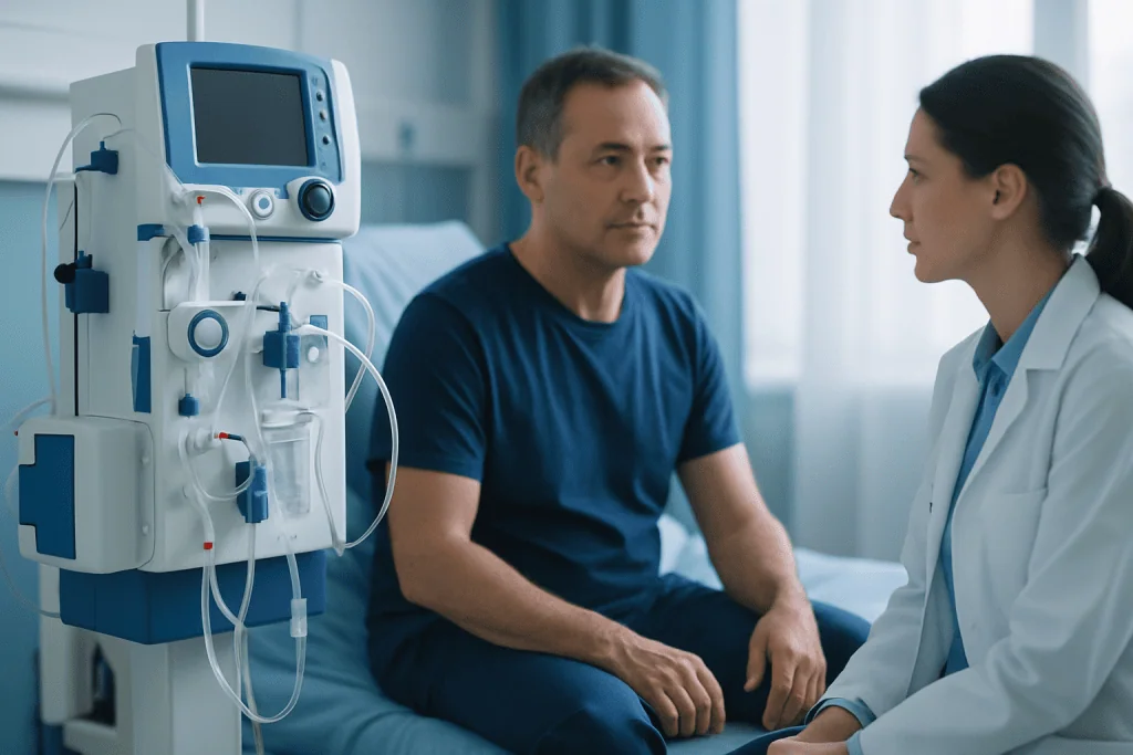
A clinician may recommend removal when the catheter has produced little or no fluid during several drainage sessions and the person remains comfortable without worsening breathlessness. Chest X-ray or ultrasound may be used to check for a significant return of fluid. The exact drainage threshold and timing vary because the right decision depends on the reason the catheter was placed and the person’s overall health.
Removal may also be advised if the catheter becomes blocked, displaced, damaged, painful, or infected. In these situations, the clinical team will first assess whether the catheter should be removed, exchanged, treated with medication, or left in place temporarily. If infection is suspected, prompt assessment is especially important because management may include antibiotics and drainage planning.
Before the appointment, patients should tell the team about blood-thinning medicines, medication allergies, bleeding disorders, fever, new cough, chest discomfort, or changes in drainage. The clinician will provide individualized instructions about medicines, eating and drinking, and whether someone should accompany the patient home. Underlying pleural fluid problems may be assessed alongside related conditions such as pleural effusion.
PleurX catheter removal procedure: step by step
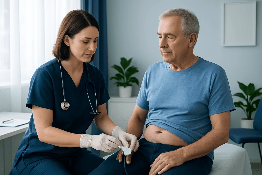
The procedure is often completed in a clinic, procedure room, or hospital bedside setting. The patient is positioned comfortably, usually sitting partly upright or lying on one side. The skin around the catheter is cleaned carefully, sterile drapes are used, and local anesthetic is injected to numb the area. Most people remain awake throughout the procedure.
The clinician makes a small incision, if needed, near the catheter’s entry site. The tissue around the cuff is gently freed, allowing the catheter to be withdrawn slowly. Mild pulling or pressure can be felt, but sharp pain should be reported so that additional local anesthetic or comfort measures can be considered.
After the catheter is removed, the site is closed with stitches, skin-closure strips, or a dressing depending on the technique and skin condition. The team checks that bleeding is controlled and explains how to keep the area clean and dry. A chest X-ray is not required after every removal, but it may be ordered if there are symptoms, a difficult removal, or concern about fluid or air around the lung.
A clinical record may refer to a “pleurx catheter removal procedure note,” “pleurx catheter removal note,” or “pleurx removal procedure note.” This documentation commonly records the reason for removal, anesthetic used, how the catheter was removed, the condition of the catheter and skin site, complications if any, and aftercare instructions.
What happens after pleural catheter removal?
After pleural catheter removal, patients are usually observed briefly and can often return home the same day. The site is covered with a sterile dressing, and there may be mild tenderness, bruising, or a small amount of clear or blood-tinged drainage. The care team will explain when the dressing can be changed and when showering is safe; instructions vary according to the closure method and the condition of the skin.
It is common to be asked to avoid soaking in a bath or swimming until the wound has closed fully. Heavy lifting and strenuous upper-body exercise may need to be limited for a short time to protect the site. Normal gentle movement, short walks, and regular breathing are usually encouraged unless the treating clinician gives different advice.
Follow-up may include a wound check, stitch removal if non-dissolvable stitches were used, review of breathing symptoms, and imaging when clinically appropriate. The goal is not only to ensure the skin heals but also to identify early whether fluid is returning. Ongoing management may involve a respiratory physician, interventional radiologist, thoracic surgeon, oncologist, cardiologist, or another specialist depending on the cause of the effusion.
How long does it take to heal after removing a catheter?
The small skin opening often begins sealing within days, while complete surface healing may take around one to two weeks. If sutures are used, they may be removed at a follow-up visit or dissolve on their own, depending on the type. Healing can take longer in people with fragile skin, diabetes, poor nutrition, immune suppression, ongoing infection, or cancer treatment.
Comfort usually improves steadily over the first few days. Simple pain relief may be recommended by the clinician if needed, taking into account other medicines and medical conditions. The dressing should be kept clean and changed as instructed; patients should not apply creams, powders, or adhesive products unless their care team recommends them.
Healing of the skin does not always mean the pleural condition itself is fully resolved. A person may feel well after removal yet need continued follow-up for the disease that caused the fluid buildup. Any new breathlessness, chest pressure, or a noticeable decline in exercise tolerance should be discussed with the care team rather than assumed to be part of normal wound healing.
How long does it take to recover from pleural effusion drainage?
Recovery from pleural effusion drainage varies widely. Some people notice easier breathing within hours or days after fluid is drained, while others recover more gradually because the lung, chest muscles, or underlying illness needs additional time to stabilize. A PleurX catheter is often used when repeated drainage is needed, so recovery may be tied more closely to the cause of the fluid than to the removal procedure itself.
Fatigue can persist after pleural drainage, particularly when the effusion was large, recurrent, infection-related, or associated with cancer, heart disease, kidney disease, or another long-term condition. The clinical team may recommend follow-up imaging, blood tests, medication review, rehabilitation, nutrition support, or treatment for the underlying condition.
Breathing exercises, gentle activity, adequate fluids where medically appropriate, and gradual return to usual routines can support recovery. However, patients should follow condition-specific advice, especially if they have heart, kidney, or lung disease. Persistent or worsening breathlessness should always be medically assessed because it may indicate recurrent fluid, infection, anemia, blood clotting problems, or another cause.
Is pain after PleurX drainage normal?
Mild discomfort during or after PleurX drainage can be normal, especially when fluid is removed quickly or when the pleural space is becoming less expandable as fluid volume decreases. People may describe pressure, pulling, aching near the catheter site, or a brief cough. Drainage should generally be slowed or stopped if pain becomes significant, and the care team should be informed.
After catheter removal, mild site pain or tenderness is also expected for a short period. It should gradually improve rather than intensify. Severe pain, new sharp chest pain, pain with marked breathlessness, faintness, or pain accompanied by fever requires timely medical assessment.
Drainage plans should always be individualized. Patients should not alter the drainage schedule, use unapproved solutions, or attempt to remove a catheter themselves. For ongoing pleural care, clinicians may discuss options such as thoracentesis or other interventions based on the cause, recurrence pattern, lung expansion, and overall treatment goals.
Benefits, risks and when to seek medical care
The main benefit of removing a PleurX catheter is avoiding the ongoing care burden and potential complications of a device that is no longer necessary. Removal can reduce skin irritation, dressing changes, drainage supplies, and the possibility of catheter blockage or infection. It also allows the skin to heal and can make daily activities more comfortable.
Pleurx catheter removal complications are uncommon but may include bleeding, pain, skin infection, difficulty removing the cuff, catheter breakage, wound separation, fluid leakage, recurrent pleural effusion, or a small pneumothorax (air around the lung). The care team takes steps to reduce these risks and will advise whether observation or imaging is needed after removal.
When to seek medical care: Patients should contact their clinical team urgently for fever, chills, increasing redness or warmth around the wound, swelling, pus-like or foul-smelling drainage, persistent bleeding, worsening pain, or a wound that opens. Emergency care is appropriate for severe or rapidly worsening shortness of breath, severe chest pain, blue or gray lips, confusion, or fainting.
Acibadem International’s multidisciplinary specialists and JCI-accredited hospitals provide assessment and treatment for pleural conditions for international patients. Care plans may involve respiratory medicine, thoracic surgery, oncology, imaging, and supportive care, with follow-up tailored to the reason the catheter was originally needed.
Frequently asked questions
Can a PleurX catheter be removed at home?
Removal should be performed by a qualified healthcare professional using sterile technique and appropriate equipment. In selected situations it may be done in a home-care setting by an authorized clinician, but patients and family members should not attempt removal themselves. The treating team will decide the safest location.
Why is my PleurX catheter not draining before removal?
Low or absent drainage can mean that the pleural fluid has reduced, which may be a positive reason to consider removal. It can also occur if the catheter is blocked, kinked, misplaced, or if fluid is present in a separate pocket. The care team may review drainage records, symptoms, and imaging before deciding what to do.
Can pleural fluid return after a PleurX catheter is removed?
Yes, fluid can return, particularly if the condition causing the pleural effusion remains active. Some people have durable control of fluid after catheter removal, while others need further assessment or treatment. New breathlessness or chest discomfort after removal should be reported promptly.
Will I need stitches after PleurX catheter removal?
Some removal sites are closed with stitches, while others are managed with skin-closure strips and a dressing. The approach depends on the removal technique, the size and condition of the wound, and the clinician’s judgment. Patients should follow the instructions given for dressing care and stitch removal.
Can I shower after PleurX catheter removal?
Showering is often possible after a short period, but the exact timing depends on the type of dressing and closure used. The site usually needs to be kept dry until the clinician says otherwise. Swimming, bathing, and hot tubs should generally be avoided until the wound is fully closed.
What should I bring to my catheter removal appointment?
Patients may be asked to bring their drainage log, a current medication list, and details of any recent symptoms such as fever, pain, or breathlessness. Comfortable clothing that allows access to the catheter site can be helpful. It is also useful to arrange support for the trip home if the care team recommends it.
References
- American Thoracic Society
- British Thoracic Society
- National Cancer Institute
- Cleveland Clinic
This article is for general information only and is not a substitute for professional medical advice. Please consult a qualified doctor about your individual situation.
Oncology care in Turkey — second opinion and treatment plan
JCI-accredited · board-certified surgeons · reply within 24h
Add us as a Preferred Source to see more of our trusted health content across Google Search, AI Overviews and Discover.