

Proximal Phalanx — Explained by Medical Evidence, Not Myths
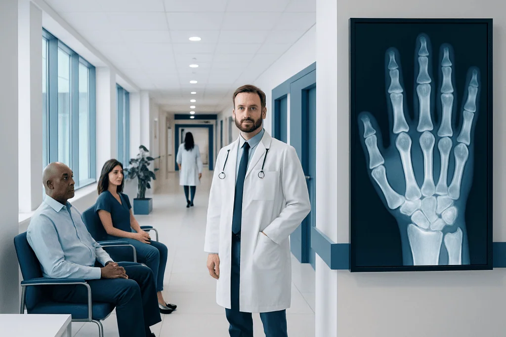
The proximal phalanx is the bone closest to the hand or foot in each finger or toe, except the thumb and big toe, which have fewer phalanges. Common proximal phalanx problems include fractures, sprains, dislocations, tendon injuries, and stiffness after trauma.
Key Takeaways
- The proximal phalanx is the bone closest to the hand or foot in each finger or toe, except the thumb and big toe, which have fewer phalanges.
- Common proximal phalanx problems include fractures, sprains, dislocations, tendon injuries, and stiffness after trauma.
- Pain, swelling, bruising, deformity, and difficulty bending or straightening a digit may suggest a proximal phalanx injury.
- X-rays are the main test for suspected fractures, while clinical examination helps assess joint stability, tendon function, and circulation.
- Treatment ranges from splinting and buddy taping to hand therapy and, in some cases, surgery for unstable or displaced fractures.
- Early medical assessment can help reduce the risk of long-term stiffness, poor healing, or loss of function.
The proximal phalanx is the first bone of a finger or toe after the knuckle, and it plays a major role in grip, balance, and fine movement. Most questions about the proximal phalanx relate to anatomy, fractures, joint injuries, and how these problems are diagnosed and treated.
What the proximal phalanx is
The proximal phalanx is the first small long bone in a finger or toe, located just beyond the knuckle where the digit meets the hand or foot. In the fingers, it sits between the metacarpal bone and the middle phalanx. In the toes, it lies between the metatarsal and the next toe bone. The thumb and the big toe are slightly different because they have only two phalanges instead of three.
This bone may seem small, but it has an important role in movement and stability. In the hand, the proximal phalanx helps with gripping, pinching, typing, writing, and other precise tasks. In the foot, it contributes to balance, push-off, and walking mechanics. Because it connects to joints and tendons at both ends, an injury can affect not only the bone itself but also alignment and function.
When people search for “proximal phalanx,” they are often looking for an explanation after seeing the term on an X-ray report or after an injury. In medical practice, the term most commonly comes up in the setting of a hand fracture or toe fracture, though sprains, dislocations, and tendon problems can also involve this area.
How it works in the hand and foot
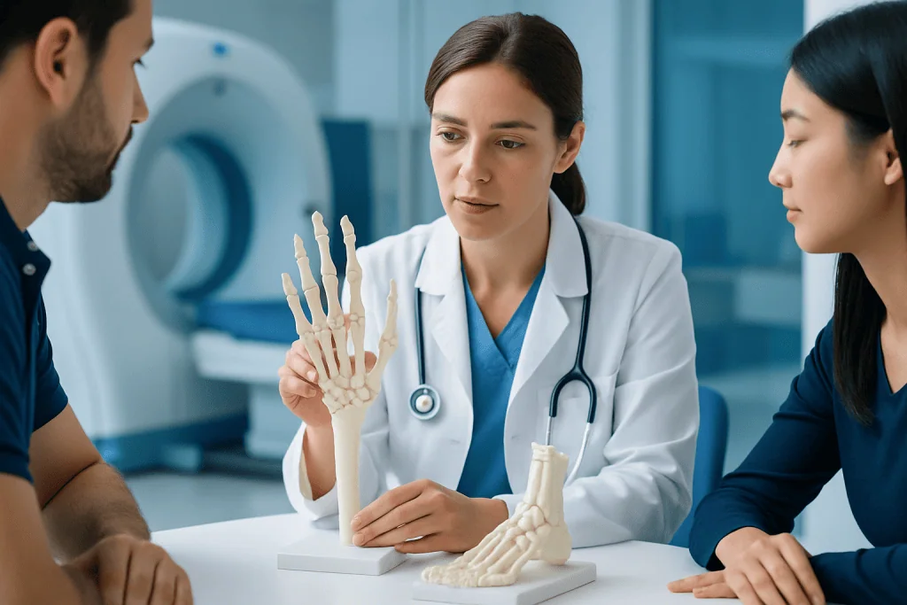
The proximal phalanx forms a joint at its base and another at its head. In the fingers, the base meets the metacarpal at the metacarpophalangeal joint, often called the knuckle. The head connects to the middle phalanx at the proximal interphalangeal joint. These joints allow bending and straightening, while surrounding ligaments and tendons guide movement and keep the finger stable.
In the foot, the same general arrangement helps the toes flex and extend during standing and walking. Although toe motion is less delicate than finger motion, proper alignment still matters. Even a small injury to the proximal phalanx can make everyday activities uncomfortable, especially if swelling limits movement or shoes place pressure on the area.
Because the proximal phalanx is closely linked with tendons, joint surfaces, and soft tissues, doctors do not view it as “just a small broken bone.” A thorough assessment looks at the whole digit: bone position, joint alignment, skin condition, circulation, nerve sensation, and whether the tendons still allow normal bending and straightening.
Common symptoms of proximal phalanx problems
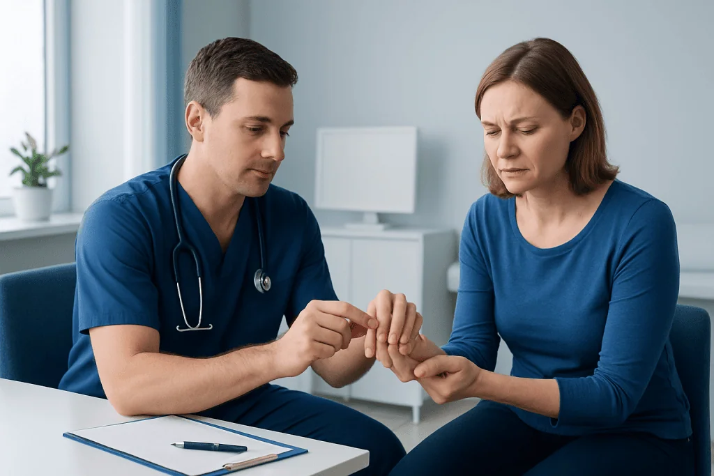
Symptoms depend on whether the issue is a fracture, sprain, dislocation, tendon injury, or overuse problem. The most common signs are pain, swelling, bruising, and tenderness around the affected finger or toe. Some people notice stiffness right away, while others can move the digit but find that motion is painful.
A proximal phalanx fracture may cause visible deformity, rotation, or shortening of the digit. For example, a finger may appear crooked or overlap a neighboring finger when making a loose fist. A toe may be swollen, difficult to move, and painful with weight-bearing. If the joint is involved, the range of motion may be limited long after the initial swelling starts to settle.
Other warning signs include numbness, tingling, skin discoloration, or an open wound over the injury. These findings may suggest damage beyond a simple bruise. In some cases, the symptoms overlap with a sprain or trigger finger-related tendon irritation, which is why a physical examination and imaging can be important when the diagnosis is not clear.
- Pain with gripping, pinching, or walking
- Swelling and bruising around the knuckle or toe joint
- Reduced range of motion
- Crooked appearance or finger overlap
- Tenderness when pressing on the bone
- Difficulty straightening or bending the digit
Causes and risk factors
Most proximal phalanx injuries happen after trauma. Common causes include falls, sports injuries, a finger being struck by a ball, a hand getting caught in a door, workplace accidents, or stubbing a toe against a hard surface. In the hand, twisting or direct impact can produce fractures or dislocations. In the foot, repetitive pressure or sudden force may injure the bone or nearby joint.
Fractures can be simple and stable, or they can be displaced, angulated, rotated, or extend into the joint. Joint involvement is particularly important because uneven healing at the joint surface may increase the risk of stiffness or later arthritis. Soft-tissue damage often occurs at the same time, including ligament sprains or tendon injuries.
Risk may be higher in contact sports, occupations involving machinery or repetitive hand use, and situations where protective footwear or gloves are not used. Bone health also matters. Osteoporosis, low bone density, older age, and certain medical conditions can make fractures more likely. Children can injure the growth plate near the proximal phalanx, which deserves careful assessment because growing bone behaves differently from adult bone.
How doctors diagnose a proximal phalanx injury
Diagnosis begins with a medical history and physical examination. A clinician will ask how the injury happened, where the pain is located, whether the digit can still move, and whether there are symptoms such as numbness or color change. The examination usually includes checking swelling, tenderness, rotation, joint stability, circulation, skin integrity, and tendon function.
X-rays are the main imaging test for suspected proximal phalanx fractures. They show whether the bone is broken, whether the pieces have shifted, and whether the fracture reaches the joint. Different views are often needed because some fractures are easier to see from one angle than another. If the injury pattern is complex or surgery is being considered, additional imaging may sometimes be recommended.
Not every painful finger or toe needs advanced imaging, but a doctor may look more closely if symptoms seem out of proportion, the examination suggests tendon damage, or the injury is not improving as expected. In specialist settings, assessment may involve orthopedics or hand surgery expertise when alignment, joint function, or tendon balance is at risk.
Treatment options and recovery
Treatment depends on which proximal phalanx is affected, the type of injury, whether the bone is stable, and whether the joint or surrounding tendons are involved. Many minor, stable injuries can be treated without surgery using rest, ice, elevation, splinting, or buddy taping. These approaches help control pain and swelling while the bone or soft tissue heals in a protected position.
If a fracture is displaced, rotated, unstable, open, or extends into the joint, reduction or surgery may be needed to restore alignment and protect function. This is especially important in the hand, where even a small rotational deformity can interfere with grip. Depending on the pattern, care may include splints, pins, screws, or other fixation methods, followed by supervised exercises to regain movement.
Rehabilitation is often a key part of recovery. The biggest challenge after a finger fracture is not always pain but stiffness. Guided physical therapy and rehabilitation or hand therapy may help restore range of motion, strength, and everyday function. Recovery time varies widely, so the safest plan is individualized follow-up with a qualified clinician rather than comparing one injury with another.
When surgical care is appropriate, patients may also need coordinated orthopedic surgery follow-up to monitor healing and prevent complications. Near the end of the care pathway, Acibadem International’s multidisciplinary specialists and JCI-accredited hospitals diagnose and treat bone and hand injuries for international patients when specialist evaluation is needed.
Self-care, prevention, and protecting function
Early self-care after a minor injury usually focuses on protecting the area and limiting swelling. Resting the affected hand or foot, applying ice wrapped in cloth for short periods, and elevating the limb may help in the first day or two. Rings should be removed early after a finger injury if swelling is starting, because they can become difficult to take off later.
People should avoid forcing a painful digit through full movement soon after an injury, especially if a fracture is possible. At the same time, prolonged unnecessary immobilization can contribute to stiffness, particularly in the fingers. This is one reason a prompt, accurate diagnosis matters: the best balance between protection and early movement depends on the exact injury.
Prevention includes using protective sports equipment, safe work practices, and well-fitting footwear. Maintaining bone health through weight-bearing activity, adequate nutrition, and medical care for osteoporosis may also reduce fracture risk. For those recovering from an injury, following splint instructions, attending follow-up visits, and completing exercises as advised can support better long-term function.
When to seek medical care
Medical assessment is important if there is severe pain, significant swelling, a visible deformity, inability to move the finger or toe normally, or pain that does not improve after a short period of home care. An open wound, bleeding near the bone, or a nail-bed injury should also be evaluated promptly. These findings can signal a more serious injury that needs treatment beyond rest alone.
Urgent care is especially important if the digit looks pale or blue, feels numb, becomes cold, or appears rotated or out of place. People should also seek care after high-impact trauma, crush injuries, or if a child injures a finger or toe near a growth plate. Delayed treatment can make alignment problems and stiffness harder to correct.
Even when symptoms seem mild, a professional evaluation can be helpful if daily tasks are affected or if the diagnosis is uncertain. A doctor can distinguish between a bruise, sprain, dislocation, tendon problem, and fracture, and can advise whether simple support or specialist treatment is the safer option.
Frequently asked questions
What is the proximal phalanx?
The proximal phalanx is the first bone in a finger or toe after the knuckle. It helps the digit bend, straighten, and stay aligned during movement.
Is a proximal phalanx fracture serious?
Some proximal phalanx fractures are minor and heal well with splinting or buddy taping. Others are more complex, especially if the bone is displaced, rotated, or extends into a joint, and these may need specialist care.
How do doctors know if the proximal phalanx is broken?
Doctors usually begin with an examination and then order X-rays. The images help show whether the bone is broken, how well it is aligned, and whether the joint is involved.
How long does a proximal phalanx injury take to heal?
Healing time varies depending on the type of injury, the person’s age, and whether treatment is surgical or non-surgical. Pain and swelling often improve before stiffness fully resolves, so recovery can continue beyond the initial bone healing period.
Can a proximal phalanx injury heal without surgery?
Yes, many stable fractures and soft-tissue injuries can heal without surgery. The key is proper alignment, protection during healing, and follow-up to make sure movement and function return safely.
When should someone see a doctor for finger or toe pain after an injury?
A doctor should be consulted if there is severe pain, marked swelling, deformity, numbness, discoloration, or trouble moving the digit. Medical care is also important if symptoms persist, worsen, or interfere with normal daily use.
References
- American Academy of Orthopaedic Surgeons
- American Society for Surgery of the Hand
- National Institute of Arthritis and Musculoskeletal and Skin Diseases
- MedlinePlus
- Mayo Clinic
This article is for general information only and is not a substitute for professional medical advice. Please consult a qualified doctor about your individual situation.
Joint and spine care in Turkey — expert assessment & treatment
JCI-accredited · board-certified surgeons · reply within 24h
Add us as a Preferred Source to see more of our trusted health content across Google Search, AI Overviews and Discover.

More from the Health Library


Related Specialists

Dr. Nisa Pekcan Bölükbaşı
Pediatrics
Dr. Bülent Yücel
Orthopedic Surgery & Traumatology
Ody. Betül Yıldızlı
Audiology
Prof. Dr. Özgür Çetik
Orthopedic Surgery & Traumatology