Retina Detachment — Explained by Medical Evidence, Not Myths
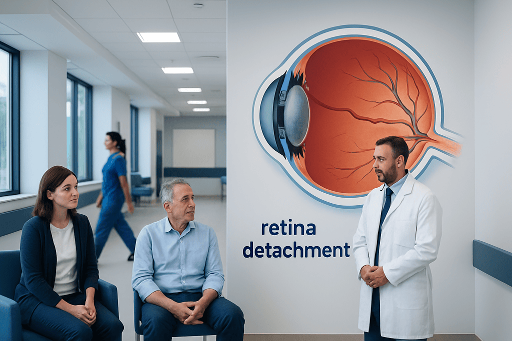
Retina detachment usually causes sudden flashes, new floaters, blurred vision, or a shadow like a curtain over part of the vision. It is often linked to a retinal tear, aging changes in the gel inside the eye, eye injury, severe nearsightedness, or previous eye surgery.
Key Takeaways
- Retina detachment usually causes sudden flashes, new floaters, blurred vision, or a shadow like a curtain over part of the vision.
- It is often linked to a retinal tear, aging changes in the gel inside the eye, eye injury, severe nearsightedness, or previous eye surgery.
- Diagnosis requires a prompt dilated eye examination, and sometimes ultrasound or retinal imaging.
- Treatment may include laser or freezing therapy for tears, or surgery such as pneumatic retinopexy, scleral buckle, or vitrectomy for a detachment.
- Anyone with sudden visual symptoms should seek urgent eye care the same day.
Retina detachment is a serious eye condition in which the light-sensitive retina separates from the tissue that nourishes it. It needs urgent medical assessment because prompt treatment can help preserve vision, while delays increase the risk of permanent sight loss.
What retina detachment is
Retina detachment means the retina, the thin layer of tissue at the back of the eye that senses light, has lifted away from the underlying tissue that supplies it with oxygen and nutrients. When this separation happens, the retina cannot work properly. Vision may become distorted, dim, or partly blocked, and if the detachment is not treated quickly, some vision loss can become permanent.
This condition is different from ordinary age-related eye changes and should not be dismissed as harmless. A person may first develop a retinal tear or hole, which allows fluid to pass underneath the retina. In other cases, scar tissue pulls the retina away, or fluid collects under it because of another eye disease. Understanding this difference matters, because a simple tear may be treated before it progresses to a full detachment.
Retina detachment is usually painless. That can make the condition easy to underestimate, especially if symptoms begin in only one eye or involve the side vision first. The key message is straightforward: sudden visual changes, especially flashes, floaters, or a curtain-like shadow, need urgent professional assessment rather than watchful waiting.
Symptoms and early warning signs

The most common warning signs are sudden flashes of light, a new shower of floaters, blurred vision, or the feeling that a gray shadow or curtain is moving across part of the visual field. Some people also notice reduced side vision or a patch of missing sight. These symptoms often appear quickly, over minutes to hours, although in some cases they develop over a day or two.
Floaters are very common with aging and are often harmless, but a sudden increase is different from long-standing occasional specks. Flashes may be more noticeable in dim light or when moving the eyes. If the central part of the retina becomes involved, straight-ahead vision may drop sharply, reading may become difficult, and faces may look distorted.
Symptoms alone cannot confirm retina detachment, because similar complaints can occur with a posterior vitreous detachment, migraine aura, bleeding in the eye, or other retinal problems. Even so, sudden onset is the important clue. When in doubt, it is safer to treat new flashes, floaters, and field loss as urgent until an eye specialist says otherwise.
- Sudden flashes of light
- New or rapidly increased floaters
- Blurred or distorted vision
- Loss of side vision
- A curtain, veil, or shadow over vision
Why it happens: causes and risk factors
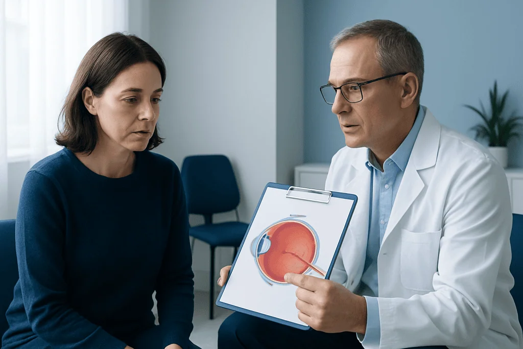
The most common type is rhegmatogenous retinal detachment. This begins with a tear or hole in the retina. As people age, the vitreous gel inside the eye naturally becomes more liquid and may pull away from the retina. This process, called posterior vitreous detachment, is common and often harmless, but in some eyes it creates enough traction to tear the retina. Fluid can then pass through the tear and separate the retina from the tissue beneath it.
Other forms occur for different reasons. Tractional retinal detachment develops when scar tissue on the retinal surface pulls the retina away. This can happen in advanced diabetic eye disease, after inflammation, or with certain vascular disorders. Exudative retinal detachment happens when fluid collects under the retina without a tear, sometimes because of inflammation, tumors, or diseases affecting the blood vessels of the eye.
Risk is higher in people with severe nearsightedness, a previous retinal tear or detachment, prior cataract or other eye surgery, significant eye injury, a family history of retinal detachment, or conditions such as diabetic retinopathy. Having a detachment in one eye also increases the chance of retinal problems in the other eye. Related retinal disease may overlap with conditions such as diabetic retinopathy, which can create traction and bleeding inside the eye.
How doctors diagnose retina detachment
Diagnosis begins with a careful history of the symptoms: when they started, whether they are getting worse, and whether one or both eyes are affected. An eye doctor then examines visual acuity, the pupils, eye pressure, and the front of the eye. The most important step is a dilated retinal examination, which allows the specialist to look for tears, holes, bleeding, and areas where the retina has lifted.
Special lenses and imaging tools may be used to inspect the far edges of the retina, where many tears start. If blood, cataract, or another problem blocks the view, ocular ultrasound can help show whether the retina is detached. In selected cases, retinal photography or optical coherence tomography may add useful detail, especially when the central retina is involved or when another condition is suspected.
Rapid diagnosis is important because timing affects the chance of preserving vision. A retinal tear without detachment can often be treated promptly to reduce the risk of progression. Once a larger detachment develops, especially if the macula, the central vision area, becomes affected, treatment is usually more urgent and recovery of vision may be less complete.
Treatment options and what they aim to do
Treatment depends on whether there is only a retinal tear, a small localized detachment, or a larger detachment involving important visual areas. The goal is to seal tears, relieve traction, and place the retina back against the underlying tissue so it can function again. Treatment choice is individualized based on the type of detachment, the location of the tear, overall eye health, and the person’s age and medical history.
For a retinal tear or very limited detachment, laser photocoagulation or cryopexy may be used to create a scar around the tear and secure the retina. For established detachments, doctors may recommend procedures such as injecting a gas bubble into the eye to help close the break, placing a silicone band around the eye wall, or removing the vitreous gel and repairing the retina internally. These approaches are commonly known as retinal laser treatment when sealing tears, and more advanced surgery may involve vitrectomy or other retinal procedures.
Recovery varies. Some people need to keep the head in a certain position for a period after treatment, especially when a gas bubble is used. Vision often improves gradually over weeks to months, but it may not return fully, particularly if the detachment was large or the macula was detached before repair. Follow-up visits are essential because new tears or repeat detachment can occur and may need further care. In complex cases, evaluation by specialists in eye surgery can help determine the most appropriate plan.
Prevention and self-care after symptoms or treatment
Not all cases can be prevented, but early detection makes a meaningful difference. People at higher risk, such as those with severe myopia, previous retinal problems, or diabetic eye disease, should keep regular eye appointments and follow their doctor’s advice about dilated retinal examinations. Protective eyewear during sports, home repairs, or work with flying debris can reduce the risk of eye injury, which is another preventable cause.
Self-care does not replace treatment, but it does support eye health and recovery. Good control of conditions such as diabetes and high blood pressure may lower the risk of retinal complications. After retinal treatment or surgery, patients should follow activity restrictions, positioning instructions, and medication guidance carefully. If a gas bubble has been placed in the eye, doctors may advise against flying or traveling to high altitudes until it has fully resolved.
It is also helpful to monitor both eyes for any new flashes, floaters, shadows, or sudden visual blur. One retinal problem can increase the chance of another later on. People should not drive themselves if vision has suddenly changed. Arranging urgent assessment with an eye specialist is safer than waiting to see whether symptoms settle on their own.
When to seek medical care
Urgent medical care is needed the same day if there are sudden flashes, a burst of new floaters, a dark curtain or shadow over any part of vision, sudden reduced side vision, or a rapid drop in sight in one eye. These symptoms do not always mean retina detachment, but they can signal a retinal tear or bleeding that also needs prompt treatment. Speed matters because earlier treatment often offers the best chance of protecting vision.
Routine eye appointments are not enough when symptoms are sudden. Emergency assessment is especially important after eye trauma, recent eye surgery, or in people with high myopia or diabetes. If an eye clinic is not immediately available, an emergency department can help direct urgent ophthalmic care.
For international patients who need specialist evaluation, Acibadem International’s multidisciplinary eye teams in JCI-accredited hospitals diagnose and treat retinal conditions with modern imaging and surgical care. Whatever the setting, the safest next step is prompt examination by a qualified eye doctor rather than trying home remedies or waiting for the symptoms to pass.
Frequently asked questions
Is retina detachment an emergency?
Yes. Retina detachment is considered an eye emergency because the retina needs a normal blood supply and support to function well. The faster it is diagnosed and treated, the better the chance of preserving vision.
Can retina detachment heal on its own?
No, a true retinal detachment does not usually reattach on its own. A retinal tear or detachment generally needs prompt treatment from an eye specialist to reduce the risk of lasting vision loss.
Are flashes and floaters always a sign of retina detachment?
Not always. Flashes and floaters can happen with normal age-related vitreous changes, but a sudden new onset or a large increase can also signal a retinal tear or detachment. Because symptoms can overlap, urgent eye examination is the safest approach.
What is the difference between a retinal tear and retina detachment?
A retinal tear is a break in the retina, while retina detachment means the retina has lifted away from the tissue beneath it. A tear can lead to a detachment if fluid passes underneath, so treating a tear early may prevent a more serious problem.
Can vision return after treatment?
Many people have meaningful visual improvement after treatment, but results vary. Recovery depends on how long the retina was detached, whether the macula was involved, and whether other eye diseases are present.
Who is more likely to develop retina detachment?
Risk is higher in older adults, people with severe nearsightedness, those who have had eye injury or surgery, and anyone with a history of retinal tears or detachment. Some retinal diseases, including advanced diabetic eye disease, can also increase risk.
References
- American Academy of Ophthalmology
- National Eye Institute
- National Health Service
- Mayo Clinic
- Merck Manual
This article is for general information only and is not a substitute for professional medical advice. Please consult a qualified doctor about your individual situation.
Explore treatments in Turkey — costs, top hospitals & a free quote
JCI-accredited · board-certified surgeons · reply within 24h
Add us as a Preferred Source to see more of our trusted health content across Google Search, AI Overviews and Discover.

More from the Health Library



Related Specialists

Dr. Ömer Faruk Karakoç
Emergency Service
Fzt. Mustafa Özcan
Physical Medicine & Rehabilitation
Prof. Dr. Tugan Beşe
Gynecology & Obstetrics
Dr. Sinem Altunyuva Usta
Pediatric Cardiology