Retinal Vein Occlusion: Sudden Vision Changes and Care

Retinal vein occlusion is sometimes described as an eye stroke because blood flow out of the retina becomes blocked. Common symptoms include sudden painless blurred vision, dark spots, distorted vision, or reduced central vision in one eye.
Key Takeaways
- Retinal vein occlusion is sometimes described as an eye stroke because blood flow out of the retina becomes blocked.
- Common symptoms include sudden painless blurred vision, dark spots, distorted vision, or reduced central vision in one eye.
- High blood pressure, diabetes, high cholesterol, glaucoma, smoking, and increasing age can raise the risk.
- Treatment often focuses on complications such as macular edema or abnormal new blood vessels, commonly with eye injections or laser therapy.
- Prompt diagnosis and long-term control of cardiovascular risk factors help protect vision and overall health.
Retinal vein occlusion is a blockage in one of the veins that drains blood from the retina, the light-sensitive tissue at the back of the eye. It can cause sudden blurred or reduced vision and should be assessed promptly by an eye specialist.
Overview
Retinal vein occlusion occurs when a vein that carries blood away from the retina becomes partly or completely blocked. The retina needs a steady supply of oxygen and nutrients to send clear visual signals to the brain. When drainage is interrupted, blood and fluid can leak into retinal tissue, leading to swelling, bleeding, and changes in vision.
There are two main types. Central retinal vein occlusion, or CRVO, affects the main retinal vein and can involve a large area of the retina. Branch retinal vein occlusion, or BRVO, affects a smaller branch vein and may cause vision loss in a more limited area, depending on which part of the retina is involved.
People sometimes call retinal vein occlusion an eye stroke. This term can help explain the vascular nature of the condition, but it is not the same as a stroke in the brain. Still, it often shares similar risk factors, so care usually includes both eye treatment and attention to general vascular health.
Symptoms
Retinal vein occlusion usually affects one eye and is often painless. The most common symptom is a sudden or rapidly developing change in vision. Some people notice blurred vision on waking, while others recognize a dark or missing area in their field of view during daily activities such as reading or driving.
Symptoms can vary depending on the location and severity of the blockage. A small branch occlusion away from the macula may cause mild symptoms, while swelling in the macula, the central part of the retina used for detailed vision, can make faces, text, and fine work appear blurred or distorted.
- Sudden painless blurred or reduced vision in one eye
- Dark spots, shadows, or missing areas in the field of vision
- Distorted vision, where straight lines may look wavy
- Difficulty reading or recognizing faces
- Floaters if bleeding occurs inside the eye
Any sudden vision loss should be treated as urgent, even when there is no pain. Early assessment helps identify the cause and allows timely treatment for complications that may threaten sight.
Causes and Risk Factors

A retinal vein blockage most often develops when blood flow slows or a vein is compressed by a nearby hardened retinal artery. Over time, vascular changes related to high blood pressure, diabetes, or high cholesterol can make blood vessels less flexible. This can increase the chance of clot formation or impaired drainage within retinal veins.
Risk factors are more common with age, but retinal vein occlusion can also occur in younger adults, especially when there are clotting disorders, inflammatory conditions, or other medical problems. Glaucoma is an important eye-related risk factor, particularly for central retinal vein occlusion, because increased eye pressure may affect blood flow around the optic nerve.
- High blood pressure
- Diabetes or poor blood sugar control
- High cholesterol or other lipid disorders
- Smoking
- Glaucoma or increased eye pressure
- Cardiovascular disease or a history of blood clots
- Rare inherited or acquired clotting conditions
In many patients, retinal vein occlusion is not caused by a single factor. It is often the result of several vascular risks acting together. Identifying and managing these risks is important not only for the affected eye but also for overall cardiovascular health.
Diagnosis
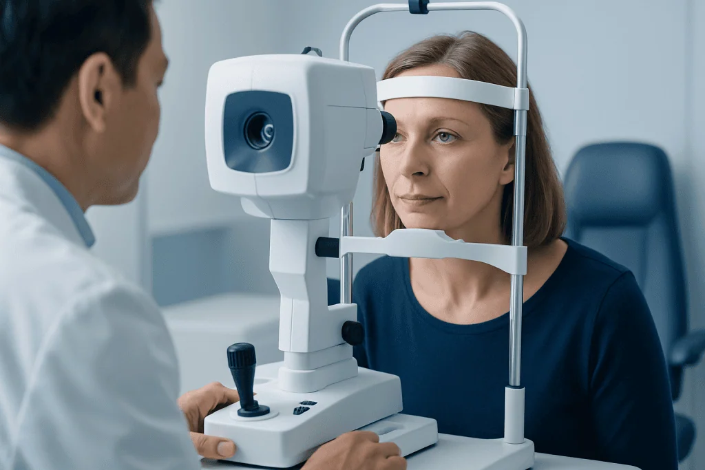
An ophthalmologist diagnoses retinal vein occlusion through a detailed eye examination. This usually includes checking visual acuity, measuring eye pressure, and examining the retina after dilating the pupils. The doctor may see retinal bleeding, dilated or twisted veins, cotton-wool spots, swelling, or other signs that suggest reduced blood flow.
Specialized imaging helps confirm the diagnosis and guide treatment. Optical coherence tomography, often called OCT, creates detailed cross-sectional images of the retina and is especially useful for detecting macular edema. Fluorescein angiography may be used to show blood flow in the retinal vessels and identify areas with leakage or poor circulation.
The eye specialist may also recommend a general medical evaluation. Blood pressure measurement, blood sugar testing, cholesterol assessment, and review of cardiovascular history can help identify treatable risk factors. In selected patients, especially younger people or those with unusual findings, additional blood tests may be considered to look for clotting or inflammatory conditions.
Treatment Options
Treatment depends on the type of retinal vein occlusion, the level of vision affected, and whether complications are present. There is usually no simple way to remove the blockage itself. Instead, treatment aims to reduce retinal swelling, control abnormal blood vessel growth, preserve vision, and reduce the risk of further vascular problems.
The most common treatment for vision loss caused by macular edema is injection of anti-VEGF medicine into the eye. These medicines reduce leakage and swelling by blocking a signal that promotes abnormal vessel leakage. Some patients may need repeated injections over time, and response is monitored with vision tests and OCT scans. Steroid injections or implants may be considered in certain situations, but they require monitoring for side effects such as increased eye pressure or cataract progression.
Laser treatment may be recommended for specific complications. In branch retinal vein occlusion, focal or grid laser may be used in selected cases of persistent macular swelling. If poor circulation causes abnormal new blood vessels, panretinal laser treatment can help lower the risk of bleeding inside the eye or painful pressure problems.
Treatment plans are individualized. Some mild cases may be monitored closely without immediate procedures, while others require active treatment soon after diagnosis. Follow-up is essential because retinal vein occlusion can change over weeks or months, and complications may develop even after symptoms seem stable.
Prevention and Self-care
Not every retinal vein occlusion can be prevented, but controlling vascular risk factors can lower the chance of future problems. Patients should work with their primary care doctor, cardiologist, endocrinologist, or other specialists to manage blood pressure, diabetes, cholesterol, and any known cardiovascular disease. These steps support both eye health and general health.
Healthy daily habits also matter. Stopping smoking, maintaining a balanced diet, staying physically active as advised by a doctor, and taking prescribed medicines consistently can help protect blood vessels. People with glaucoma should attend regular eye appointments and use eye pressure-lowering medicines as directed.
- Monitor blood pressure and keep follow-up appointments
- Manage diabetes with an individualized care plan
- Check cholesterol and discuss treatment if levels are high
- Avoid smoking and seek support to quit if needed
- Report any new vision changes promptly
- Keep scheduled retinal imaging visits after diagnosis
Self-care cannot replace specialist treatment, but it can improve long-term outcomes. Patients should avoid starting blood thinners, supplements, or alternative therapies specifically for retinal vein occlusion unless recommended by a qualified doctor, because the right approach depends on the person’s overall medical condition.
When to See a Doctor
A person should seek urgent eye care for sudden blurred vision, a new dark area in vision, sudden distortion, new floaters, or any unexplained loss of vision in one eye. Prompt evaluation is important because several eye conditions can cause similar symptoms, including retinal detachment, bleeding inside the eye, optic nerve problems, and retinal artery occlusion.
After retinal vein occlusion is diagnosed, regular follow-up with a retina specialist is important even if vision improves. The doctor will monitor for macular edema, abnormal new blood vessels, increased eye pressure, and changes in the other eye. Follow-up intervals vary according to the severity of the condition and the treatment being used.
International patients who need evaluation or treatment can access multidisciplinary eye and medical care at Acibadem International, where JCI-accredited hospitals provide diagnosis and management of retinal conditions. As with any medical decision, patients should discuss their individual situation, expected benefits, and possible risks with a qualified ophthalmologist.
Frequently asked questions
Is retinal vein occlusion a medical emergency?
Sudden vision changes should be assessed urgently by an eye specialist. Retinal vein occlusion itself may not always require emergency surgery, but early diagnosis helps rule out other serious causes and allows timely treatment for retinal swelling or bleeding.
Can vision return after retinal vein occlusion?
Vision recovery varies from person to person. Some people improve, especially when macular swelling responds well to treatment, while others may have lasting changes if the retina has been significantly damaged. Regular follow-up gives the best chance to manage complications early.
What is the difference between branch and central retinal vein occlusion?
Branch retinal vein occlusion affects one smaller vein branch in the retina, so symptoms may involve only part of the visual field. Central retinal vein occlusion affects the main retinal vein and can cause more widespread retinal changes. Both types require ophthalmic evaluation and monitoring.
Are eye injections painful?
Most patients tolerate eye injections well because numbing drops and antiseptic preparation are used before the procedure. A person may feel pressure, mild discomfort, or irritation afterward. The ophthalmology team explains aftercare and warning signs to report.
Does retinal vein occlusion mean a person will have a stroke?
Retinal vein occlusion does not mean a brain stroke will definitely occur. However, it shares risk factors such as high blood pressure, diabetes, and high cholesterol. For this reason, doctors often recommend a general health check and careful control of vascular risks.
Can retinal vein occlusion happen in the other eye?
It can occur in the other eye, although many people have only one affected eye. The risk depends on underlying factors such as blood pressure, diabetes, cholesterol, glaucoma, and clotting tendency. Managing these risks and attending regular eye examinations can help protect both eyes.
This article is for general information only and is not a substitute for professional medical advice. Please consult a qualified doctor about your individual situation.
Explore treatments in Turkey — costs, top hospitals & a free quote
JCI-accredited · board-certified surgeons · reply within 24h
Add us as a Preferred Source to see more of our trusted health content across Google Search, AI Overviews and Discover.
Check your numbers in seconds
BMI, calories, due date, blood pressure and 30+ more clinical calculators — free, instant, doctor-reviewed ranges.

Related Treatments
More from the Health Library

Contact Lenses for Dry Eyes: Are They Still an Option?

Is LASIK Permanent? How Stable Are the Results?

How Much Is LASIK Laser Eye Surgery? Cost Breakdown

Does Insurance Cover LASIK? Coverage and Exceptions

Dry Eye Disease: Burning, Gritty Eyes, and Treatment Options