Skin Cancer Images: What Different Types Can Look Like

Skin cancer can appear in different ways, including a changing mole, a non-healing sore, a shiny bump, or a scaly patch. Images are helpful for awareness, but many harmless skin changes can look similar, so diagnosis requires a medical exam.
Key Takeaways
- Skin cancer can appear in different ways, including a changing mole, a non-healing sore, a shiny bump, or a scaly patch.
- Images are helpful for awareness, but many harmless skin changes can look similar, so diagnosis requires a medical exam.
- The main types are basal cell carcinoma, squamous cell carcinoma, and melanoma, each with typical but variable appearances.
- The ABCDE rule can help identify warning signs of melanoma: asymmetry, border, color, diameter, and evolving change.
- People with fair skin, heavy sun exposure, tanning bed use, or a personal or family history of skin cancer have higher risk.
- Any new, changing, bleeding, painful, or non-healing skin lesion should be checked by a qualified doctor.
Skin cancer images can be useful for learning what suspicious spots may look like, but they cannot confirm a diagnosis. Understanding common visual patterns, along with regular skin checks and timely medical evaluation, can help people recognize changes early.
Overview: why skin cancer images matter
Skin cancer images are often used to help people learn what suspicious skin changes may look like. They can show examples of warning signs such as a mole that changes shape or color, a sore that does not heal, or a pearly bump on sun-exposed skin. Seeing these patterns can make self-checks more meaningful and may encourage earlier medical advice.
At the same time, images have limits. Skin cancers do not always look textbook, and harmless conditions such as seborrheic keratoses, eczema, psoriasis, cysts, or benign moles can sometimes look concerning in photos. Lighting, skin tone, camera quality, and the stage of the lesion can also change how a spot appears.
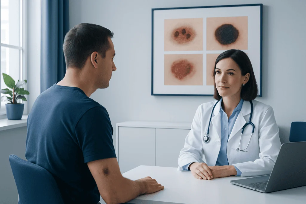
For that reason, skin cancer images should be treated as educational tools rather than a way to self-diagnose. A dermatologist or other qualified doctor may use skin examination, dermoscopy, and sometimes a biopsy to determine whether a spot is cancerous. This is especially important because early skin cancer is often easier to treat.
What different types of skin cancer can look like
The three main types of skin cancer are basal cell carcinoma, squamous cell carcinoma, and melanoma. Each has some common visual features, but appearances vary widely from person to person. They can occur on lighter or darker skin and may look different depending on body location.
Basal cell carcinoma often appears as a shiny or pearly bump, a pink growth, or a sore that crusts, bleeds, and returns. Some look like a flat scar-like area that is pale, waxy, or slightly firm. These lesions are common on sun-exposed areas such as the face, ears, scalp, neck, and shoulders.
Squamous cell carcinoma may look like a rough, scaly patch, a thickened wart-like bump, or an open sore that does not heal. It may become tender, crusted, or bleed. This type also often develops on areas with long-term sun exposure, including the lips, face, hands, forearms, and scalp.
Melanoma often begins as a new dark spot or a changing mole. It may be uneven in shape, have irregular borders, show more than one color, or grow over time. Some melanomas are pink, red, skin-colored, or even nearly colorless, which is one reason visual assessment alone can be difficult.
Common warning signs seen in skin cancer images
Many educational resources use the ABCDE rule to describe possible signs of melanoma. This can be a practical starting point during self-exams, though not every melanoma follows these rules exactly.
- A – Asymmetry: one half does not match the other.
- B – Border: edges are irregular, ragged, blurred, or notched.
- C – Color: more than one color is present, such as tan, brown, black, red, white, or blue.
- D – Diameter: the spot is larger than about 6 mm, although melanomas can be smaller.
- E – Evolving: the lesion changes in size, shape, color, or symptoms.
Another useful idea is the “ugly duckling” sign. This means a mole or spot looks noticeably different from a person’s other moles. A lesion that stands out, even if it does not fit all ABCDE features, deserves attention.
Beyond moles, skin cancer images often show warning signs such as a sore that does not heal within several weeks, repeated bleeding or crusting, itching or pain in a skin lesion, a new bump that grows, or a patch that becomes firmer, redder, scalier, or more tender over time. These changes are not always cancer, but they should not be ignored.
Causes and risk factors
The main cause of most skin cancers is damage to skin cell DNA, often related to ultraviolet (UV) radiation from sunlight or tanning beds. This damage can build up over time. That is why skin cancers are common on areas that receive frequent sun exposure, though they can also occur on less exposed parts of the body.
Several factors can increase risk. These include fair skin, light eyes, blond or red hair, frequent sunburns, a history of intense sun exposure, tanning bed use, older age, and a large number of moles. A personal or family history of skin cancer also raises risk, especially for melanoma.
People with weakened immune systems can be more vulnerable to certain skin cancers. Some skin cancers are also linked to chronic skin injury, certain precancerous lesions, or exposure to particular chemicals. Even so, anyone can develop skin cancer, including people with darker skin tones, where it may be diagnosed later if warning signs are overlooked.
Because skin cancer can resemble other conditions, a suspicious lesion may initially be confused with acne, eczema, a wart, a fungal rash, or a harmless mole. Educational images can raise awareness, but they do not replace a professional assessment for skin cancer.
How doctors diagnose suspicious skin lesions
Diagnosis begins with a medical history and careful skin examination. The doctor may ask when the lesion first appeared, whether it has changed, and if there are symptoms such as itching, pain, crusting, or bleeding. They will also ask about personal risk factors, including sun exposure, tanning bed use, and previous skin cancers.
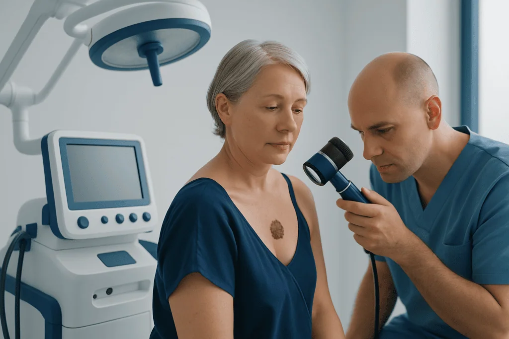
Many specialists use a dermatoscope, a handheld device that helps them view structures beneath the skin surface more clearly. This can improve evaluation of pigmented spots and other suspicious lesions. In some cases, total body photography or digital mole mapping may be used to follow people at higher risk.
If a lesion looks suspicious, the most important next step is a biopsy. During a biopsy, all or part of the lesion is removed and examined in a laboratory. This is the only way to confirm the diagnosis and determine the exact type of skin cancer.
Depending on the findings, additional imaging or lymph node assessment may sometimes be needed, especially for more advanced disease. Patients who are being evaluated for a concerning skin lesion may also need a broader cancer assessment if melanoma is suspected, which can be part of comprehensive cancer screening and follow-up planning.
Treatment options and what happens next
Treatment depends on the type of skin cancer, its size, depth, location, and whether it has spread. Many skin cancers are treated successfully when found early. The care plan may involve dermatologists, surgeons, pathologists, and oncology specialists working together.
For many basal cell and squamous cell cancers, the main treatment is surgical removal. This may include standard excision or a tissue-sparing approach in selected cases. Some superficial lesions may be managed with other local treatments, such as topical medicines, cryotherapy, curettage with electrosurgery, or light-based methods, depending on the diagnosis and clinical setting.
Melanoma usually requires surgical removal with an appropriate margin of normal skin. In some cases, nearby lymph nodes may need assessment. If melanoma or another skin cancer is more advanced, treatment may include medical oncology care such as immunotherapy, targeted therapy, or other systemic treatments. Some patients may also need radiation oncology as part of their treatment plan.
After treatment, regular follow-up is important because people who have had one skin cancer may develop another in the future. Near the end of the care journey, some international patients choose evaluation and treatment through centers such as Acibadem International, where multidisciplinary specialists in JCI-accredited hospitals diagnose and treat skin cancers.
Prevention and self-care
Although not every case can be prevented, daily sun protection can lower risk. Practical steps include using a broad-spectrum sunscreen, wearing protective clothing and a wide-brimmed hat, seeking shade during strong midday sun, and avoiding tanning beds. Sunscreen is especially important on commonly missed areas such as the ears, scalp line, lips, neck, and backs of the hands.
Monthly self-exams can help people notice change earlier. It can be useful to check the entire body in a good mirror, including the scalp, soles of the feet, under the nails, and the back. Taking clear photos over time may help track a spot that is being monitored, but any concerning change should still be reviewed by a doctor rather than judged from images alone.
People at higher risk may benefit from regular professional skin checks. This can include those with many moles, a prior history of melanoma, strong family history, significant past sun damage, or immune suppression. Preventive care matters not only for skin cancer but also for related concerns that may be evaluated alongside melanoma in specialist practice.
When to see a doctor
A doctor should assess any new or changing skin lesion that seems unusual. This includes a mole that changes in color, size, or shape; a sore that does not heal; a spot that repeatedly bleeds or crusts; or a bump or patch that keeps growing. Persistent itching, tenderness, or pain in a skin lesion are also reasons to seek medical advice.
Prompt evaluation is especially important for people with higher risk factors, including previous skin cancer, many atypical moles, strong family history, heavy sun exposure, or tanning bed use. Even if a lesion turns out to be benign, reassurance from a professional exam can be valuable.
Images found online can be useful for awareness, but they can never provide certainty. When there is any doubt, an in-person examination is the safest next step. Early assessment can lead to earlier treatment if needed and may help avoid unnecessary worry when a spot is harmless.
Frequently asked questions
Can skin cancer be identified from images alone?
No. Skin cancer images are helpful for learning common warning signs, but they cannot confirm whether a spot is cancerous. Many harmless skin conditions can look similar, so a medical examination and sometimes a biopsy are needed for diagnosis.
What does melanoma usually look like?
Melanoma often appears as a new dark spot or a changing mole with uneven shape, irregular borders, or more than one color. However, some melanomas are pink, red, or skin-colored, so not all look dark or fit the classic pattern.
What is the difference between basal cell and squamous cell carcinoma in appearance?
Basal cell carcinoma often looks like a pearly or shiny bump, a pink patch, or a sore that heals and returns. Squamous cell carcinoma more often appears as a rough, scaly patch, a crusted sore, or a thickened wart-like growth.
Should a non-healing sore always be checked?
Yes. A sore that does not heal within several weeks, or that repeatedly bleeds, crusts, or returns, should be assessed by a doctor. It may not be skin cancer, but it is important to find the cause.
Can people with darker skin get skin cancer?
Yes. Skin cancer can affect people of all skin tones. In darker skin, it may be less expected and therefore noticed later, which is why any unusual or changing lesion should be evaluated.
How often should someone check their skin?
Many doctors advise regular self-exams, often about once a month, especially for people with many moles or higher risk. Professional skin checks may also be recommended based on personal history, family history, and overall risk.
References
- World Health Organization
- American Academy of Dermatology
- National Cancer Institute
- American Cancer Society
- British Association of Dermatologists
This article is for general information only and is not a substitute for professional medical advice. Please consult a qualified doctor about your individual situation.
Skin Cancer in Turkey — costs, top hospitals & a free quote
JCI-accredited · board-certified surgeons · reply within 24h
Add us as a Preferred Source to see more of our trusted health content across Google Search, AI Overviews and Discover.

More from the Health Library



Related Specialists

Ezgi Nur Çiçek
Physical Medicine & Rehabilitation
Dr. Fatma Erbay Apaydın
Infectious Diseases & Clinical Microbiology
Prof. Dr. Arzu Akçay
Pathology
Dt. Müge Tiralı Türkkan
Oral & Dental Health