Sonogram — Explained by Medical Evidence, Not Myths

A sonogram and an ultrasound are commonly used to mean the same imaging test. Sonograms use sound waves, not X-rays, so they do not expose the body to ionizing radiation.
Key Takeaways
- A sonogram and an ultrasound are commonly used to mean the same imaging test.
- Sonograms use sound waves, not X-rays, so they do not expose the body to ionizing radiation.
- They are used in pregnancy and in many other areas, including the abdomen, heart, thyroid, blood vessels, and soft tissues.
- Preparation depends on the body part being examined and may include fasting or arriving with a full bladder.
- Most sonograms are painless, quick, and done without sedation.
- Results should be interpreted by a qualified doctor in the context of symptoms and other tests.
A sonogram is an imaging test that uses high-frequency sound waves to create pictures of organs, tissues, blood flow, or a developing baby. It is widely used because it is noninvasive, does not use ionizing radiation, and can provide real-time information that helps guide diagnosis and care.
What a sonogram is
A sonogram is a medical imaging test that uses high-frequency sound waves to produce pictures from inside the body. In everyday use, people often use the words sonogram and ultrasound interchangeably. More precisely, ultrasound is the technology, and a sonogram is the image it creates, but both terms usually refer to the same examination.
This test helps doctors look at organs, soft tissues, blood vessels, and movement in real time. Unlike X-rays or CT scans, a sonogram does not use ionizing radiation. That is one reason it is commonly chosen when doctors need safe, repeatable imaging, including during pregnancy.
Sonograms are used for many purposes beyond obstetric care. They can help assess abdominal pain, swelling, lumps, thyroid problems, urinary symptoms, heart function, and circulation. In some settings, ultrasound also helps guide procedures such as fluid drainage, needle biopsies, or placement of vascular access.
How a sonogram works and what it can show
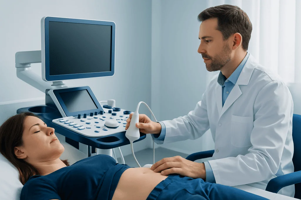
During a sonogram, a handheld device called a transducer sends sound waves into the body. These waves bounce off tissues and return as echoes. A computer processes the echoes into moving or still images that can be viewed immediately on a screen.
Different tissues reflect sound in different ways, which allows the test to show structures such as the liver, gallbladder, kidneys, uterus, ovaries, testicles, thyroid gland, muscles, tendons, and fluid-filled areas. Doppler ultrasound is a special technique that can show how blood is flowing through vessels or the heart.
Because it produces live images, a sonogram can be especially helpful when doctors need to observe motion. Examples include the heartbeat, movement of heart valves, fetal movement, or blood flow through narrowed vessels. In some cases, more advanced studies such as echocardiography or targeted vascular ultrasound may be used for a closer look at the heart and circulation.
Common types of sonogram and why they are ordered

There is not just one kind of sonogram. The type depends on the body area being examined and the clinical question. A doctor may request an abdominal ultrasound for pain, bloating, abnormal liver tests, or suspected gallstones. A pelvic ultrasound may be used for menstrual concerns, pelvic pain, ovarian cysts, fibroids, infertility assessment, or pregnancy-related evaluation.
Pregnancy ultrasound is one of the best-known uses. It can help confirm a pregnancy, estimate gestational age, check fetal growth, evaluate the placenta, and monitor amniotic fluid. A pregnancy sonogram can also help assess symptoms such as pain or bleeding, including situations where doctors need to rule out an ectopic pregnancy.
Other common studies include:
-
Thyroid sonogram for nodules, enlargement, or inflammation
-
Breast sonogram to examine a lump or clarify findings from other imaging
-
Scrotal sonogram for pain, swelling, or a suspected torsion or mass
-
Vascular sonogram to assess blood clots, narrowed arteries, or poor circulation
-
Musculoskeletal sonogram for tendon, ligament, or soft-tissue problems
In emergency and inpatient care, ultrasound may also be used quickly at the bedside to answer focused questions. For example, it can help identify fluid in the abdomen, assess the bladder, or support the work-up of urinary blockage such as kidney stones.
What happens during the test
Most sonograms are simple outpatient tests. A healthcare professional applies a clear gel to the skin so the transducer can move smoothly and transmit sound waves well. The transducer is then placed over the area being examined and moved gently to obtain images from different angles.
The test is usually painless, although there may be mild pressure if the area is tender or if the examiner needs a better view. Some sonograms are done internally for more detailed images. For example, a transvaginal ultrasound may be used to assess the uterus and ovaries, and a transrectal ultrasound may be used in certain prostate evaluations.
The length of the examination varies, but many are completed within 15 to 45 minutes. In some cases, ultrasound is combined with another service or used to guide a procedure, such as a tissue sample from a lump. When a doctor needs more information about a suspicious mass, ultrasound may be part of the planning for biopsy.
How to prepare and whether a sonogram is safe
Preparation depends on the body part being scanned. For an abdominal sonogram, fasting for several hours may improve visibility by reducing gas in the intestines and helping the gallbladder stay full. For some pelvic studies, patients may be asked to drink water and arrive with a full bladder, which can make structures easier to see.
Many other sonograms require little or no preparation. Wearing comfortable clothing can help, especially if the area scanned is the neck, abdomen, groin, or limbs. If preparation instructions are unclear, it is best to check with the imaging center beforehand so the exam is not delayed or repeated unnecessarily.
In general, a sonogram is considered very safe when used appropriately by trained professionals. It does not use ionizing radiation, which makes it different from X-rays and CT scans. It is important, however, that ultrasound be used for medical purposes and interpreted in context, rather than relied on for nonmedical reassurance alone.
Understanding sonogram results and their limits
A sonogram can provide valuable information, but it does not always give a complete diagnosis on its own. The images must be interpreted together with symptoms, the physical examination, medical history, and sometimes blood tests or other scans. A normal ultrasound does not automatically rule out every condition, and an abnormal result may need further clarification.
For example, ultrasound is excellent for identifying cysts, gallstones, fluid collections, or blood flow problems, but it may be less informative if bowel gas blocks the view or if a structure lies deep in the body. Some conditions are better assessed with MRI, CT, endoscopy, or laboratory testing depending on the organ involved.
If a sonogram shows a finding such as a nodule, cyst, blockage, or inflammation, the next step may include follow-up imaging, referral to a specialist, or treatment planning. In digestive conditions, ultrasound findings may be part of the work-up before additional studies such as endoscopy are considered.
For patients, the most helpful question is often not just “Was the ultrasound normal?” but “What does this result mean for my symptoms and what happens next?” Asking for that context can make the report easier to understand and more useful in decision-making.
When to seek medical care
A sonogram itself is not a condition, but the symptoms leading to it may need prompt assessment. Medical care should be sought if there is severe or worsening abdominal pain, chest symptoms, sudden swelling in one leg, heavy vaginal bleeding, fainting, high fever, or difficulty breathing. In pregnancy, urgent care is especially important for strong pain, bleeding, reduced fetal movement later in pregnancy, or signs of possible complications.
It is also wise to contact a doctor if a lump grows, pain does not improve, urinary symptoms persist, or a previously ordered sonogram has not been reviewed with a clinician. The timing depends on the symptoms and the reason for the test, but unexplained or ongoing symptoms should not be ignored simply because an ultrasound was performed.
At the end of the diagnostic pathway, some patients may need coordinated specialist care. Acibadem International’s multidisciplinary specialists and JCI-accredited hospitals diagnose and treat conditions evaluated with sonography for international patients.
Frequently asked questions
Is a sonogram the same as an ultrasound?
In common use, yes. People usually use sonogram and ultrasound to mean the same test. Technically, ultrasound is the sound-wave technology, while a sonogram is the image produced.
Does a sonogram use radiation?
No. A sonogram uses sound waves, not ionizing radiation. That is why it is often preferred when repeat imaging is needed and is widely used during pregnancy.
Is a sonogram painful?
Most sonograms are not painful. Patients may feel cool gel on the skin and mild pressure from the transducer, especially if the area is tender. Internal ultrasound exams can feel uncomfortable for some people, but they are usually brief.
How long does a sonogram take?
Many sonograms take about 15 to 45 minutes, depending on the body part and the complexity of the study. Some targeted bedside scans are shorter, while more detailed examinations may take longer.
Do patients need to fast or drink water before a sonogram?
Sometimes. Abdominal sonograms may require fasting, while some pelvic ultrasounds are easier to perform with a full bladder. The imaging center or doctor usually gives specific instructions based on the type of exam.
Can a sonogram detect all health problems?
No single test can detect everything. A sonogram is very useful for many soft-tissue, fluid, and blood-flow questions, but some conditions are better evaluated with CT, MRI, lab tests, or other procedures. Doctors interpret the results together with symptoms and other findings.
This article is for general information only and is not a substitute for professional medical advice. Please consult a qualified doctor about your individual situation.
Explore treatments in Turkey — costs, top hospitals & a free quote
JCI-accredited · board-certified surgeons · reply within 24h
Add us as a Preferred Source to see more of our trusted health content across Google Search, AI Overviews and Discover.

More from the Health Library

Man Breasts: A Complete Medical Overview

Austedo: A Complete Medical Overview

Forced Vital Capacity — Explained by Medical Evidence, Not Myths

Symbravo: What Patients Need to Know

Exfoliate — Explained by Medical Evidence, Not Myths