Spot Sun: What Patients Need to Know

"Spot sun" is not a medical diagnosis; it can describe several different sun-related skin changes. Flat brown sunspots are often harmless, while rough scaly patches may be actinic keratosis and need evaluation.
Key Takeaways
- "Spot sun" is not a medical diagnosis; it can describe several different sun-related skin changes.
- Flat brown sunspots are often harmless, while rough scaly patches may be actinic keratosis and need evaluation.
- Any spot that changes in size, shape, color, or starts bleeding or not healing should be checked promptly.
- Daily sun protection helps prevent new sun spots and reduces further skin damage.
- A dermatologist may diagnose a spot by skin examination and sometimes a biopsy.
Spot sun commonly refers to a visible skin mark linked to sun exposure, including freckles, solar lentigines (age spots), and rough precancerous patches called actinic keratoses. Many sun-related spots are harmless, but a new, changing, bleeding, or non-healing spot should be assessed by a qualified doctor.
Overview: what “spot sun” usually means
“Spot sun” is a non-medical phrase people often use when they notice a mark on the skin after years of sun exposure. In practice, this can refer to several different conditions, including freckles, solar lentigines (often called sunspots or age spots), melasma, actinic keratosis, and, less commonly, skin cancer.
Most sun-related spots are benign, especially if they are stable, flat, and evenly colored. However, the skin can also develop abnormal areas from ultraviolet (UV) damage that need treatment or closer follow-up. This is why it is helpful not to assume every dark or rough spot is simply cosmetic.
Sun-exposed areas are the most common places for these marks to appear, especially the face, scalp, ears, chest, shoulders, forearms, and hands. People with lighter skin tones often notice sun-related pigment changes earlier, but anyone can develop UV-related skin damage.
If the spot is new, changing, painful, itchy, crusting, bleeding, or not healing, medical assessment is important. A dermatologist can help identify whether the lesion is harmless sun damage, a precancerous change such as actinic keratosis, or another skin condition that needs treatment.
Common types of sun-related spots
Several skin findings may be described as a spot caused by the sun. Freckles are small flat brown marks that often darken with sun exposure and are usually harmless. Solar lentigines are larger, sharply defined flat brown spots that develop over time on sun-exposed skin and are also generally benign.
Actinic keratoses are different because they are caused by cumulative UV damage and are considered precancerous. They often feel rough, dry, or scaly and may be easier to feel than to see at first. These patches can resemble persistent dry skin, but unlike ordinary dryness, they do not fully go away and may slowly thicken.
Other possibilities include melasma, which causes larger darker patches, and post-inflammatory hyperpigmentation, which can appear after irritation or acne and become more noticeable with sun exposure. Certain harmless growths, such as seborrheic keratoses, may also be mistaken for sunspots even though they are not caused only by sun.
Because the terms sound similar, people may confuse benign sunspots with skin cancers. Basal cell carcinoma, squamous cell carcinoma, and melanoma can sometimes start as a spot that seems minor at first. When there is uncertainty, a clinical skin examination is the safest way to clarify the cause.
Symptoms and warning signs to watch for

A harmless sunspot is often flat, painless, evenly colored, and stable over time. It may be tan, light brown, or dark brown and usually appears in places that receive regular sun. Many people notice them gradually rather than suddenly.
Features that deserve more attention include a spot that changes in color, becomes irregular at the border, grows noticeably, or looks different from nearby spots. Itching, tenderness, crusting, bleeding, or a sore that does not heal are also important warning signs.
Rough texture matters as much as color. A persistent scaly or sandpaper-like patch may suggest actinic keratosis rather than a simple pigment spot. A pearly bump, a pink patch, or a dark lesion with several colors may point to other diagnoses and should not be self-diagnosed.
- New spot after age 30 to 40 that behaves differently from others
- Asymmetry or an uneven outline
- More than one shade of color within the same spot
- Bleeding, crusting, ulceration, or persistent irritation
- A lesion that remains despite moisturizers or routine skin care
Causes and risk factors
The main cause of many sun spots is ultraviolet radiation from sunlight and tanning devices. UV exposure increases pigment production in some cases and causes cumulative cellular damage in others. Over the years, that damage can show up as brown spots, rough patches, premature aging, and a higher risk of skin cancer.
Repeated sun exposure, especially without sunscreen or protective clothing, increases the chance of developing visible changes. History matters too: people who spend significant time outdoors for work, sport, or leisure often develop more sun-related spots on the face, neck, chest, forearms, and backs of the hands.
Risk is influenced by skin type, age, family history, and immune status. People with fair skin, light eyes, or a tendency to burn may notice damage sooner, but darker skin tones are not immune to UV injury. Older adults are more likely to have cumulative damage simply because of longer lifetime exposure.
Certain medicines and health conditions can also make skin more sun-sensitive. A doctor may review medications, previous severe sunburns, tanning bed use, and any personal or family history of skin cancer when assessing a suspicious lesion.
How doctors diagnose a suspicious sun spot
Diagnosis usually begins with a careful skin examination and a discussion of how long the spot has been present, whether it has changed, and whether there are symptoms such as bleeding or itching. Many lesions can be recognized clinically by an experienced dermatologist, especially when texture, color, and location fit a common pattern.
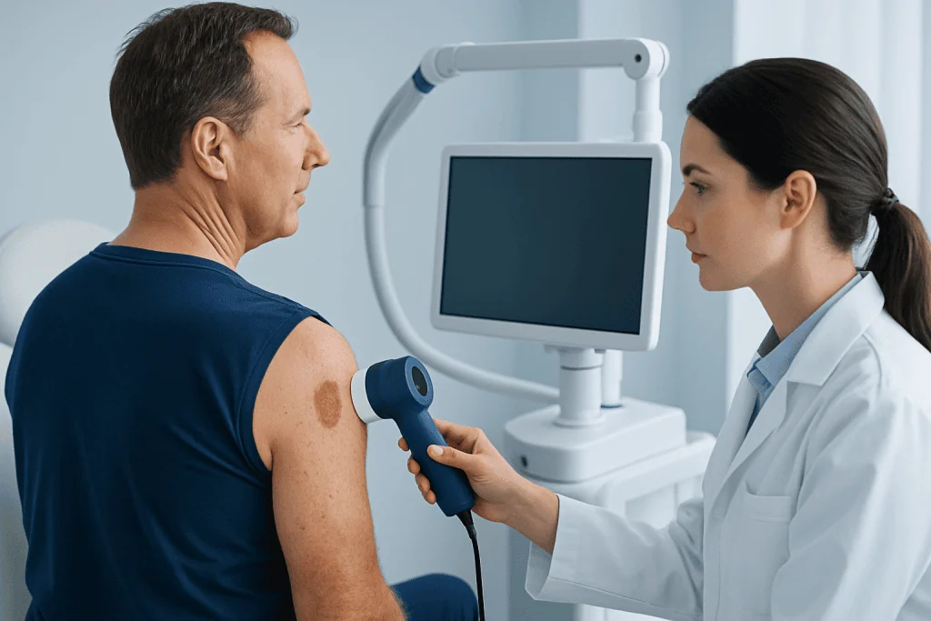
A dermatoscope may be used to examine the lesion more closely. This handheld device magnifies the skin and helps reveal structures that are not visible to the naked eye. It can improve distinction between benign lesions, precancerous lesions, and cancers, although it does not replace pathology when there is doubt.
If the spot looks atypical or concerning, the doctor may recommend a skin biopsy. In a biopsy, a small sample or the entire lesion is removed and examined in a laboratory. This is the most reliable way to confirm whether the spot is benign, precancerous, or malignant.
When a lesion raises concern for skin cancer, further treatment planning depends on the biopsy result. For some patients, evaluation may also include discussion of skin cancer warning signs elsewhere on the body and advice on how to monitor the skin safely at home.
Treatment options
Treatment depends entirely on what the spot turns out to be. Benign solar lentigines may not need treatment at all unless the patient wishes to address their appearance. In those cases, dermatologists may discuss options such as topical therapies, chemical peels, cryotherapy, or selected light- and laser-based procedures.
Actinic keratosis should be assessed and usually treated because it can progress to squamous cell carcinoma in some cases. Common treatments include cryotherapy, prescription creams, curettage, and photodynamic therapy for suitable lesions or field treatment of sun-damaged skin.
If the lesion is confirmed to be cancerous, treatment may involve surgical removal or other specialized approaches depending on the type, size, and location. For selected cases, clinicians may discuss skin cancer treatment options as part of a broader plan that prioritizes complete removal and cosmetic outcome where possible.
Cosmetic treatment should never come before diagnosis. A spot that is changing or suspicious should not be treated as a simple beauty concern without proper evaluation first. Near the end of the care pathway, patients who need specialist assessment may consider centers such as Acibadem International, where multidisciplinary specialists in JCI-accredited hospitals diagnose and treat skin conditions for international patients.
Prevention and self-care
The most effective prevention is daily sun protection. Broad-spectrum sunscreen, shade, protective clothing, hats, and UV-protective sunglasses all help reduce further skin damage. Protection matters year-round, not only on hot or sunny days, because UV exposure accumulates over time.
People with a history of sunspots or actinic keratoses benefit from making skin checks part of routine self-care. It helps to look at the skin monthly in good light and notice any lesion that is new, enlarging, darkening, roughening, or failing to heal. Taking clear dated photographs can make changes easier to track.
Self-care should also include avoiding tanning beds and limiting intentional sun exposure during peak UV hours when possible. If a patch feels persistently rough or dry despite moisturizers, it is wise to seek medical advice rather than repeatedly treating it as ordinary irritation.
For cosmetic sun spots that have already formed, over-the-counter products may have limited benefit. They should be used cautiously, especially if the diagnosis is uncertain. Before trying peels, bleaching agents, or device-based treatments, professional assessment is the safer approach.
When to seek medical care
Medical care is appropriate any time a sun spot is new and unexplained, or when it begins to change in visible or noticeable ways. A doctor should assess a spot that bleeds, crusts, hurts, itches persistently, or does not heal within a few weeks.
Prompt review is especially important for people with a personal history of skin cancer, a strong family history of melanoma, many atypical moles, immune suppression, or extensive past sun exposure. In these groups, even subtle changes may deserve a lower threshold for examination.
Urgent evaluation is sensible if a lesion grows quickly, becomes very dark or multicolored, or develops an irregular shape. When in doubt, a dermatology visit can provide reassurance or early treatment if needed. Early assessment is often simpler than waiting for a lesion to become more obvious.
People who need diagnosis of a suspicious lesion, imaging when appropriate, or procedural care may be referred for specialist services such as dermatology evaluation. If removal is recommended, the best method depends on the lesion type, size, and location.
Frequently asked questions
Is a spot sun always a sign of skin cancer?
No. Many sun-related spots, such as freckles and solar lentigines, are harmless. The concern rises when a spot changes, becomes rough, bleeds, or does not heal, because those features can suggest a precancerous or cancerous lesion.
What is the difference between a sunspot and actinic keratosis?
A sunspot usually refers to a flat brown mark caused by sun exposure and is often benign. Actinic keratosis is usually rough, scaly, or sandpaper-like and is considered a precancerous change that should be evaluated and typically treated.
Can sunscreen make existing sun spots disappear?
Sunscreen does not usually remove spots that are already present. Its main role is to prevent further UV damage, reduce darkening of existing marks, and lower the chance of developing new sun-related skin changes.
Should a rough patch that looks like dry skin be checked?
Yes, especially if it persists despite moisturizers or keeps returning in the same place. Some actinic keratoses are easier to feel than to see, so a persistent rough patch on sun-exposed skin should not be ignored.
How do doctors confirm whether a sun spot is dangerous?
Doctors start with a skin examination and may use dermoscopy to look more closely at the lesion. If the spot appears suspicious, a biopsy can confirm the diagnosis by examining tissue under a microscope.
Are darker skin tones protected from sun spots and sun damage?
Darker skin tones may have more natural protection against some UV effects, but they are not fully protected from sun damage. Pigment changes, actinic damage, and skin cancer can still occur, so sun protection and attention to changing lesions remain important.
References
- American Academy of Dermatology
- National Cancer Institute
- Centers for Disease Control and Prevention
- World Health Organization
- British Association of Dermatologists
This article is for general information only and is not a substitute for professional medical advice. Please consult a qualified doctor about your individual situation.
Explore treatments in Turkey — costs, top hospitals & a free quote
JCI-accredited · board-certified surgeons · reply within 24h
Add us as a Preferred Source to see more of our trusted health content across Google Search, AI Overviews and Discover.

More from the Health Library

Hydrocortisone Suppository: A Complete Medical Overview

Endemic: An Evidence-Based Guide for Patients

Muskmelon: A Complete Medical Overview

Armpit Fat: A Complete Medical Overview

Symbicort Generic: An Evidence-Based Guide for Patients