Stroke Diagnosis: Brain Scans, Blood Tests, and How Doctors Find the Cause
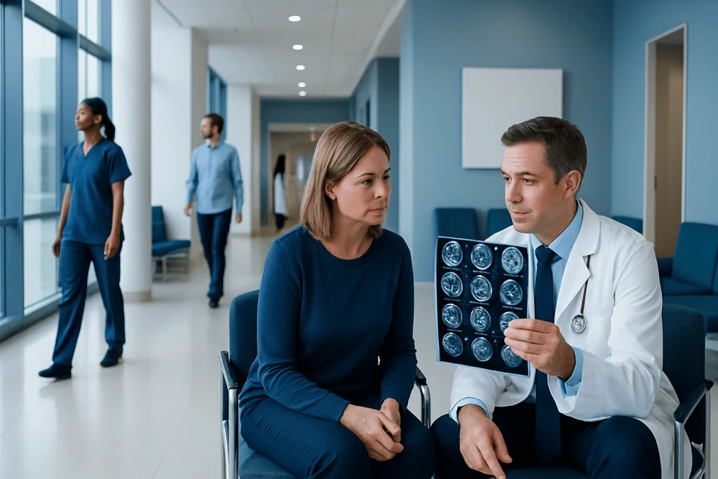
Stroke diagnosis must happen urgently because treatment depends on how quickly the stroke is identified. Brain imaging helps doctors tell the difference between ischemic stroke and hemorrhagic stroke.
Key Takeaways
- Stroke diagnosis must happen urgently because treatment depends on how quickly the stroke is identified.
- Brain imaging helps doctors tell the difference between ischemic stroke and hemorrhagic stroke.
- Blood tests do not diagnose stroke alone, but they help rule out similar problems and guide safe treatment.
- Doctors often check the heart and blood vessels to find why a stroke happened and reduce the risk of another one.
- A person with sudden facial drooping, arm weakness, speech trouble, or other stroke symptoms needs emergency medical care immediately.
Stroke diagnosis is a rapid process that helps doctors confirm whether a stroke has happened, identify its type, and start the right treatment as quickly as possible. It usually involves a neurological exam, brain imaging, blood tests, and additional checks of the heart and blood vessels to look for the underlying cause.
Overview: How Stroke Diagnosis Works
Stroke diagnosis is the process doctors use to confirm whether a person is having, or has recently had, a stroke. It also helps determine the type of stroke and whether another condition could be causing similar symptoms. This distinction matters because different stroke types need different treatments, and some treatments that help one type can be harmful in another.
The first priority is speed. When someone arrives with sudden weakness, trouble speaking, facial drooping, vision loss, severe dizziness, or confusion, the medical team quickly gathers information about when symptoms began and how they developed. Doctors then combine a focused neurological examination with urgent tests, especially brain imaging, to make decisions as safely and quickly as possible.
There are two main types of stroke: ischemic stroke, caused by a blocked blood vessel, and hemorrhagic stroke, caused by bleeding in or around the brain. A temporary episode called a transient ischemic attack, or TIA, can cause stroke-like symptoms that resolve, but it still needs urgent evaluation because it may be a warning sign of a future stroke. In some cases, doctors also consider related conditions such as brain hemorrhage when evaluating symptoms and imaging findings.
What Doctors Look for During the First Assessment
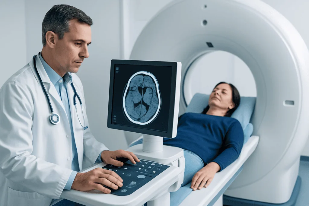
The initial assessment begins with a medical history and a neurological exam. Doctors ask when the person was last known to be well, what symptoms started first, whether they came on suddenly, and whether there is a history of stroke, high blood pressure, diabetes, heart rhythm problems, smoking, or recent surgery or bleeding. If the person cannot answer, family members or witnesses can provide vital details.
During the exam, the clinician checks alertness, speech, facial movement, arm and leg strength, sensation, balance, coordination, and vision. They may use a standardized scoring system, such as the National Institutes of Health Stroke Scale, to estimate the severity of neurological symptoms and track changes over time. This structured approach helps guide decisions about urgent treatment and monitoring.
Doctors also measure blood pressure, pulse, oxygen level, temperature, and blood glucose. This is important because low blood sugar, very high blood pressure, seizures, migraine with neurological symptoms, infections, or other conditions can sometimes mimic stroke. The early assessment is therefore both a search for stroke and a careful effort to rule out look-alike conditions.
Brain Scans Used in Stroke Diagnosis
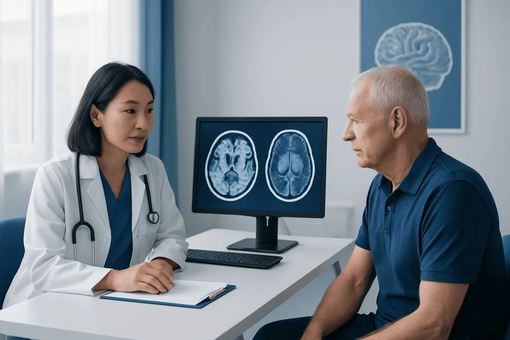
Brain imaging is central to stroke diagnosis. In most emergency settings, the first test is a non-contrast computed tomography, or CT, scan of the head. This scan is fast and widely available, and it is particularly useful for showing bleeding in the brain. If bleeding is present, treatment plans change immediately, so this first distinction is one of the most important steps in stroke care.
A CT scan may be normal in the very earliest hours of an ischemic stroke, especially if the blocked area is small. For that reason, doctors interpret the scan together with the patient’s symptoms and exam findings. Depending on the situation, they may also use CT angiography to look at the arteries in the head and neck, or CT perfusion to assess how blood is flowing through brain tissue and whether there is brain at risk that may still be saved.
Magnetic resonance imaging, or MRI, can show ischemic stroke earlier and in greater detail than a standard CT in many cases. Diffusion-weighted MRI is especially sensitive for detecting areas of recent brain injury. MRI may also help evaluate small strokes, strokes in the back of the brain, or uncertain cases. In some patients, imaging tests may be part of a broader diagnostic pathway that also includes advanced interventional neuroradiology evaluation when doctors suspect a blocked major artery or another complex vascular problem.
Imaging may also include studies of the blood vessels, such as MR angiography or carotid ultrasound. These tests help identify narrowing, blockage, dissection, aneurysm, or other abnormalities in the arteries supplying the brain. Finding the exact vascular problem helps doctors plan treatment and future prevention more precisely.
Why Blood Tests Matter in Stroke Evaluation
Blood tests are an important part of stroke diagnosis, although they do not by themselves prove that a stroke has happened. Instead, they help identify conditions that can mimic stroke, reveal medical problems that may have contributed to it, and show whether certain treatments can be given safely. They are usually ordered quickly, often at the same time as brain imaging.
Common tests include blood glucose, a complete blood count, kidney function, electrolytes, and clotting studies. Blood glucose is checked right away because very low or very high sugar levels can cause weakness, confusion, or speech problems that resemble stroke. A complete blood count can show anemia, infection, or problems with platelets, while clotting tests help doctors assess bleeding risk, especially if emergency clot-busting treatment is being considered.
Doctors may also order tests such as cardiac enzymes, inflammatory markers, or lipid and diabetes screening depending on the clinical picture. If a person is taking blood thinners, special tests may be needed to understand how strongly the medication is affecting clotting. In younger patients or in strokes without an obvious cause, the team may investigate less common contributors such as autoimmune conditions or inherited clotting disorders.
How Doctors Find the Cause of a Stroke
Confirming that a stroke occurred is only the first step. The next goal is to find out why it happened. This is especially important because preventing another stroke often depends on treating the underlying cause, such as narrowing in a neck artery, an abnormal heart rhythm, uncontrolled blood pressure, or a problem with blood clotting.
One common cause of ischemic stroke is a clot that forms in the heart and travels to the brain. To look for this, doctors may perform an electrocardiogram to check the heart rhythm and may monitor the heartbeat over time to detect atrial fibrillation or other rhythm disturbances. Echocardiography, an ultrasound of the heart, can help identify clots, valve problems, structural defects, or weak heart pumping that may increase stroke risk.
Doctors also assess the blood vessels that supply the brain. Carotid ultrasound can detect narrowing in the carotid arteries of the neck. CT angiography or MR angiography may show narrowing, blockage, dissection, or unusual vessel anatomy in the neck or brain. In some situations, more detailed vascular assessment and treatment planning may involve interventional neurology teams, particularly when a large vessel blockage is suspected.
If a person has symptoms that suggest a brief warning event rather than a completed stroke, doctors may consider the diagnosis of transient ischemic attack and still recommend urgent evaluation. Even when symptoms improve, a TIA can signal a high risk of a future stroke, so identifying its cause remains very important.
How Diagnosis Guides Treatment Decisions
Stroke treatment depends directly on the results of the diagnostic workup. If brain imaging shows an ischemic stroke and the patient arrives within an appropriate time window, doctors may consider treatments that restore blood flow. These can include clot-dissolving medicine in selected patients or a procedure to remove a clot from a large artery. If imaging shows bleeding instead, treatment focuses on controlling the bleed, lowering risk factors such as very high blood pressure, and managing complications.
When a blocked major artery is found, some patients may be evaluated for mechanical thrombectomy to reopen the vessel. The decision depends on several factors, including symptom severity, the location of the blockage, brain imaging findings, and the time since symptoms began or the last known well time. This is why prompt transport to emergency care is so important.
Diagnosis also shapes longer-term treatment. A person with atrial fibrillation may need medication to reduce clot formation. Someone with significant carotid artery disease may need specific medical treatment or a procedure. If the stroke is related to high blood pressure, diabetes, cholesterol problems, smoking, or sleep apnea, those conditions become key parts of the care plan.
Rehabilitation planning often begins soon after diagnosis. Depending on the symptoms, this may include speech therapy, physical therapy, occupational therapy, and swallowing assessment. At Acibadem International, multidisciplinary specialists in JCI-accredited hospitals diagnose and treat stroke for international patients, with care coordinated across emergency medicine, neurology, imaging, and rehabilitation services.
Prevention, Follow-up, and Self-care After Diagnosis
After stroke diagnosis, preventing another event becomes a major focus. Doctors usually review blood pressure, cholesterol, blood sugar, heart rhythm, and lifestyle factors. They may prescribe medications such as antiplatelet drugs, cholesterol-lowering treatment, blood pressure medicines, or anticoagulants, depending on the stroke type and cause. The exact plan should always be individualized by the treating physician.
Healthy daily habits support recovery and prevention. These include not smoking, limiting alcohol, following a balanced eating plan, being physically active within medical advice, taking medicines as directed, and attending all follow-up appointments. If swallowing is affected, nutrition and hydration plans may need to be adjusted until swallowing improves.
Family support and education can make a meaningful difference. Loved ones can help notice changes in speech, walking, memory, or mood that may need medical attention. They can also encourage adherence to medications and rehabilitation. Because emotional changes are common after stroke, asking for support with mood, cognition, or fatigue is an important part of recovery, not a sign of weakness.
When to Seek Emergency Help
A possible stroke is always a medical emergency. A person should seek immediate emergency care if sudden symptoms appear, especially facial drooping, arm weakness, speech difficulty, confusion, vision loss, severe dizziness, loss of coordination, or a sudden severe headache unlike usual headaches. Even if symptoms improve quickly, urgent evaluation is still needed because a transient episode may be a warning sign.
It is safest not to wait to see if symptoms go away. Delays can reduce treatment options and may increase the chance of lasting brain injury. Calling emergency services is usually better than traveling by private car because paramedics can begin assessment early and alert the hospital stroke team.
After a stroke has already been diagnosed, urgent reassessment is also important if new weakness, new speech problems, worsening headache, fainting, chest pain, breathing difficulty, or sudden confusion occurs. Any sudden change in neurological function should be taken seriously and discussed with a qualified doctor right away.
Frequently asked questions
What is the first test doctors usually do for stroke diagnosis?
In many emergency departments, the first major test is a non-contrast CT scan of the head. It is fast and helps doctors quickly see whether there is bleeding in the brain, which is essential for choosing the right treatment.
Can blood tests show if someone is having a stroke?
Blood tests cannot confirm a stroke on their own. However, they are very useful for ruling out similar conditions, identifying risk factors, and checking whether emergency treatments can be given safely.
Why do doctors need to know when symptoms started?
The time symptoms began helps determine which treatments may still be effective and safe. Some stroke therapies are most helpful only within specific time windows, so accurate timing can directly affect care.
What is the difference between a CT scan and an MRI for stroke?
A CT scan is often used first because it is fast and good at detecting bleeding. An MRI can be more sensitive for early ischemic stroke and may provide more detail, but it is not always the first test in an emergency.
Do all people with stroke need heart tests?
Many people do, especially when doctors suspect the stroke may have started with a clot from the heart. Tests such as an ECG, heart rhythm monitoring, and echocardiography can help find causes like atrial fibrillation or structural heart problems.
Can stroke symptoms come and go and still be serious?
Yes. Symptoms that improve quickly may be caused by a transient ischemic attack, which is a warning sign that a larger stroke could happen later. This still requires urgent medical assessment.
This article is for general information only and is not a substitute for professional medical advice. Please consult a qualified doctor about your individual situation.
Explore treatments in Turkey — costs, top hospitals & a free quote
JCI-accredited · board-certified surgeons · reply within 24h
Add us as a Preferred Source to see more of our trusted health content across Google Search, AI Overviews and Discover.
Check your numbers in seconds
BMI, calories, due date, blood pressure and 30+ more clinical calculators — free, instant, doctor-reviewed ranges.

More from the Health Library
Stroke Diagnosis: CT, MRI, and Other Tests Doctors Use in the First Hours
Ischemic Stroke: Causes, Clot Blockage, and Emergency Treatment

Recurrent Transient Ischemic Attacks: When a Second Stroke Opinion Matters
Speech Problems After Stroke: Aphasia, Slurred Speech, and What Therapy Can Improve
Ischemic Stroke Explained: How a Blocked Brain Artery Is Diagnosed and Treated