Stroke Diagnosis: CT, MRI, and Other Tests Doctors Use in the First Hours

A brain CT scan is often the first test because it is fast and helps detect bleeding. MRI can show brain tissue changes in more detail and may detect very early ischemic stroke.
Key Takeaways
- A brain CT scan is often the first test because it is fast and helps detect bleeding.
- MRI can show brain tissue changes in more detail and may detect very early ischemic stroke.
- Doctors also use blood tests, heart tests, and vessel imaging to identify the cause and safest treatment.
- The main goal in the first hours is to distinguish ischemic stroke from hemorrhagic stroke.
- Rapid stroke diagnosis helps determine whether clot-busting treatment or a procedure is appropriate.
Stroke diagnosis must happen quickly because treatment depends on whether the stroke is caused by a blocked blood vessel or bleeding in the brain. In the first hours, doctors use brain imaging, blood tests, heart tests, and neurological examination to confirm the diagnosis and guide urgent care.
Overview: Why stroke diagnosis is so time-sensitive
Stroke diagnosis is an emergency process designed to answer a few critical questions as quickly as possible. Doctors need to know whether symptoms are truly caused by a stroke, what type of stroke is present, when the symptoms began, and which treatment can be given safely. Because brain tissue can be injured very quickly when blood flow is interrupted, early testing is central to improving outcomes.
There are two main types of stroke. Ischemic stroke happens when a blood vessel in the brain is blocked, usually by a clot. Hemorrhagic stroke happens when a blood vessel bleeds into or around the brain. Although the symptoms may look similar, the treatments are very different, so doctors cannot rely on symptoms alone.
In the first hours, stroke diagnosis usually combines a focused neurological examination with imaging tests such as CT or MRI. Blood tests, heart rhythm checks, and imaging of the blood vessels in the head and neck may also be done. Together, these tests help the team decide whether urgent therapies such as stroke treatment or interventional neuroradiology procedures are appropriate.
The first assessment in the emergency setting
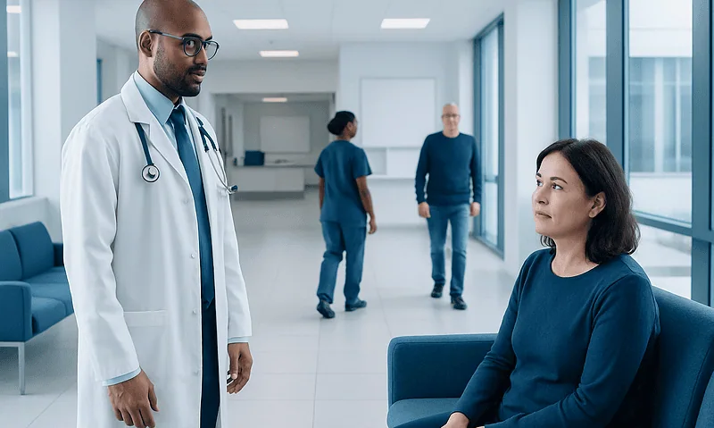
When someone arrives with possible stroke symptoms, doctors first stabilize breathing, blood pressure, blood sugar, and other immediate concerns. At the same time, they gather a brief history. Important details include the exact time symptoms started or the last time the person was known to be well, current medications such as blood thinners, recent surgery or bleeding, and prior stroke history.
A neurological examination follows right away. The clinician checks speech, facial movement, arm and leg strength, sensation, coordination, alertness, vision, and balance. Many hospitals use a standardized stroke scale to measure the severity of symptoms and track changes over time.
Doctors also consider conditions that can mimic stroke. Low blood sugar, seizures, migraine, brain tumors, infections, and certain metabolic problems can all cause stroke-like symptoms. This is one reason blood glucose is checked immediately and brain imaging is done without delay. The goal is not only to confirm a stroke, but also to avoid giving a treatment that may be unsafe if the diagnosis is something else.
CT scan: often the first imaging test
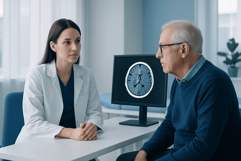
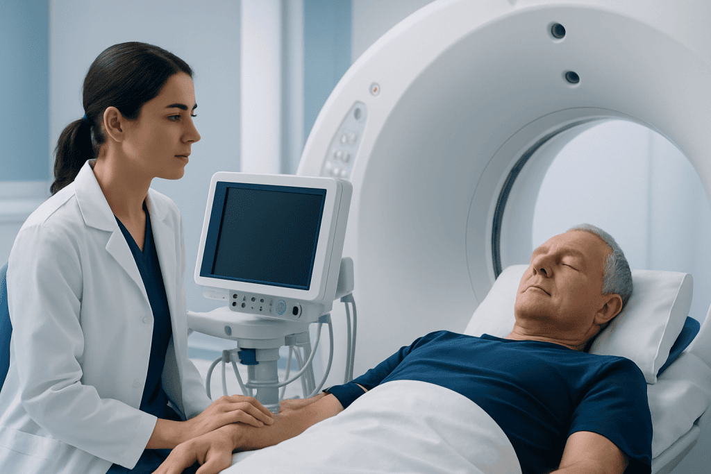
A non-contrast CT scan of the brain is commonly the first imaging test used in stroke diagnosis. It is widely available, fast, and very effective at showing bleeding in the brain. This matters because a person with hemorrhagic stroke needs a different treatment plan than someone with ischemic stroke.
In the first minutes to hours after an ischemic stroke, a standard CT scan may look normal or show only subtle early changes. Even so, it remains extremely useful because it can quickly rule out hemorrhage and help doctors decide whether clot-dissolving treatment may be possible. In emergency care, speed is essential, and CT is often the fastest way to get that answer.
Doctors may also request CT angiography, which uses contrast dye to look at the blood vessels in the brain and neck. This can show whether a large artery is blocked. In some cases, CT perfusion is added to estimate how much brain tissue is already injured and how much may still be saved. These advanced CT techniques can help identify patients who may benefit from minimally invasive clot-removal procedures.
MRI: detailed imaging for early ischemic stroke
MRI is another important tool in stroke diagnosis, especially for ischemic stroke. Certain MRI sequences, particularly diffusion-weighted imaging, can detect changes in brain tissue very early after blood flow is reduced. This can make MRI more sensitive than CT for some small or early strokes.
MRI can also help clarify difficult cases, such as strokes in the brainstem, very small strokes, or symptoms that may have another explanation. It gives detailed information about the location and extent of injury and may help estimate how recent the stroke is. In some patients, this information helps guide treatment decisions when the exact timing of symptom onset is uncertain.
Despite these advantages, MRI is not always the first test in the emergency department. It takes longer, may be less available in some settings, and is not suitable for every patient. For example, some implanted devices, severe claustrophobia, or medical instability may limit its use. Doctors choose between CT and MRI based on urgency, availability, and the clinical situation, sometimes using both.
Other tests doctors use in the first hours
Brain imaging is central, but it is only one part of stroke diagnosis. Blood tests are usually done at the same time to look for problems that may mimic stroke or affect treatment safety. Common tests include blood sugar, blood count, kidney function, electrolytes, and blood clotting measures, especially if the person may be taking anticoagulant medication.
Heart tests are also important because some strokes are caused by blood clots that form in the heart and travel to the brain. An electrocardiogram, or ECG, helps detect abnormal rhythms such as atrial fibrillation. Continuous heart monitoring may be started in the emergency department because some rhythm problems come and go.
Doctors may also use ultrasound or angiographic imaging to examine the carotid arteries and other vessels supplying the brain. Depending on the case, this may include carotid Doppler ultrasound, CT angiography, or MR angiography. These tests help identify narrowing, blockage, or other vascular problems that can contribute to stroke and influence immediate and long-term treatment planning.
- Blood glucose check for stroke mimic screening
- Complete blood count and clotting studies
- Kidney function tests before some contrast studies
- ECG and cardiac monitoring for arrhythmias
- Vessel imaging of the head and neck
How the results guide treatment decisions
The most important early question is whether the stroke is ischemic or hemorrhagic. If imaging shows a blocked artery without bleeding, doctors may consider clot-busting medication if the person arrives within the appropriate time window and meets safety criteria. If a large vessel blockage is seen, a catheter-based procedure to remove the clot may also be considered in selected patients.
If bleeding is found, treatment focuses on controlling blood pressure, managing the effects of the hemorrhage, and addressing the source of bleeding. In some cases, neurosurgical or neurointerventional care may be needed. Doctors also review medications such as blood thinners because these may need urgent reversal depending on the situation.
Imaging and test results can also reveal causes that shape prevention after the emergency has passed. For example, a narrowed carotid artery, atrial fibrillation, or a disorder affecting the blood vessels may require specific follow-up care. This is one reason early diagnosis is not only about the first treatment, but also about reducing the risk of another event such as a transient ischemic attack or recurrent stroke.
What patients and families should know in the first hours
For patients and families, the first hours can feel fast and overwhelming. It is common for the care team to repeat questions, perform frequent examinations, and move quickly between tests. This helps them track changes in symptoms and make time-sensitive decisions safely.
Families can be especially helpful by sharing the time symptoms started, a list of medications, allergies, recent surgeries, and previous medical problems. If the person was last seen well at a certain time before symptoms were noticed, that detail is often very important. Bringing pill bottles or a medication list can save valuable time.
After the emergency evaluation, doctors usually explain what the scans showed, whether a blood vessel is blocked or bleeding is present, and what treatment options are available. In complex cases, specialists in neurology, radiology, neurosurgery, cardiology, and rehabilitation may all be involved. At Acibadem International, multidisciplinary specialists in JCI-accredited hospitals diagnose and treat stroke for international patients, with care plans based on imaging findings and individual needs.
When to seek urgent medical care
Possible stroke symptoms should never be watched at home to see if they pass. Emergency care is needed right away for sudden facial drooping, arm weakness, speech difficulty, confusion, vision loss, severe dizziness, trouble walking, or a sudden severe headache unlike usual headaches. Even symptoms that improve quickly can be a warning sign.
Calling emergency services is safer than trying to drive to the hospital because treatment can begin sooner and the person can be monitored on the way. Rapid transport also helps the hospital prepare imaging and the stroke team before arrival. The earlier stroke diagnosis begins, the more treatment options may be available.
Anyone who has had a stroke or mini-stroke should also follow up closely after discharge. Doctors may recommend additional heart testing, blood vessel imaging, rehabilitation, or risk factor treatment such as blood pressure control, diabetes management, smoking cessation, and cholesterol-lowering therapy. These steps support recovery and help lower the chance of future events.
Frequently asked questions
Why is a CT scan usually done before an MRI in suspected stroke?
A CT scan is fast, widely available, and very good at showing bleeding in the brain. Doctors need that information immediately because bleeding and clot-related strokes are treated differently. MRI may be added later or used first in some hospitals when it is available and appropriate.
Can a CT scan miss a stroke?
A standard CT scan may not show very early or very small ischemic strokes right away. However, it is still extremely useful because it can quickly identify bleeding and other urgent problems. If the diagnosis remains uncertain, doctors may use MRI or additional vessel imaging.
What is the difference between ischemic and hemorrhagic stroke on tests?
Ischemic stroke is caused by a blocked blood vessel, while hemorrhagic stroke involves bleeding in or around the brain. Imaging tests, especially CT and MRI, help doctors tell these apart. This distinction is essential because treatments that help one type may be harmful in the other.
What blood tests are done during stroke diagnosis?
Doctors commonly check blood sugar, blood count, kidney function, electrolytes, and clotting tests. These tests can identify stroke mimics, show whether contrast imaging is safe, and help determine if clot-busting medicine may be appropriate. They are usually done alongside imaging rather than instead of it.
Do all patients with stroke symptoms need an MRI?
Not always. Many patients are first assessed with CT because it is faster in emergency settings. MRI is often used when more detail is needed, when CT findings are unclear, or when doctors suspect a small or very early ischemic stroke.
How do doctors know if someone is eligible for clot-removal treatment?
Doctors combine the neurological exam with brain and vessel imaging to see whether a large artery is blocked and whether brain tissue may still be saved. They also consider timing, general health, and bleeding risk. This decision is made quickly by the stroke team based on established medical criteria.
This article is for general information only and is not a substitute for professional medical advice. Please consult a qualified doctor about your individual situation.
Explore treatments in Turkey — costs, top hospitals & a free quote
JCI-accredited · board-certified surgeons · reply within 24h
Add us as a Preferred Source to see more of our trusted health content across Google Search, AI Overviews and Discover.
Check your numbers in seconds
BMI, calories, due date, blood pressure and 30+ more clinical calculators — free, instant, doctor-reviewed ranges.

More from the Health Library
What Is a Stroke? Types, Brain Effects, and Why Every Minute Matters
What Causes a Stroke? Common Triggers and Modifiable Risk Factors

Cerebral Infarction: Symptoms, Diagnosis, and How It Is Treated
Stroke Mimics: Conditions That Can Look Like a Stroke

Signs of Stroke: Early Symptoms You Should Never Ignore