Transvaginal Ultrasound: A Complete Medical Overview

Transvaginal ultrasound uses a slim ultrasound probe placed gently into the vagina to view pelvic organs in detail. It does not use radiation and is widely used for gynecologic evaluation and early pregnancy assessment.
Key Takeaways
- Transvaginal ultrasound uses a slim ultrasound probe placed gently into the vagina to view pelvic organs in detail.
- It does not use radiation and is widely used for gynecologic evaluation and early pregnancy assessment.
- The test can help identify causes of pelvic pain, abnormal bleeding, ovarian cysts, fibroids, and some fertility concerns.
- Most people feel pressure rather than pain, and the scan is usually brief.
- Results need to be interpreted alongside symptoms, examination findings, and sometimes blood tests or other imaging.
Transvaginal ultrasound is an internal pelvic imaging test that uses sound waves to create detailed pictures of the uterus, ovaries, cervix, and surrounding structures. It is commonly used to evaluate pelvic symptoms, investigate abnormal bleeding, and assess early pregnancy because it often provides clearer detail than an abdominal ultrasound.
Overview: what a transvaginal ultrasound is
Transvaginal ultrasound is a medical imaging test that allows a doctor to examine the pelvic organs from inside the vagina using high-frequency sound waves. The scan produces real-time images of structures such as the uterus, endometrium, cervix, ovaries, fallopian tube regions, and nearby pelvic tissues. Because the probe is positioned close to these organs, the images are often more detailed than those from a standard abdominal pelvic ultrasound.
This test is commonly used in gynecology, reproductive medicine, and early pregnancy care. It can help doctors investigate symptoms such as pelvic pain, irregular bleeding, missed periods, pain during intercourse, or unexplained fertility difficulties. In pregnancy, it may be used to confirm an early intrauterine pregnancy, estimate gestational age, or assess bleeding and pain in the first trimester.
Although the name may sound intimidating, the procedure is usually straightforward and brief. A covered, lubricated ultrasound probe is inserted gently into the vagina while the patient lies on an examination table. The scan does not use radiation, and most people describe it as uncomfortable rather than painful, though individual experiences vary depending on symptoms and personal sensitivity.
When and why doctors recommend it
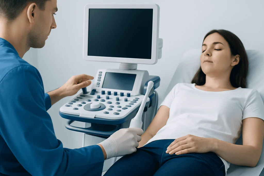
A transvaginal ultrasound is often requested when a closer look at the female pelvis is needed. It may be used after symptoms begin or as part of follow-up care for a known condition. Compared with abdominal imaging, this approach can better show the thickness of the uterine lining, the appearance of ovarian follicles or cysts, and subtle changes in pelvic anatomy.
Common reasons for ordering the test include abnormal uterine bleeding, pelvic or lower abdominal pain, suspected ovarian cysts, concern for uterine fibroids, infertility evaluation, and monitoring during fertility treatment. It can also help assess the position of an intrauterine device and support investigation of conditions such as endometriosis or uterine fibroids when imaging findings are considered together with symptoms.
In early pregnancy, transvaginal ultrasound can be especially helpful because very early structures may not yet be visible through the abdomen. Doctors may use it to look for a gestational sac, yolk sac, embryo, or fetal heartbeat at the appropriate stage. It may also assist in evaluating vaginal bleeding, pelvic pain, or concern for pregnancy developing outside the uterus.
- Assessing abnormal bleeding or postmenopausal bleeding
- Investigating pelvic pain or pressure
- Evaluating ovarian cysts or masses
- Checking the uterus and endometrial lining
- Monitoring ovulation or fertility treatment
- Examining early pregnancy and first-trimester symptoms
What the scan can show

Transvaginal ultrasound gives detailed information about pelvic anatomy, but it is best understood as one part of a broader medical assessment. The scan may show the size and shape of the uterus, whether fibroids are present, how thick the endometrium appears, and whether the ovaries contain normal follicles, cysts, or more complex findings. It may also identify free fluid in the pelvis or features that prompt further testing.
In gynecologic care, the test can help support diagnosis or follow-up of several common conditions. These include ovarian cysts, adenomyosis, uterine polyps, pelvic inflammatory changes, and masses that need further evaluation. Sometimes the scan is paired with Doppler ultrasound, which looks at blood flow patterns that may add useful information in selected cases.
During fertility assessment, transvaginal ultrasound may be used to count ovarian follicles, monitor ovulation, and assess the uterus before treatment. In some settings, it is part of preparation for IVF treatment or a wider fertility evaluation and treatment plan. In early pregnancy, it may help confirm viability, estimate gestational age, and identify signs that require closer observation.
How to prepare and what happens during the procedure
Preparation for a transvaginal ultrasound is usually simple. Patients are often asked to empty their bladder before the scan, since a full bladder can make the internal images less comfortable and less helpful. Clothing from the waist down is removed or adjusted, and a drape is typically provided for privacy. The healthcare team explains the process before starting and should answer questions at any stage.
During the examination, the patient lies on the table, usually with knees bent. A slim ultrasound probe is covered with a protective sheath and lubricating gel, then inserted gently into the vagina. The sonographer or doctor moves the probe slightly to capture images from different angles. The process commonly takes only a short time, though the exact length depends on the reason for the scan.
Most people feel pressure, fullness, or mild discomfort rather than significant pain. If there is active pelvic pain, infection, or anxiety about the exam, it may feel more uncomfortable. Patients should say so during the procedure; adjustments can often be made to improve comfort. In many centers, a chaperone can be requested, and clear communication is encouraged throughout.
After the scan, there is generally no recovery time, and normal activities can usually be resumed immediately. A small amount of gel or light spotting may occur occasionally, especially if the tissues are sensitive, but this is usually mild and short-lived.
Understanding the results
The images from a transvaginal ultrasound are interpreted by a radiologist, gynecologist, or another trained specialist. Results are not based only on what is seen on the scan. Doctors also consider age, menstrual cycle timing, menopausal status, pregnancy status, symptoms, medical history, examination findings, and sometimes blood tests such as hormone levels or pregnancy tests.
A normal result may show a uterus and ovaries with expected size and appearance for the person’s age and cycle stage. In reproductive-age patients, normal findings can vary from one part of the menstrual cycle to another. For example, the endometrial lining naturally changes in thickness, and ovarian follicles may be seen before ovulation.
An abnormal result does not automatically mean a serious problem. Some findings, such as simple ovarian cysts, may be common and may only need observation. Other findings, including complex masses, marked endometrial thickening, or signs of ectopic pregnancy, need prompt medical review and sometimes further testing. Depending on the result, the next step may include repeat imaging, blood tests, office procedures, or referral to a specialist.
Benefits, limits, and possible next steps
The main benefit of transvaginal ultrasound is image clarity. Because the probe is close to the pelvic organs, it can reveal details that are harder to see through the abdominal wall. The test is also widely available, does not use ionizing radiation, and can be performed relatively quickly. These features make it an important first-line tool in many gynecologic and early pregnancy situations.
At the same time, no single scan can answer every question. Ultrasound may not always determine exactly what a mass is, whether symptoms are caused by microscopic disease, or how severe a condition may be. Some problems are better assessed with blood tests, MRI, hysteroscopy, laparoscopy, or tissue sampling, depending on the clinical situation. For example, a suspicious ovarian finding may lead to further review in a center offering specialist gynecologic oncology care when appropriate.
Another limitation is that results can depend on timing. A scan done at one point in the menstrual cycle may look different from a scan done later, and early pregnancy findings can evolve over days rather than weeks. That is why doctors sometimes recommend repeat imaging instead of making a conclusion from a single examination.
When to seek medical care
A transvaginal ultrasound itself is generally safe, but the symptoms leading to the scan may need medical attention. Prompt assessment is important if pelvic pain is severe, if there is heavy vaginal bleeding, fainting, fever, or worsening symptoms during pregnancy. These symptoms do not always signal an emergency, but they should not be ignored.
Medical advice should also be sought for ongoing abnormal bleeding between periods, bleeding after menopause, repeated pelvic pain, trouble becoming pregnant, or pain during intercourse that does not improve. Early evaluation can help identify common causes and guide the right treatment plan.
People who feel unsure about the procedure because of pain history, prior trauma, cultural concerns, or anxiety should discuss this in advance with their healthcare team. Alternative approaches or extra support may sometimes be available. Near the end of the care pathway, patients may benefit from a multidisciplinary review; Acibadem International’s specialists in JCI-accredited hospitals diagnose and treat pelvic and reproductive conditions for international patients.
Frequently asked questions
Is a transvaginal ultrasound painful?
Most people feel pressure or mild discomfort rather than pain. The experience can be more uncomfortable if there is existing pelvic pain, infection, or anxiety, but the examiner can usually adjust the procedure to improve comfort.
How is a transvaginal ultrasound different from an abdominal ultrasound?
An abdominal ultrasound is performed over the lower abdomen, while a transvaginal ultrasound uses a probe placed inside the vagina. The internal approach usually gives clearer, more detailed images of the uterus, ovaries, cervix, and early pregnancy.
Does transvaginal ultrasound use radiation?
No. It uses sound waves to create images, so it does not expose the body to ionizing radiation. This is one reason it is commonly used in gynecology and early pregnancy assessment.
Can a transvaginal ultrasound detect pregnancy?
It can often confirm an early intrauterine pregnancy sooner than an abdominal scan. It may also help evaluate first-trimester bleeding or pain, but findings must be interpreted with the pregnancy stage and blood test results.
Do patients need to prepare for the test?
Preparation is usually minimal. Patients are often asked to empty their bladder before the scan, and they should tell the healthcare team about pregnancy, symptoms, infections, or any concerns about the exam.
What conditions can a transvaginal ultrasound help investigate?
It can help assess abnormal bleeding, pelvic pain, ovarian cysts, uterine fibroids, endometrial changes, infertility concerns, and some early pregnancy problems. It is an important imaging tool, but doctors may still recommend other tests to confirm a diagnosis.
References
- World Health Organization
- American College of Obstetricians and Gynecologists
- NHS
- Radiological Society of North America
- International Society of Ultrasound in Obstetrics and Gynecology
This article is for general information only and is not a substitute for professional medical advice. Please consult a qualified doctor about your individual situation.
Ultrasound in Turkey — costs, top hospitals & a free quote
JCI-accredited · board-certified surgeons · reply within 24h
Add us as a Preferred Source to see more of our trusted health content across Google Search, AI Overviews and Discover.
Check your numbers in seconds
BMI, calories, due date, blood pressure and 30+ more clinical calculators — free, instant, doctor-reviewed ranges.

Check-up & Preventive Medicine
Comprehensive health screenings designed for early detection and prevention.
1 specialists in this unit