Understanding Leg Early Stage Skin Cancer: A Complete Patient Guide

Skin cancer can develop anywhere on the leg, including areas that receive intermittent sun exposure. A spot that changes, bleeds, does not heal, or looks different from surrounding marks should be assessed.
Key Takeaways
- Skin cancer can develop anywhere on the leg, including areas that receive intermittent sun exposure.
- A spot that changes, bleeds, does not heal, or looks different from surrounding marks should be assessed.
- Basal cell carcinoma, squamous cell carcinoma, and melanoma can have different appearances and levels of urgency.
- A skin examination and, when needed, a small biopsy are used to confirm the diagnosis.
- When found early, many skin cancers on the leg can be treated effectively with procedures that remove or destroy the abnormal cells.
Leg early stage skin cancer can look like a changing mole, a persistent sore, a rough scaly patch, or a new growth on the lower or upper leg. Most suspicious skin changes are not cancer, but a prompt examination by a qualified clinician is the only reliable way to identify cancer early and arrange appropriate care.
Overview: What Does Early Stage Skin Cancer on the Leg Mean?
Leg early stage skin cancer refers to cancerous changes in skin cells that are limited to the skin or have not spread beyond the original area. It may occur on the thigh, knee, shin, calf, ankle, foot, or toes. Early lesions can be subtle, which is why regular awareness of the skin on the legs is useful, including areas that are not frequently noticed.
The three main forms are basal cell carcinoma, squamous cell carcinoma, and melanoma. Basal cell carcinoma often grows slowly and rarely spreads, while squamous cell carcinoma can be more likely to grow deeper if left untreated. Melanoma is less common but requires timely assessment because it may spread if not detected early.
It is not possible to diagnose skin cancer based on appearance alone. Infections, eczema, psoriasis, scars, warts, benign moles, and harmless age-related skin changes can resemble cancer. A dermatologist or another trained clinician can examine a concerning spot closely and recommend testing when appropriate.
Changes to Look For on the Leg
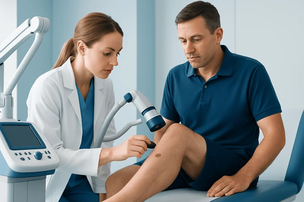
A new mark or an existing spot that changes over weeks or months deserves attention. Changes may involve size, shape, color, texture, elevation, or symptoms such as itching, tenderness, crusting, or bleeding. Taking note of when a lesion first appeared and whether it has changed can be helpful at a medical appointment.
Melanoma is often discussed using the ABCDE guide: asymmetry, irregular border, uneven color, diameter that is increasing, and evolution or change. However, melanomas do not always follow every part of this guide. Some may be small, pink, red, skin-colored, or resemble a bruise or a slowly changing dark streak.
Basal cell carcinoma on the leg may appear as a pearly or shiny bump, a pink patch, a sore that repeatedly heals and reopens, or a scar-like pale area. Squamous cell carcinoma may look like a firm red bump, a rough scaly plaque, a crusted growth, or a nonhealing sore. On the lower legs, these appearances may sometimes be confused with dry skin, injury, or poor wound healing.
- A mole or spot that is noticeably different from others, sometimes called the “ugly duckling” sign
- A wound or scab that has not healed after several weeks
- A lesion that bleeds with little contact or repeatedly forms a crust
- A growing, thickened, painful, or tender scaly patch
- A new dark line under or around a toenail, particularly if it widens or changes
Why Skin Cancer Can Develop on the Legs

Ultraviolet, or UV, radiation from sunlight is a leading cause of many skin cancers. UV exposure can damage the genetic material in skin cells over time. The legs may receive substantial exposure during outdoor activities, walking, swimming, or wearing shorts and skirts. Intense intermittent sun exposure and episodes of sunburn can be particularly important risk factors for melanoma.
Indoor tanning devices also expose the skin to UV radiation and increase skin cancer risk. Skin cancer may still arise on areas that are usually covered, so sun exposure is not the only factor. It is important not to assume that a lesion is harmless simply because it is on the inner thigh, lower leg, or another less sun-exposed site.
Other factors that can increase risk include fair or easily sunburned skin, a personal or family history of skin cancer, numerous moles, atypical moles, increasing age, and a weakened immune system. Long-standing wounds, scars from burns, chronic inflammation, and previous radiation treatment can also be relevant in certain cases. People with darker skin can develop skin cancer too, sometimes in less expected locations such as the soles, nails, or between the toes.
How a Suspicious Leg Lesion Is Diagnosed
Diagnosis usually begins with a medical history and a full skin examination. The clinician may ask when the spot appeared, whether it has changed, whether it causes symptoms, and whether there is a history of sun exposure, tanning bed use, skin cancer, or immune suppression. Examining the whole skin surface is often useful because more than one concerning lesion may be present.
A dermatologist may use a dermatoscope, a handheld device that magnifies the skin and reveals patterns not easily seen with the naked eye. Dermoscopy can help decide whether a lesion needs close monitoring, biopsy, or removal, but it does not replace laboratory confirmation when cancer is suspected.
A biopsy is the standard way to confirm a diagnosis. A clinician removes all or part of the lesion using local anesthetic, and a pathologist examines the tissue under a microscope. If melanoma is diagnosed, the pathology report provides important details, such as the thickness of the tumor, that guide next steps. Further imaging or lymph node evaluation is not needed for every early lesion; it is considered based on the specific cancer type and pathology findings.
Treatment Options for Early Skin Cancer on the Leg
Treatment depends on the confirmed diagnosis, the size and depth of the lesion, its exact location, whether it has returned after prior treatment, and the person’s overall health. The aim is to remove or destroy the cancer while preserving as much healthy tissue and function as possible. A clinician can explain the expected scar, wound care needs, and follow-up plan before treatment.
For many basal cell and squamous cell carcinomas, surgical excision is a common approach. The doctor removes the lesion along with a margin of normal-looking skin, and the tissue is checked in the laboratory. In selected situations, other options may include curettage and cautery, cryotherapy, topical medicines, photodynamic therapy, or radiation therapy. The most suitable option depends on the lesion and should not be chosen without medical assessment.
Early melanoma is generally treated with surgery to remove the melanoma and an appropriate area of surrounding skin. Depending on the melanoma’s characteristics, the care team may discuss wider excision and, for some patients, sentinel lymph node biopsy. Prompt specialist-led care is important, but treatment decisions should be individualized rather than based on photographs or online descriptions.
After treatment on the lower leg, healing can occasionally take longer than on some other body areas because of swelling, circulation issues, or tension on the skin. Following wound-care instructions, keeping follow-up appointments, and reporting increasing redness, pain, drainage, or fever can support safe recovery.
Protecting the Skin and Checking the Legs Regularly
Sun protection lowers the risk of further UV-related skin damage. Practical measures include seeking shade when possible, wearing protective clothing, using a broad-spectrum sunscreen with an appropriate SPF, and reapplying sunscreen according to the product instructions, especially after swimming or sweating. Sunscreen is helpful but should be used alongside, not instead of, shade and clothing.
A monthly skin check can help people become familiar with what is normal for them. In a well-lit room, they can inspect the front and back of both legs, calves, shins, ankles, feet, soles, and the spaces between the toes. A hand mirror can help view the backs of the legs, and a partner may assist with difficult-to-see areas.
Photographs can be useful for monitoring a spot over time if they are taken in similar lighting and from a similar distance. However, photography should not delay an appointment for a lesion that is changing or concerning. People who have previously had skin cancer or have a higher-risk history may need regular professional skin checks at intervals recommended by their clinician.
When to Seek Medical Care
A person should arrange a non-urgent medical appointment soon for a new or changing lesion on the leg, especially if it has persisted for more than a few weeks. Assessment is also appropriate for a mole that is enlarging, has irregular edges or several colors, or simply looks unlike the person’s other moles. Early assessment is a sensible precaution and does not mean that cancer is likely.
More prompt medical advice is appropriate for a lesion that is rapidly growing, bleeding without a clear injury, very painful, ulcerated, or associated with a new lump in the groin. People with a weakened immune system, a past melanoma, or a strong family history of melanoma should have a lower threshold for requesting an examination.
Acibadem International’s multidisciplinary specialists and JCI-accredited hospitals can assess suspicious skin lesions and coordinate dermatology, pathology, surgical, and oncology care for international patients when needed. A qualified clinician should always evaluate persistent or changing skin findings in person.
Frequently asked questions
What does early stage skin cancer on the leg look like?
It can look like a changing mole, a new dark or unevenly colored spot, a persistent pink patch, a pearly bump, a rough scaly area, or a sore that does not heal. There is no single appearance that confirms skin cancer. A clinician should assess any lesion that changes, bleeds, grows, or remains present.
Can skin cancer develop on the lower leg?
Yes. Skin cancer can develop on any part of the lower leg, including the shin, calf, ankle, foot, sole, and around the toenails. Although sun exposure is an important risk factor, some skin cancers arise in areas that are usually covered.
Is a nonhealing sore on the leg always skin cancer?
No. A nonhealing sore may result from injury, infection, circulation problems, diabetes, inflammatory skin conditions, or other causes. However, a sore that repeatedly scabs, bleeds, or does not improve should be examined, particularly if there is no clear explanation.
How is skin cancer on the leg tested?
A clinician first examines the lesion and may use a dermatoscope to view it in greater detail. If cancer is suspected, a biopsy is usually performed. A pathologist examines the sample to determine whether cancer is present and identify its type.
Can early skin cancer on the leg be treated?
Many early skin cancers can be treated effectively, often with a procedure to remove or destroy the abnormal cells. The best treatment depends on whether the cancer is basal cell carcinoma, squamous cell carcinoma, melanoma, or another type. Early diagnosis helps provide the widest range of treatment options.
Should a person monitor a suspicious mole before seeing a doctor?
Briefly noting changes can be helpful, but monitoring should not replace medical assessment when a mole is new, evolving, bleeding, or clearly different from other moles. A person should arrange an appointment rather than waiting for additional changes. This is especially important for people with a personal or family history of melanoma.
References
- World Health Organization
- American Academy of Dermatology Association
- National Cancer Institute
- American Cancer Society
- European Academy of Dermatology and Venereology
This article is for general information only and is not a substitute for professional medical advice. Please consult a qualified doctor about your individual situation.
Skin Cancer in Turkey — costs, top hospitals & a free quote
JCI-accredited · board-certified surgeons · reply within 24h
Add us as a Preferred Source to see more of our trusted health content across Google Search, AI Overviews and Discover.