Ventricles of the Brain — Explained by Medical Evidence, Not Myths
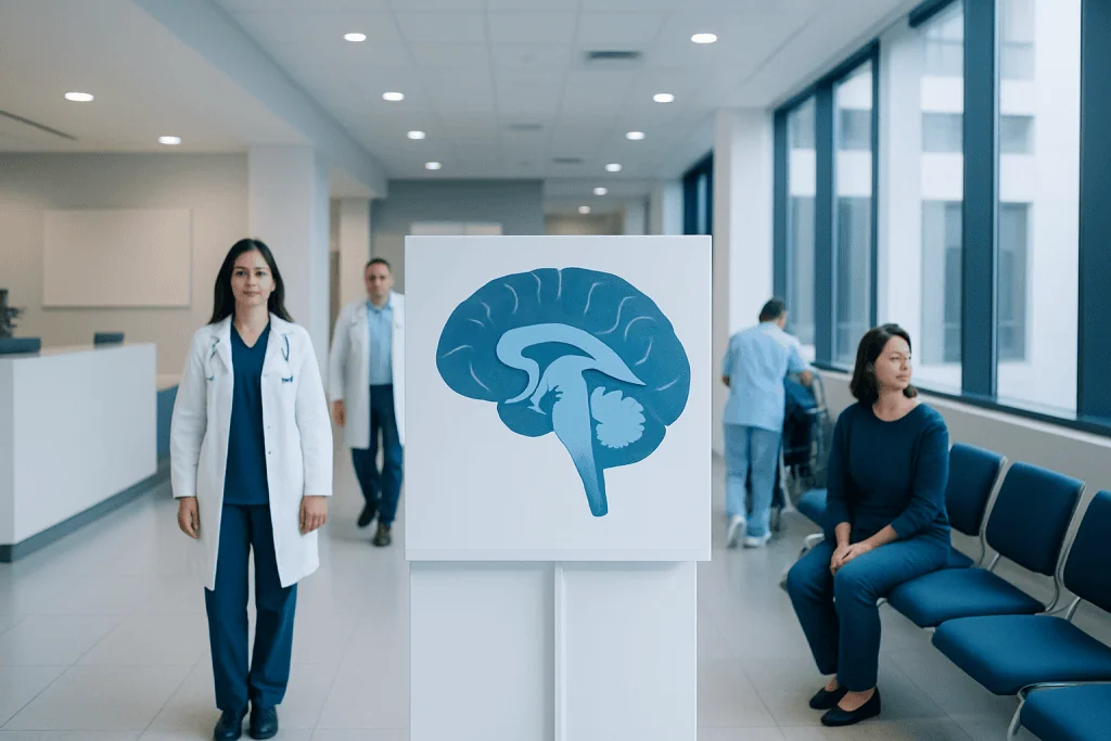
The ventricles of the brain are four connected chambers filled with cerebrospinal fluid. Their main role is to produce, move, and help regulate cerebrospinal fluid around the brain and spinal cord.
Key Takeaways
- The ventricles of the brain are four connected chambers filled with cerebrospinal fluid.
- Their main role is to produce, move, and help regulate cerebrospinal fluid around the brain and spinal cord.
- Problems arise when fluid flow is blocked, fluid is not absorbed properly, or pressure changes affect the brain.
- Symptoms of ventricular disorders can include headache, nausea, balance problems, confusion, vision changes, or rapid head growth in infants.
- Diagnosis usually involves neurological assessment and brain imaging such as CT or MRI.
- Treatment depends on the cause and may include observation, medicines for symptoms, or procedures to drain or redirect fluid.
The ventricles of the brain are connected, fluid-filled spaces that produce and circulate cerebrospinal fluid. They help protect the brain and spinal cord, support normal brain chemistry, and can be involved in conditions such as hydrocephalus, bleeding, infection, or tumors.
Overview: what the ventricles of the brain are
The ventricles of the brain are four connected cavities inside the brain that contain cerebrospinal fluid, often called CSF. This clear fluid cushions the brain and spinal cord, carries nutrients, helps remove waste, and supports a stable environment for nerve cells. In simple terms, the ventricular system is part of the brain’s built-in fluid circulation network.
There are two lateral ventricles, one on each side of the brain, plus the third ventricle and the fourth ventricle in the midline and lower part of the brain. These spaces are connected by narrow channels that allow CSF to flow in an organized direction. The fluid is mainly produced by the choroid plexus, a specialized tissue lining parts of the ventricles.
The ventricles themselves are normal anatomy, not a disease. However, they become clinically important when they enlarge, are compressed, or are affected by bleeding, infection, inflammation, tumors, or poor CSF drainage. Understanding the ventricles of the brain helps explain why certain neurological symptoms happen and how doctors evaluate them.
How the ventricular system works
CSF is produced continuously and circulates through the ventricles before flowing around the brain and spinal cord. From the lateral ventricles, it passes through small openings into the third ventricle, then through a narrow channel called the cerebral aqueduct into the fourth ventricle. From there, it exits into the spaces surrounding the brain and spinal cord, where it is eventually reabsorbed into the bloodstream.
This circulation serves several purposes. CSF helps reduce the effect of sudden movements by acting as a protective cushion. It also supports pressure balance inside the skull, transports some nutrients and signaling molecules, and helps remove metabolic waste products from the central nervous system.
Because the skull is a closed space, even small disruptions in fluid production, flow, or absorption can matter. If too much fluid builds up, the ventricles may enlarge and pressure may rise. If nearby brain tissue shrinks for another reason, the ventricles may also appear larger on imaging, but that does not always mean there is a blockage or dangerous pressure increase.
Common conditions that affect the ventricles

One of the best-known ventricular disorders is hydrocephalus, a condition in which CSF accumulates abnormally. This can happen because the fluid is blocked, not absorbed well, or, less commonly, produced in excess. Enlarged ventricles may put pressure on brain tissue and lead to symptoms that vary by age and cause.
The ventricles can also be affected by bleeding, especially after head injury, stroke, or complications in premature infants. Infection and inflammation, such as meningitis or ventriculitis, may disturb normal CSF flow and absorption. Tumors or cysts near the ventricular pathways may narrow or block important channels.
In older adults, doctors may evaluate enlarged ventricles in the context of walking difficulty, bladder symptoms, and cognitive changes, sometimes to assess for normal pressure hydrocephalus. In other situations, enlarged ventricles reflect loss of brain tissue rather than a primary CSF disorder. Imaging findings must therefore be interpreted together with symptoms, examination, and medical history.
- Hydrocephalus
- Intraventricular hemorrhage or other brain bleeding
- Brain infections affecting CSF pathways
- Tumors or cysts near the ventricles
- Congenital structural differences present from birth
- Age-related or disease-related brain volume loss
Symptoms linked to ventricular problems
The ventricles themselves do not cause symptoms unless a related condition affects fluid flow, pressure, or nearby brain structures. In adults, possible symptoms can include headache, nausea, vomiting, blurred or double vision, balance problems, drowsiness, confusion, and difficulty concentrating. Some people may also develop changes in walking, urinary urgency or incontinence, or slowed thinking, depending on the pattern and cause.
In infants and young children, signs can be different because the skull bones have not fully fused. Parents may notice a rapidly increasing head size, a bulging soft spot, poor feeding, irritability, vomiting, sleepiness, or eyes that appear to look downward. Developmental delay may also be a clue in some children.
These symptoms are not specific to ventricular disorders and can overlap with many other neurological conditions. For that reason, sudden or progressive symptoms should be assessed by a qualified doctor rather than self-diagnosed from anatomy images or online descriptions.
How doctors diagnose ventricular disorders
Diagnosis begins with a detailed medical history and neurological examination. The doctor asks about headache pattern, gait changes, memory or attention problems, vision symptoms, falls, fever, injury, and timing of symptom onset. In infants and children, growth measurements, developmental milestones, and head circumference are especially important.
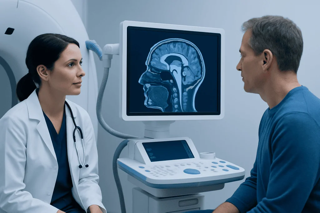
Brain imaging is central to diagnosis. CT can quickly show enlarged ventricles, bleeding, or acute pressure-related problems, while MRI provides more detailed views of brain tissue, fluid pathways, and possible structural causes. When needed, doctors may use specialized imaging or flow studies to better understand how CSF is moving.
Additional tests depend on the clinical situation. These may include eye examination for signs of raised pressure, blood tests, or lumbar puncture in selected cases. If a tumor, structural blockage, or complex hydrocephalus is suspected, the person may be referred to specialists in neurosurgery or neurology for a more focused evaluation.
Treatment options and what they aim to do
Treatment depends entirely on the cause. If imaging shows normal ventricles and no evidence of disease, no treatment may be needed. If the ventricles are enlarged because of brain tissue loss rather than excess CSF pressure, management focuses on the underlying neurological condition rather than fluid drainage.
When there is symptomatic hydrocephalus or a blocked CSF pathway, treatment often aims to restore normal fluid movement or relieve pressure. This may involve a shunt, which diverts CSF to another part of the body, or an endoscopic procedure that creates a new route for fluid to flow. In some cases, treatment is directed at the cause, such as removing a mass or treating infection.
Supportive care also matters. People may need rehabilitation for balance, mobility, or cognitive difficulties, and follow-up imaging may be used to monitor changes over time. If surgery is being considered, doctors explain the expected benefits, limitations, and possible risks in the context of the individual’s overall health and diagnosis, sometimes alongside brain tumor surgery or other condition-specific care when relevant.
At experienced centers, care is often coordinated across neurology, radiology, rehabilitation, and surgery. Acibadem International’s multidisciplinary specialists and JCI-accredited hospitals diagnose and treat ventricular disorders for international patients when advanced assessment or treatment planning is needed.
Prevention, self-care, and living with a ventricular condition
There is no single way to prevent all disorders involving the ventricles of the brain, because causes vary widely. Some are congenital, some follow injury or infection, and others are linked to tumors or age-related neurological disease. Even so, general brain health measures remain helpful: using seat belts and helmets, managing blood pressure, seeking prompt care for severe infections, and following up on new neurological symptoms.
For people already diagnosed with a ventricular condition, self-care mainly means staying engaged with medical follow-up and noticing changes early. It can help to keep a record of headaches, walking changes, bladder symptoms, cognition, falls, or vision problems. Family members often notice subtle changes first, especially in older adults.
After treatment such as a shunt or endoscopic procedure, patients should follow their care team’s advice about activity, wound care, and monitoring. Any return of previous symptoms, new fever, persistent vomiting, severe headache, or sudden change in alertness should be reported quickly. Rehabilitation, sleep, hydration, and medication review can also support recovery and day-to-day functioning.
When to seek medical care
Medical review is appropriate for persistent headaches, new balance problems, unexplained cognitive changes, repeated vomiting, vision changes, or urinary symptoms combined with gait or memory decline. Infants with rapidly increasing head size, poor feeding, unusual sleepiness, or a bulging soft spot should be assessed promptly.
Urgent care is needed for sudden severe headache, reduced consciousness, new seizures, acute weakness, major confusion, or symptoms after head injury. These signs can reflect pressure changes, bleeding, infection, or another serious neurological problem and should not be ignored.
If a doctor suspects a structural cause such as a mass, referral may include advanced imaging and treatment planning for conditions like brain tumor or procedures such as MRI to clarify the diagnosis. Early evaluation often makes management more straightforward and can help prevent complications.
Frequently asked questions
What are the ventricles of the brain in simple terms?
They are four connected spaces inside the brain that contain cerebrospinal fluid. This fluid cushions the brain and spinal cord, helps transport nutrients, and supports a stable environment for normal brain function.
Are enlarged ventricles always dangerous?
No. Enlarged ventricles can occur for different reasons, and not all of them mean there is harmful pressure or a blocked fluid pathway. Doctors interpret ventricular size together with symptoms, examination findings, and imaging details.
What is the difference between ventricles and cerebrospinal fluid?
The ventricles are the spaces or chambers within the brain. Cerebrospinal fluid is the clear fluid inside them that circulates around the brain and spinal cord.
Can problems with the ventricles cause headaches?
Yes, they can, especially if cerebrospinal fluid flow is blocked or pressure rises. However, headaches are common and have many possible causes, so imaging and a medical evaluation may be needed to understand the reason.
How is hydrocephalus related to the ventricles of the brain?
Hydrocephalus happens when cerebrospinal fluid builds up abnormally, usually causing the ventricles to enlarge. This may happen because fluid flow is blocked, absorption is reduced, or, less commonly, production is abnormal.
Which test shows the ventricles of the brain best?
Both CT and MRI can show the ventricles clearly. CT is often used quickly in urgent situations, while MRI usually provides more detailed information about brain tissue and fluid pathways.
This article is for general information only and is not a substitute for professional medical advice. Please consult a qualified doctor about your individual situation.
Joint and spine care in Turkey — expert assessment & treatment
JCI-accredited · board-certified surgeons · reply within 24h
Add us as a Preferred Source to see more of our trusted health content across Google Search, AI Overviews and Discover.

More from the Health Library

Cauliflorini: A Complete Medical Overview

Butterfly Stretch: A Complete Medical Overview

Newborn Bitten by Mosquito: A Complete Medical Overview

Lymph Nodes Enlarge: A Complete Medical Overview

Nasal Spots: A Complete Medical Overview