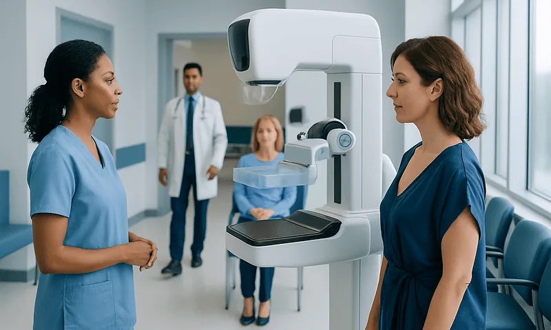
What Is a Diagnostic Mammogram Result Called Back For?

A callback after a screening mammogram is common and usually means more images are needed. Diagnostic mammography uses extra views and sometimes breast ultrasound to examine a specific area.
Key Takeaways
- A callback after a screening mammogram is common and usually means more images are needed.
- Diagnostic mammography uses extra views and sometimes breast ultrasound to examine a specific area.
- Many callbacks are caused by overlapping tissue, benign cysts, or technical factors rather than cancer.
- Breast density, prior imaging, and individual risk factors can affect how results are interpreted.
- Following up promptly helps radiologists compare findings and decide whether monitoring or biopsy is needed.
A diagnostic mammogram callback happens when a screening mammogram shows an area that needs a closer look. In most cases, this means more imaging is needed for clarity, not that breast cancer has been diagnosed.
Overview: What a diagnostic mammogram callback means
A diagnostic mammogram callback means a person has been asked to return after a screening mammogram so the radiology team can take a closer look at one area of the breast. Screening mammograms are designed to look for early changes in people who may have no symptoms. Because they are broad screening tests, they sometimes show findings that are not fully clear on the first set of images.
When this happens, the next step is usually a diagnostic mammogram. This is not a different disease or a diagnosis by itself. It is a more focused imaging exam that takes additional pictures of the area in question. The goal is to clarify whether the finding is simply normal overlapping tissue, a benign change such as a cyst, or something that needs further evaluation.
Being called back can understandably cause worry, but a callback does not mean cancer has been found. In many cases, the additional images show that the finding is harmless. The most important step is to attend the follow-up appointment so the radiologist can make the most accurate assessment possible.
Why people are called back after a screening mammogram
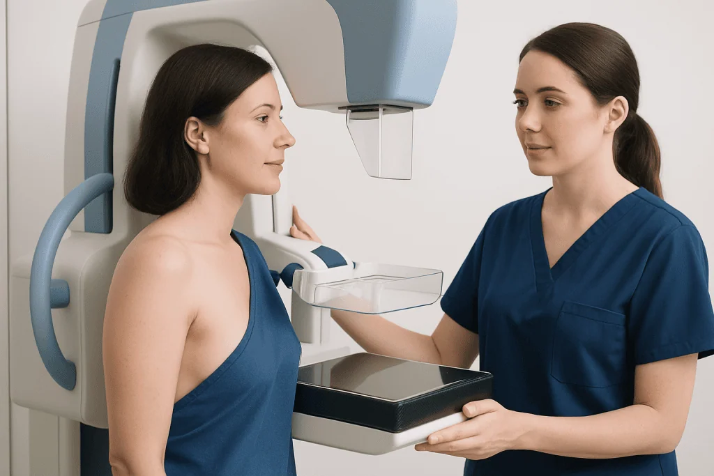
There are several common reasons for being called back after a screening mammogram. One of the most frequent is that breast tissue can overlap on the images, especially in dense breasts. This overlap may create an area that looks unusual on screening views but disappears or becomes clearly benign when extra images are taken.
Radiologists may also request further imaging if they see an asymmetry, a mass, calcifications, or an area of architectural distortion that needs better characterization. Sometimes the images from the initial exam are limited by motion, positioning, or technical factors, and additional views are needed to complete the study properly.
Another reason for callback is that the radiologist may not yet have prior mammograms for comparison. Comparing current images with earlier studies often helps determine whether a finding has been stable over time. Stable findings are more reassuring, while a new or changing area may need closer evaluation.
- Overlapping normal breast tissue
- Dense breast tissue that makes interpretation harder
- A possible mass or focal asymmetry
- Calcifications that need magnified views
- Technical or positioning issues on the screening exam
- No prior imaging available for comparison
What happens during a diagnostic mammogram
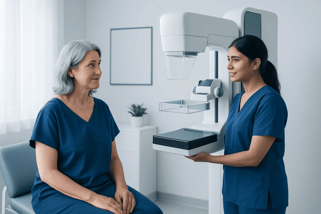
A diagnostic mammogram is similar to a screening mammogram, but it is more targeted. The technologist takes extra images focused on the specific area the radiologist wants to assess. These may include spot compression views, magnification views, or images from different angles. Compression may feel briefly uncomfortable, but it helps spread the tissue and improve image detail.
In many cases, the radiologist reviews the images while the person is still in the imaging center. This allows additional pictures to be taken right away if needed. Depending on the finding, the radiologist may also recommend a breast ultrasound to tell whether an area is solid or fluid-filled and to better assess tissue that is difficult to evaluate on mammography alone.
Sometimes further imaging is still not enough to fully explain the finding. If that happens, the radiologist may recommend short-interval follow-up imaging, often in several months, or a biopsy to obtain a tissue sample. In selected situations, advanced imaging such as breast MRI may be helpful, especially in people with very dense breast tissue or higher individual risk.
Possible results and what they may indicate
After a diagnostic mammogram callback, there are several possible outcomes. Often, the additional images show no suspicious abnormality, and the person can return to routine screening. In other cases, the finding may be categorized as benign, meaning it is not cancer and does not need treatment.
Some findings are considered probably benign. This means they are very unlikely to be cancer, but the safest plan is to repeat imaging after a short interval to confirm that the area remains stable. This follow-up approach helps avoid unnecessary procedures while still monitoring carefully.
If the radiologist sees features that remain concerning, a biopsy may be recommended. A biopsy does not mean cancer is certain; it is the best way to determine exactly what the tissue shows. Results may reveal a benign condition such as breast cyst changes, a noncancerous growth, or less commonly a breast cancer that needs treatment planning.
Breast imaging reports often use a standardized system called BI-RADS. This system helps communicate whether a finding is incomplete, benign, probably benign, suspicious, or highly suggestive of malignancy. The radiologist or referring doctor can explain what the category means for the next steps.
Risk factors, breast density, and personal history
Some people are more likely to be called back because their mammograms are harder to interpret, not necessarily because they are more likely to have cancer. Dense breast tissue is one of the most common reasons. Dense tissue and some abnormalities both appear white on a mammogram, which can make small changes more difficult to distinguish on screening images.
Age, hormonal influences, previous breast procedures, and scar tissue can also affect how the breast looks on imaging. People who have had surgery, implants, or prior biopsies may have changes that require careful comparison over time. If prior mammograms from another center are available, bringing them to the appointment can be very helpful.
Personal and family history still matter. A history of breast cancer, inherited risk, prior chest radiation, or certain high-risk lesions may affect how findings are interpreted and whether additional imaging is advised. In some cases, doctors may also discuss risk-based screening plans that include mammography together with other tests.
Treatment options if more than imaging is needed
A diagnostic mammogram callback itself is part of the evaluation process, not a treatment. If the imaging shows a benign finding, no treatment may be needed at all. The plan may simply be routine annual screening or a short-term follow-up examination to confirm stability.
If a suspicious area remains after diagnostic imaging, the next step is often a biopsy. Depending on the location and appearance of the finding, this may be an image-guided needle biopsy using mammography, ultrasound, or another imaging method. A biopsy helps determine whether the change is benign, high-risk, or cancerous.
If cancer is diagnosed, care is planned according to the type, size, stage, and biological features of the tumor, as well as the person’s overall health and preferences. Treatment may involve surgery, medication, radiation therapy, or a combination of approaches. Related benign conditions, such as fibroadenoma, may only need observation unless they are large, symptomatic, or uncertain in diagnosis.
Near the end of the diagnostic pathway, some people seek coordinated care in specialized centers. Acibadem International’s multidisciplinary specialists and JCI-accredited hospitals diagnose and treat breast conditions for international patients when further evaluation or treatment is required.
How to prepare and cope with the waiting period
Waiting for follow-up imaging can be stressful, but a few practical steps may help. It is useful to schedule the callback as soon as possible, bring prior breast imaging if it was done elsewhere, and ask whether deodorant, powders, or creams should be avoided on the day of the exam. Wearing a two-piece outfit may also make the visit easier.
During the appointment, many people find it reassuring to ask what type of images are being taken and when results are expected. Some centers provide same-day feedback from the radiologist, while others send results to the referring doctor soon afterward. Knowing the timeline can reduce uncertainty.
Emotional support matters as well. Talking with a trusted family member, friend, or healthcare professional can help manage anxiety while waiting. It may also help to remember that callbacks are common, and many turn out to be due to benign findings rather than serious disease.
When to contact a doctor promptly
A callback for additional breast imaging is usually not an emergency, but it should not be ignored. It is best to arrange the follow-up visit promptly so any unclear finding can be fully evaluated. Delays can prolong anxiety and may postpone diagnosis if treatment is needed.
Independent of mammogram results, a person should contact a doctor sooner if they notice a new breast lump, persistent focal pain, skin dimpling, nipple inversion, bloody nipple discharge, redness that does not improve, or swelling in the underarm area. These symptoms do not always mean cancer, but they do deserve medical assessment.
If there is difficulty obtaining records, scheduling the exam, or understanding the report, it is reasonable to ask the imaging center or referring physician for clarification. Clear communication helps patients make informed decisions and continue appropriate breast screening over time.
Frequently asked questions
Does a diagnostic mammogram callback mean cancer?
No. A diagnostic mammogram callback means the radiologist needs more information about an area seen on the screening mammogram. Many callbacks are caused by normal overlapping tissue, benign cysts, or image quality factors.
What is the difference between a screening mammogram and a diagnostic mammogram?
A screening mammogram is a routine test used to look for breast changes in people without symptoms. A diagnostic mammogram is a more focused exam that takes additional images of a specific area to clarify a finding or evaluate a symptom.
Will I need an ultrasound after a diagnostic mammogram?
Sometimes. A breast ultrasound is often used if the radiologist wants to look more closely at a lump or determine whether an area is solid or fluid-filled. Not everyone needs one, and the decision depends on the imaging findings.
How long does it take to get results after a callback?
This varies by imaging center. In some facilities, the radiologist reviews the images during the visit and provides preliminary feedback the same day. In others, results are sent to the referring doctor shortly afterward.
What are BI-RADS categories?
BI-RADS is a standardized reporting system used in breast imaging. It helps describe how concerning a finding is and what follow-up is recommended, such as routine screening, short-term follow-up, or biopsy.
If the finding is probably benign, why is follow-up still needed?
Probably benign means the finding is very unlikely to be cancer, but short-interval follow-up helps confirm that it stays stable over time. This approach can safely reduce unnecessary biopsies while still monitoring carefully.
Should I be worried if I have dense breasts?
Dense breasts are common and do not mean something is wrong. However, dense tissue can make mammograms harder to interpret, which may increase the chance of a callback and sometimes leads doctors to consider additional imaging based on overall risk.
This article is for general information only and is not a substitute for professional medical advice. Please consult a qualified doctor about your individual situation.
Explore treatments in Turkey — costs, top hospitals & a free quote
JCI-accredited · board-certified surgeons · reply within 24h
Add us as a Preferred Source to see more of our trusted health content across Google Search, AI Overviews and Discover.

More from the Health Library

What Is a Diagnostic Test? A Patient Guide to Lab and Imaging Results

Quest Diagnostics Login and MyQuest: What the Patient Portal Lets You Do
Lab Test Cost: What Affects the Price of Blood and Urine Testing?

EEG Test: What Brain Wave Monitoring Can Help Diagnose
What Is a Diagnostic Test? Common Types and Why Doctors Order Them