What Is a Neuro-Ophthalmology Exam? Step by Step for Patients With Vision and Nerve Symptoms
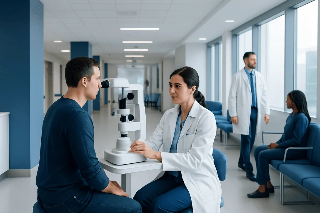
A neuro-ophthalmology exam assesses both eye health and the nervous system pathways involved in vision. The visit usually includes questions about symptoms, vision testing, pupil checks, eye movement assessment, and examination of the optic nerve.
Key Takeaways
- A neuro-ophthalmology exam assesses both eye health and the nervous system pathways involved in vision.
- The visit usually includes questions about symptoms, vision testing, pupil checks, eye movement assessment, and examination of the optic nerve.
- Additional tests such as visual fields, imaging, or blood tests may be ordered depending on the findings.
- The exam is generally noninvasive, and many parts are similar to a routine eye exam but more focused on nerve-related causes.
- Urgent evaluation is important for sudden vision loss, new double vision, severe headache, or other neurological symptoms.
A neuro-ophthalmology exam is a detailed evaluation of how the eyes, optic nerves, and brain pathways work together. It helps doctors understand symptoms such as double vision, vision loss, abnormal pupils, eye movement problems, headaches, or facial nerve-related changes.
Overview: What Is a Neuro-Ophthalmology Exam?
A neuro-ophthalmology exam is a specialized assessment for people who have visual symptoms that may be related to the brain, optic nerves, eye movement nerves, or the connection between the eyes and the nervous system. Neuro-ophthalmologists evaluate problems that are not only about seeing clearly, but also about how visual information travels and how the eyes move and respond.
This type of exam may be recommended for symptoms such as double vision, blurred vision that is not fully explained by glasses, loss of part of the visual field, drooping eyelid, unusual pupil changes, eye pain with vision changes, or episodes of temporary vision loss. It may also help in the evaluation of headaches, dizziness, facial weakness, or suspected neurological conditions affecting the eyes.
For many patients, the visit feels similar to an eye exam in some ways, but it is usually more detailed and more focused on nerve function. The goal is to understand whether symptoms come from the eye itself, the optic nerve, the muscles that move the eyes, or a part of the brain involved in vision.
Why a Patient Might Need This Exam
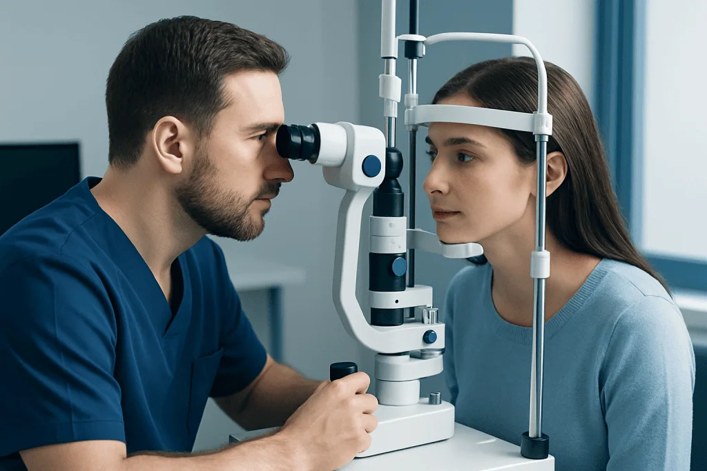
A doctor may refer a patient for a neuro-ophthalmology exam when symptoms suggest that ordinary vision testing alone may not give a complete answer. For example, someone may have reduced vision even though the front of the eye looks normal, or may develop double vision because the eye muscles are receiving abnormal nerve signals.
Common reasons for referral include sudden or gradual vision loss, unexplained visual field defects, optic nerve swelling, unequal pupils, eyelid drooping, involuntary eye movements, or difficulty moving one or both eyes. It can also be helpful after a head injury, in suspected inflammatory disease, or when an imaging test has shown a finding near the optic pathways.
Sometimes the exam helps distinguish between different causes of similar symptoms. A patient with headache and blurred vision, for example, may need evaluation for optic nerve problems, pressure-related changes, or migraine-related visual symptoms such as migraine care. In other cases, the exam may support urgent investigation for conditions such as stroke or other neurological disorders.
Step by Step: What Happens During the Visit

The appointment usually begins with a detailed medical history. The doctor asks when symptoms started, whether they are constant or intermittent, and whether they affect one eye or both. Questions may also cover headache, eye pain, weakness, numbness, balance problems, recent infections, medications, prior eye disease, and family history.
Vision testing is commonly the next step. This may include checking visual acuity with letters or symbols, color vision, contrast sensitivity, and sometimes reading performance. These tests help show whether the optic nerve and visual pathways are working as expected.
The doctor then examines the pupils and how they react to light. A pupil examination can reveal whether one optic nerve is sending a weaker signal than the other, or whether there may be a problem in the pathways that control pupil size. Eye alignment and eye movements are also checked carefully, often by asking the patient to follow a target in different directions.
Most visits also include an examination of the front and back of the eyes. Eye drops may be used to enlarge the pupils so the doctor can view the optic nerve and retina more clearly. Looking at the optic disc can provide important clues, such as swelling, pallor, or other changes that suggest inflammation, pressure changes, poor blood flow, or previous injury.
Specialized Tests That May Be Included
Some patients need additional testing during the same visit or at a later appointment. Visual field testing is one of the most common. It measures how well a person sees in the center and at the edges of vision and can detect blind spots or patterns of loss that point to a specific location in the visual pathway.
Optical coherence tomography, often called OCT, may also be used. This imaging test creates detailed pictures of the retina and optic nerve layers. It can help identify swelling, thinning, or structural changes that are not always visible during a standard examination.
Depending on the findings, the doctor may request blood tests, MRI or CT scans, or other neurological studies. Brain and orbit imaging is often arranged through neuroradiology services when there is concern about the optic nerves, eye movement nerves, inflammation, tumors, vascular problems, or pressure effects. In selected cases, other evaluations such as electrophysiology or broader neurophysiology testing may help clarify how the nervous system is functioning.
These tests are chosen based on the patient’s symptoms and examination results. Not every person needs every test, and the doctor usually explains why a particular study is or is not necessary.
What Conditions Can a Neuro-Ophthalmology Exam Help Detect?
A neuro-ophthalmology exam can help identify a wide range of conditions. These include optic neuritis, ischemic optic neuropathy, papilledema, cranial nerve palsies, myasthenia gravis affecting the eyes, thyroid eye disease, and visual pathway disorders involving the brain. It can also help detect complications from diabetes, autoimmune disease, infection, or raised intracranial pressure.
Sometimes the exam helps evaluate symptoms linked to headache disorders, including visual aura or eye movement complaints. In other cases, the findings may suggest a structural cause such as a tumor or a blood vessel problem, leading to further neurological investigation. Certain patterns can also appear in patients with meningioma or after a transient circulation problem such as transient ischemic attack.
It is important to remember that a referral for this exam does not automatically mean a serious disease is present. Many patients are evaluated to rule out dangerous causes, clarify a diagnosis, or confirm that symptoms are due to a manageable condition. A careful examination often provides reassurance as well as guidance.
After the Exam: Understanding Results and Treatment
At the end of the visit, the doctor usually explains the main findings and whether more testing is needed. In some cases, the diagnosis is clear right away. In others, the results of imaging, laboratory tests, or follow-up examinations are needed before a final conclusion is reached.
Treatment depends entirely on the cause. Some problems are treated with medication, while others require management by neurology, ophthalmology, endocrinology, or neurosurgery teams. For example, inflammatory conditions may need medical therapy, eye movement problems may improve with treatment of the underlying nerve disorder, and some patients benefit from prisms or other supportive vision measures.
If symptoms relate to complex nerve and vision disorders, patients may be referred for dedicated neuro-ophthalmology care within a broader multidisciplinary program. When headache, immune-mediated disease, or age-related neurological issues are also part of the picture, coordinated input from services such as headache medicine or other neurology subspecialties may be appropriate.
Near the end of the care pathway, some international patients choose centers such as Acibadem International, where multidisciplinary specialists and JCI-accredited hospitals evaluate and treat neuro-ophthalmic conditions with coordinated neurological and eye care.
How to Prepare and When to Seek Urgent Help
Patients can prepare for the appointment by bringing their glasses, contact lens information, a list of medications, and any prior eye exam reports, brain scans, or blood test results. It is also helpful to write down when symptoms began, what makes them better or worse, and whether they happen with headache, weakness, pain, or dizziness. Because dilating drops may blur vision for a few hours, arranging transportation can be sensible for some people.
During the visit, patients should describe symptoms as specifically as possible. For example, it helps to say whether double vision occurs only when looking in certain directions, whether vision loss is central or side vision, and whether episodes last seconds, minutes, or longer. Small details can strongly guide the diagnosis.
Urgent medical attention is important for sudden vision loss, sudden double vision, new drooping of the face or eyelid, severe eye pain with vision changes, a sudden “worst headache,” weakness, trouble speaking, or confusion. These symptoms may indicate a time-sensitive problem and should not wait for a routine appointment.
Even when symptoms are less dramatic, ongoing or unexplained visual changes should still be assessed by a qualified doctor. Early evaluation can help protect vision, identify neurological conditions sooner, and guide the right treatment plan.
Frequently asked questions
Is a neuro-ophthalmology exam the same as a regular eye exam?
No. It includes many parts of a standard eye exam, but it focuses more specifically on the optic nerve, eye movement nerves, visual pathways, and how the brain and eyes work together. It is usually ordered when symptoms suggest a neurological cause for visual problems.
Does a neuro-ophthalmology exam hurt?
Most parts of the exam are painless. Patients may feel temporary discomfort from bright lights or from keeping their eyes focused during certain tests, and dilating drops can cause brief stinging and temporary blur. In general, the exam is noninvasive and well tolerated.
How long does the appointment usually take?
The visit is often longer than a routine eye appointment because the history and examination are more detailed. Depending on the symptoms and whether extra tests are done the same day, it may take from about one hour to several hours. The care team usually explains what to expect.
Will the patient need imaging such as an MRI?
Not everyone needs imaging. MRI, CT, or other tests are ordered when the examination suggests a problem affecting the optic nerves, eye movement nerves, brain, blood vessels, or surrounding structures. The decision depends on the symptoms and the findings during the visit.
Should patients stop wearing contact lenses before the exam?
This depends on the clinic’s instructions and the type of testing planned. Many patients can attend wearing contacts, but bringing glasses is still important because some tests are easier with them. If the pupils will be dilated, vision may be blurry afterward regardless of contact lens use.
What symptoms should lead to urgent evaluation rather than a routine appointment?
Sudden vision loss, new double vision, severe headache with neurological symptoms, sudden facial drooping, trouble speaking, weakness, or confusion should be assessed urgently. These can be signs of a serious condition that needs immediate attention. If symptoms appear suddenly or worsen quickly, emergency care is appropriate.
References
- American Academy of Ophthalmology
- North American Neuro-Ophthalmology Society
- National Eye Institute
- National Institute of Neurological Disorders and Stroke
- Mayo Clinic
This article is for general information only and is not a substitute for professional medical advice. Please consult a qualified doctor about your individual situation.
Explore treatments in Turkey — costs, top hospitals & a free quote
JCI-accredited · board-certified surgeons · reply within 24h