What Is a Neuro-Ophthalmology Exam?
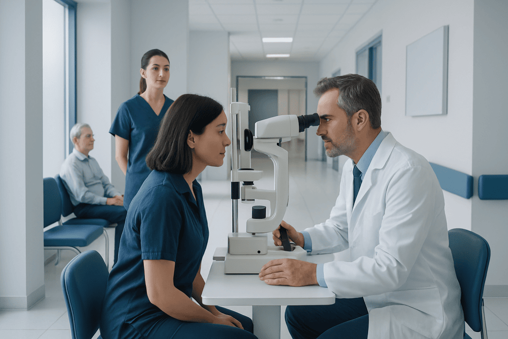
A neuro-ophthalmology exam looks at the connection between the eyes and the nervous system. It is often used for symptoms such as double vision, visual loss, drooping eyelids, and unusual pupil changes.
Key Takeaways
- A neuro-ophthalmology exam looks at the connection between the eyes and the nervous system.
- It is often used for symptoms such as double vision, visual loss, drooping eyelids, and unusual pupil changes.
- The exam may include vision testing, eye movement checks, pupil assessment, visual field testing, and a close look at the optic nerve.
- Additional tests such as blood work or brain and orbit imaging may be recommended depending on the findings.
- Many causes are treatable, so early evaluation can help protect vision and support overall neurological health.
A neuro-ophthalmology exam evaluates vision problems that may involve the eyes, optic nerves, eye movements, or the brain pathways that control sight. It helps specialists find the cause of symptoms such as unexplained vision loss, double vision, abnormal pupils, or visual field changes.
Overview: what a neuro-ophthalmology exam is
A neuro-ophthalmology exam is a detailed medical assessment of how the eyes, optic nerves, eye muscles, and brain pathways involved in vision are functioning. It is performed when a person has visual symptoms that may not be explained by a routine eye problem alone. This kind of evaluation sits at the overlap of ophthalmology and neurology.
Unlike a standard eye check that focuses mainly on eye health and glasses prescription, a neuro-ophthalmology exam looks for problems in the communication between the eyes and the brain. The specialist assesses how clearly a person sees, how the pupils react, how the eyes move together, whether the visual field is intact, and whether the optic nerve appears healthy.
The exam can help identify a wide range of conditions, from optic nerve inflammation and visual pathway disorders to eye movement problems linked to nerves or muscles. It may also help detect issues related to headache disorders, autoimmune disease, stroke, tumors, increased pressure around the brain, or systemic illness affecting vision.
When this exam may be needed
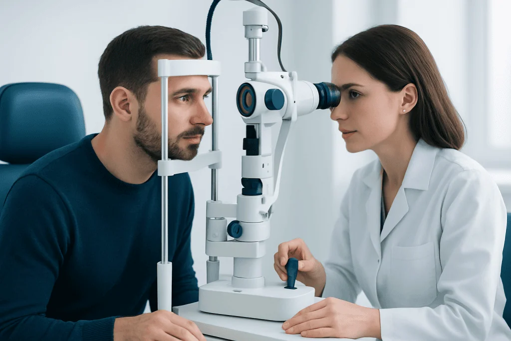
A doctor may recommend a neuro-ophthalmology exam when symptoms suggest that the problem could involve the nervous system rather than the eye surface or glasses alone. Common reasons include sudden or progressive vision loss, double vision, one eyelid drooping, unequal pupils, blurry vision that comes and goes, or loss of part of the visual field.
Some people are referred after an eye doctor notices swelling or pallor of the optic nerve, abnormal eye movements, or visual field changes. Others are referred after neurological symptoms such as headache, facial numbness, weakness, imbalance, or suspected multiple sclerosis. The exam can also be important after head injury or when imaging shows a finding near the optic nerves or visual pathways.
Typical symptoms that may lead to evaluation include:
- Unexplained decrease in vision in one or both eyes
- Double vision, especially when looking in certain directions
- Blind spots or reduced side vision
- Pain with eye movement
- Episodes of temporary visual dimming
- Abnormal pupil size or sluggish pupil response
- Drooping eyelid or difficulty moving the eyes
What happens during the exam
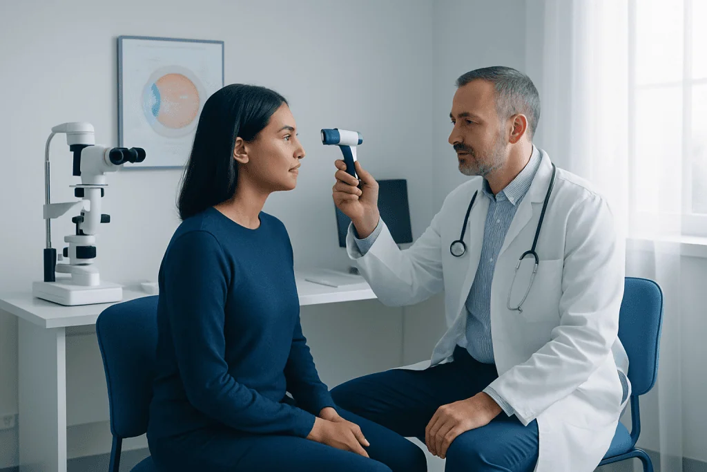
The visit usually begins with a detailed medical history. The specialist asks when the symptoms began, whether they are constant or intermittent, and whether there are associated symptoms such as headache, eye pain, weakness, numbness, or balance problems. Past eye disease, neurological conditions, medications, vascular risk factors, and autoimmune disorders are also important.
The physical examination is often more extensive than a routine eye test. Visual acuity is measured, and color vision may be checked because color changes can be an early sign of optic nerve disease. The doctor also evaluates the pupils, checks for a relative afferent pupillary defect, measures eyelid position, and watches how the eyes move in different directions.
Visual field testing is commonly used to detect areas of missing vision that may point to a problem in the optic nerve, optic chiasm, or brain. The doctor then examines the front and back of the eyes, often after dilating the pupils, to look at the optic nerve, retina, and blood vessels. In many cases, photographs or optical coherence tomography may be used to document the optic nerve and retinal nerve fiber layer in more detail.
The full appointment may take longer than a standard eye exam because several measurements are needed. Although the tests are usually not painful, dilation drops can temporarily blur near vision and increase light sensitivity for a few hours, so some patients prefer not to drive themselves home.
What the doctor is looking for
The main goal of a neuro-ophthalmology exam is to locate where the problem may be occurring. Vision can be affected at multiple points, including the optic nerve, the nerves controlling eye movement, the eye muscles, the brainstem, or the visual processing areas of the brain. The pattern of findings often gives strong clues about the cause.
For example, reduced vision with abnormal color vision and pain on eye movement can suggest optic nerve involvement. Double vision with limited movement of one eye may point to a cranial nerve palsy or an eye muscle disorder. Visual field loss on the same side in both eyes may suggest a problem in the brain’s visual pathways rather than inside the eyeball itself.
The doctor also looks for signs of swelling of the optic disc, optic nerve damage, elevated pressure around the brain, or abnormalities of the pupils. Sometimes the exam helps distinguish between a neurological condition and another eye disorder such as cataract or retinal disease. If the findings suggest another eye condition, further evaluation for related issues such as glaucoma or retinal detachment may be advised.
Possible conditions a neuro-ophthalmology exam can help diagnose
A neuro-ophthalmology exam does not diagnose just one disease; it helps identify a category of problem and guide the next steps. One common concern is optic neuropathy, which means damage or dysfunction of the optic nerve. This may be caused by inflammation, reduced blood flow, compression, infection, toxins, nutritional problems, or inherited conditions.
The exam can also help evaluate disorders that affect eye movements, such as cranial nerve palsies, myasthenia gravis, thyroid eye disease, and brainstem conditions. In people with headaches or transient visual symptoms, it may support the assessment of increased intracranial pressure, migraine-related visual phenomena, or other neurological causes.
Other conditions that may be considered include optic neuritis, idiopathic intracranial hypertension, pituitary-region lesions, stroke affecting the visual pathways, and compressive problems around the orbit or optic chiasm. Depending on the findings, the specialist may recommend further workup with MRI scanning, targeted blood tests, or other neurological investigations. If raised pressure or a structural problem is suspected, more advanced imaging or neurosurgical treatment may be discussed in selected cases.
Additional tests that may be recommended
Not everyone needs the same follow-up tests. Recommendations depend on the symptoms, examination findings, age, and overall health. If the doctor suspects a problem affecting the optic nerve, brain, or eye sockets, imaging of the brain and orbits may be ordered. MRI is often preferred because it gives detailed information about soft tissues and visual pathways.
Visual field testing may be repeated over time to monitor change. Optical coherence tomography can help measure the retinal nerve fiber layer and optic nerve swelling or thinning. In some situations, blood tests may be needed to look for inflammation, infection, autoimmune disease, vitamin deficiency, thyroid disease, or vascular risk factors.
Other evaluations may include neurological examination, blood pressure assessment, and coordination with other specialists. If a mass, aneurysm, or another structural cause is suspected, interventional neuroradiology or other specialist care may be involved. The aim is to confirm the diagnosis and start the most appropriate treatment as early as possible.
Treatment and follow-up after the exam
Treatment depends entirely on the underlying cause found during the evaluation. Some conditions need urgent attention to protect vision or overall health, while others are monitored over time. Treatment may involve medicines, management of blood pressure or blood sugar, treatment of infection or inflammation, prism glasses for double vision, or referral for neurological or surgical care.
In some cases, the best next step is observation with repeat examinations to see whether the visual function is stable, improving, or worsening. Follow-up visits may include repeat visual field testing, optic nerve imaging, and checks of eye movement and pupil function. This helps the doctor understand whether treatment is working and whether the diagnosis still fits the pattern over time.
Because neuro-ophthalmic symptoms may relate to more than one body system, care is often multidisciplinary. Ophthalmologists, neurologists, neuroradiologists, endocrinologists, and neurosurgeons may all contribute when needed. Near the end of the care pathway, some patients may seek assessment at centers such as Acibadem International, where multidisciplinary specialists in JCI-accredited hospitals diagnose and treat neuro-ophthalmic conditions for international patients.
How to prepare and when to seek medical care
Before the appointment, it helps to bring a list of symptoms, when they started, and whether anything makes them better or worse. Patients should also bring previous eye exam records, imaging reports, a list of medicines and supplements, and details of medical conditions such as diabetes, autoimmune disease, migraine, or high blood pressure. If dilation is expected, sunglasses can make the trip home more comfortable.
Some symptoms should be assessed promptly. These include sudden vision loss, new double vision, eye pain with reduced vision, a sudden drooping eyelid, severe headache with visual change, or visual symptoms together with weakness, trouble speaking, or facial drooping. These patterns may signal a problem that should not wait.
Even when symptoms are mild, persistent changes in vision deserve professional evaluation. A timely neuro-ophthalmology exam can provide reassurance when findings are benign, but it can also detect conditions that benefit from early treatment. Anyone with concerning visual symptoms should speak with a qualified eye doctor or physician rather than trying to self-diagnose.
Frequently asked questions
What is the difference between a neuro-ophthalmology exam and a regular eye exam?
A regular eye exam mainly checks vision, glasses needs, and common eye diseases. A neuro-ophthalmology exam goes further by assessing the optic nerves, pupils, eye movements, and the brain pathways involved in vision. It is usually recommended when symptoms suggest a neurological cause.
Is a neuro-ophthalmology exam painful?
Most parts of the exam are not painful. The doctor may use bright lights, ask the patient to follow targets with the eyes, and perform visual field testing, which can feel tiring but should not hurt. If dilation drops are used, they may cause temporary light sensitivity and blurred near vision.
How long does a neuro-ophthalmology exam take?
The visit often takes longer than a standard eye appointment because several different tests may be done. Depending on the symptoms and whether the pupils are dilated, the appointment may last from about an hour to longer. Extra time may also be needed if imaging or additional testing is arranged.
Why would someone be referred to a neuro-ophthalmologist?
A referral is common when there is unexplained vision loss, double vision, unusual pupil findings, optic nerve swelling, or visual field defects. Doctors may also refer patients when headaches, neurological symptoms, or brain imaging findings raise concern about the visual pathways. The goal is to find the cause and guide treatment.
Will I need an MRI after the exam?
Not everyone needs an MRI. Imaging is recommended when the history and examination suggest a problem involving the optic nerve, eye socket, brain, or surrounding structures. The decision depends on the pattern of symptoms and the doctor's findings.
Can a neuro-ophthalmology exam detect serious conditions?
Yes, it can help identify conditions that affect both vision and neurological health, including optic nerve disease, cranial nerve palsies, increased intracranial pressure, and visual pathway disorders. At the same time, not every abnormal symptom is due to a serious illness. The exam is valuable because it helps separate urgent problems from less dangerous causes.
References
- American Academy of Ophthalmology
- North American Neuro-Ophthalmology Society
- National Eye Institute
- National Institute of Neurological Disorders and Stroke
- Mayo Clinic
This article is for general information only and is not a substitute for professional medical advice. Please consult a qualified doctor about your individual situation.
Explore treatments in Turkey — costs, top hospitals & a free quote
JCI-accredited · board-certified surgeons · reply within 24h
Add us as a Preferred Source to see more of our trusted health content across Google Search, AI Overviews and Discover.