When a Lung Nodule Needs a Biopsy: How Doctors Decide
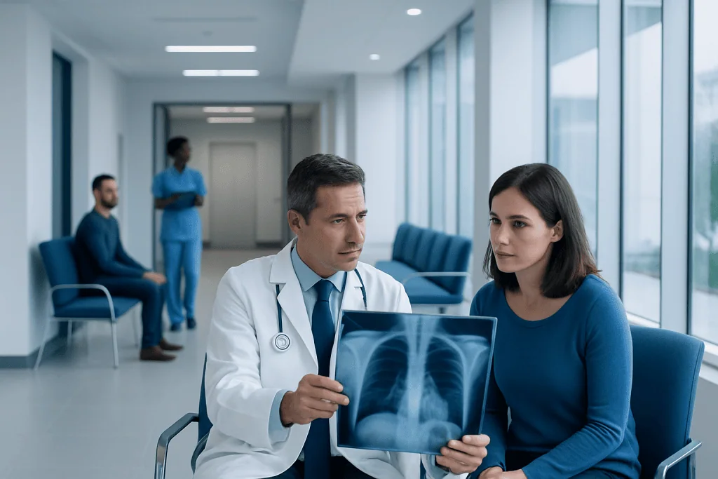
Many lung nodules are benign and can be monitored with repeat imaging instead of immediate biopsy. Biopsy is more likely when a nodule is larger, growing, irregular, or seen in someone with higher cancer risk.
Key Takeaways
- Many lung nodules are benign and can be monitored with repeat imaging instead of immediate biopsy.
- Biopsy is more likely when a nodule is larger, growing, irregular, or seen in someone with higher cancer risk.
- CT scans, PET-CT, and a patient’s age, smoking history, and medical background all help guide the decision.
- Several biopsy methods are available, and the best choice depends on the nodule’s size and location.
- A specialist can explain whether watchful waiting, biopsy, or treatment is the most appropriate next step.
Not every lung nodule needs a biopsy. Doctors weigh scan findings, personal risk factors, and the chances of infection, inflammation, or cancer before deciding whether careful follow-up or tissue sampling is the safest next step.
Overview: Why some lung nodules need a biopsy
A lung nodule is a small spot or shadow seen in the lung, often found unexpectedly during a chest X-ray or CT scan done for another reason. These nodules are common, and most are not cancer. They may develop from old infections, inflammation, scar tissue, or other noncancerous causes.
The main question after a lung nodule is found is whether it looks harmless enough to watch over time or whether it needs further testing. A lung nodule biopsy means taking a small sample of tissue so it can be examined under a microscope. This can help doctors tell the difference between benign changes, infection, inflammation, and cancer.
Doctors do not recommend biopsy for every nodule because biopsies are invasive and carry some risks, such as bleeding or a collapsed lung. Instead, the decision is based on how likely the nodule is to be cancerous and whether a biopsy result would change care. In many cases, repeat imaging at set intervals is the safest and most reasonable approach.
What doctors look at on imaging
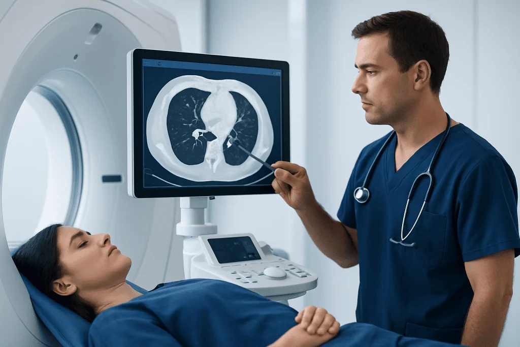
Imaging findings are central to the decision. CT scans provide much more detail than a chest X-ray and help doctors assess the nodule’s size, shape, density, and exact location. In general, larger nodules are more concerning than very small ones, although size alone does not provide a diagnosis.
Doctors also look for features linked with higher risk. A nodule with smooth, stable edges may be less concerning, while one with irregular, spiculated, or lobulated borders may deserve closer attention. Growth over time is especially important. If a nodule increases in size on follow-up scans, the chance of a serious cause becomes higher.
The internal appearance matters too. Some nodules are solid, while others are part-solid or ground-glass. Part-solid nodules can require particularly careful follow-up because some patterns are more strongly associated with early lung cancer. When a nodule cannot be clearly classified as low risk from CT alone, a doctor may recommend additional imaging such as PET-CT or referral for further evaluation of a possible lung cancer.
Symptoms and personal risk factors that influence the decision
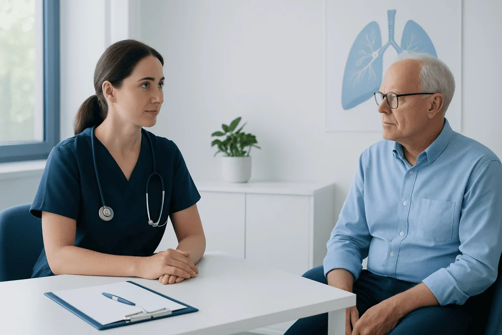
Most small lung nodules do not cause symptoms. They are often discovered incidentally during imaging for cough, chest pain, injury, or routine screening. However, the presence of symptoms may change how urgently a nodule is evaluated, especially if there is coughing up blood, unexplained weight loss, ongoing chest discomfort, or shortness of breath.
A person’s medical background is just as important as the scan itself. Doctors consider age, smoking history, secondhand smoke exposure, prior cancer, family history of lung cancer, occupational exposures such as asbestos, and any long-term lung disease. A nodule in a younger nonsmoker with no cancer history is often managed differently from a similar-looking nodule in an older person with a heavy smoking history.
Geography and immune status also matter. In some regions, fungal infections or previous tuberculosis can leave spots in the lungs that mimic cancer on imaging. People with weakened immune systems may need evaluation for infection as well as cancer. This broader clinical picture helps doctors decide whether watchful follow-up, a biopsy, or a different type of test is most appropriate.
When monitoring may be enough
If a nodule appears low risk, doctors may recommend surveillance rather than immediate biopsy. This usually means repeat CT scans over time to see whether the nodule stays stable, shrinks, or grows. Stability over an appropriate period can be reassuring and may reduce the need for invasive testing.
Monitoring is often chosen for very small nodules or for nodules with features that suggest a benign cause. It may also be preferred when the risks of biopsy outweigh the likely benefit, such as in people with severe lung disease, bleeding disorders, or other health conditions that make procedures harder to tolerate.
Careful follow-up is not the same as doing nothing. The timing of scans is planned according to established guidance and the patient’s level of risk. Keeping appointments is important because changes over time can be one of the clearest clues about whether a nodule is harmless or needs tissue diagnosis.
When a biopsy is more likely to be recommended
A biopsy becomes more likely when the overall risk of cancer is moderate to high. This may happen if the nodule is larger, has suspicious edges, is growing, or shows activity on PET-CT that suggests increased metabolic activity. Biopsy may also be advised when there is a history of cancer elsewhere in the body, because the nodule could represent spread or a new primary lung tumor.
Doctors also ask whether the biopsy result will directly influence treatment. If the answer will guide surgery, medication, or radiation planning, obtaining tissue can be very valuable. In some situations, a suspicious nodule may go straight to surgery instead of needle biopsy, especially if it is highly likely to be cancer and can be safely removed.
There is no single rule that applies to everyone. The same size nodule may lead to different decisions in different patients because cancer risk, procedure risk, and patient preference all matter. Multidisciplinary discussion among radiologists, pulmonologists, thoracic surgeons, and oncologists often helps shape the best plan.
How lung nodules are biopsied
The best biopsy method depends mainly on where the nodule is located. A nodule near the outer part of the lung may be sampled through the chest wall with a CT-guided needle biopsy. A nodule closer to the central airways may be reached with bronchoscopy, in which a doctor passes a thin tube through the nose or mouth into the lungs. Some centers use advanced navigation bronchoscopy or endobronchial ultrasound to improve access to difficult lesions.
Before recommending a method, doctors weigh the chance of getting enough tissue against the risk of complications. Needle biopsy can be very helpful, but it carries a known risk of pneumothorax, or collapsed lung, especially in people with emphysema. Bronchoscopy may be less likely to cause this problem, but it is not ideal for every nodule.
If imaging and biopsy findings remain unclear, surgery may be needed both to diagnose and treat the lesion. This is often done with minimally invasive approaches such as video-assisted thoracoscopic surgery when appropriate. In selected cases, additional procedures such as bronchoscopy or lung biopsy are used to confirm the diagnosis before a treatment plan is finalized.
What happens after the biopsy result
Biopsy results can show several possibilities. A nodule may be benign, which can mean scar tissue, a healed infection, a noncancerous growth, or an inflammatory condition. Depending on the exact result, the doctor may recommend no further action, repeat imaging, or treatment for infection or inflammation.
If the biopsy shows cancer, the next step is usually staging and treatment planning. This may include more imaging, breathing tests, blood work, and consultations with specialists. Treatment can involve surgery, radiation therapy, systemic therapies, or a combination, depending on the type and stage of disease.
Sometimes a biopsy does not give a clear answer. This does not always mean cancer is present, but it may mean the sample was too small or did not capture the right area. In that situation, doctors may advise repeat biopsy, closer imaging follow-up, or surgery. For international patients who need coordinated evaluation, Acibadem International’s multidisciplinary specialists and JCI-accredited hospitals diagnose and treat lung nodules and related conditions.
When to seek medical advice and how to prepare
Anyone told they have a lung nodule should review the finding with a qualified doctor, even if the report says it is probably benign. It helps to ask about the nodule’s size, appearance, and whether prior scans are available for comparison. Old imaging can be extremely useful because long-term stability often lowers concern.
Urgent medical advice is especially important if there are symptoms such as coughing up blood, unexplained weight loss, worsening cough, chest pain, or increasing breathlessness. People with a personal history of cancer or significant smoking exposure should also make sure follow-up is not delayed.
Preparing for a specialist visit can make the discussion clearer. Helpful steps include bringing a medication list, noting smoking and occupational exposure history, and asking whether the next step is repeat CT, PET-CT, bronchoscopy, needle biopsy, or surgery. Shared decision-making allows the patient to understand both the reasons for biopsy and the reasons a doctor may safely recommend monitoring instead.
Frequently asked questions
Does every lung nodule need a biopsy?
No. Many lung nodules are benign and can be safely monitored with repeat CT scans. Doctors usually recommend biopsy only when the nodule’s features or the person’s risk factors make cancer or another serious cause more likely.
What size lung nodule is considered concerning?
Size is one important factor, but there is no single cutoff that determines whether a nodule is dangerous. Larger nodules tend to need closer evaluation, especially if they grow or have irregular borders. Doctors always interpret size together with imaging appearance and personal risk factors.
Can a CT scan tell if a lung nodule is cancer?
A CT scan can show whether a nodule looks low risk or suspicious, but it usually cannot confirm cancer by itself. Features such as growth, shape, and density help estimate risk. A biopsy or surgery may still be needed for a definite diagnosis.
Is a lung nodule biopsy painful or risky?
Biopsy procedures are usually done with local anesthesia, sedation, or both, so discomfort is often manageable. As with any invasive test, there are risks, including bleeding or a collapsed lung, depending on the technique used. The care team chooses the method that offers the best balance of accuracy and safety.
How long does it take to get biopsy results?
Basic pathology results often come back within a few days, but timing can vary by laboratory and by whether special testing is needed. If doctors are checking for infection, inflammation, or specific tumor markers, the process may take longer. The care team will usually explain when to expect an update.
What if the biopsy is negative but the nodule still looks suspicious?
A negative biopsy can be reassuring, but it does not always end the evaluation. Sometimes the sample does not fully represent the nodule, especially if the lesion is small or hard to reach. In that case, doctors may recommend another biopsy, surgery, or repeat imaging to make sure nothing important is missed.
References
- American College of Chest Physicians
- Fleischner Society
- National Cancer Institute
- American Thoracic Society
- Radiological Society of North America
This article is for general information only and is not a substitute for professional medical advice. Please consult a qualified doctor about your individual situation.
Oncology care in Turkey — second opinion and treatment plan
JCI-accredited · board-certified surgeons · reply within 24h
Add us as a Preferred Source to see more of our trusted health content across Google Search, AI Overviews and Discover.