

Quick answer
Flash CT is an advanced computed tomography technology that captures detailed cross-sectional images in a very short time, helping reduce motion-related image blur. At Acibadem in Turkey, Flash CT examinations are planned according to the clinical question and patient needs, with images interpreted by radiology specialists.
Flash CT: Ultra-Fast, Detailed CT Imaging with a Patient-Focused Approach
Flash CT is an advanced form of computed tomography (CT) designed to capture highly detailed images of the body in a fraction of a second. It helps radiologists and specialist teams see anatomy, blood vessels, organs and tissues clearly while using scan protocols tailored to the individual patient and the clinical question.
At Acibadem International, Flash CT supports precise diagnosis, treatment planning and follow-up care. Fast image acquisition can be particularly valuable when movement is difficult to control, such as during heart imaging, in children, in patients experiencing pain, or when holding the breath for a long time is challenging.
Accurate imaging is an important foundation for personalised care. By providing high-resolution information quickly, Flash CT can help doctors make confident clinical decisions while aiming to reduce unnecessary invasive investigations and limit radiation exposure whenever medically appropriate.
What Is Flash CT?
CT scanning uses X-rays and advanced computer processing to create detailed cross-sectional images of the body. Unlike a standard X-ray, which produces a single flat image, CT can show internal structures in layers and can create three-dimensional views when needed.
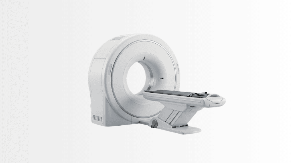
Flash CT refers to ultra-fast CT technology that can acquire images extremely quickly. The scanner rotates around the body while the patient lies on a moving examination table. Sophisticated detectors and imaging software collect information from many angles and transform it into detailed images for review by radiologists and other specialists.
The speed of Flash CT is one of its key strengths. A scan may be completed in seconds, depending on the area being examined and the purpose of the study. This can improve image quality by reducing motion blur caused by breathing, heartbeat, discomfort or difficulty remaining still.
Flash CT does not replace clinical assessment. Your doctor considers your symptoms, medical history, previous imaging, laboratory results and treatment goals before recommending any imaging examination. The most suitable scan protocol is selected individually.
How Flash CT Works
During a Flash CT examination, you lie comfortably on a padded table that moves through the circular opening of the CT scanner. The scanner does not usually enclose the entire body like some MRI systems. It is open at both ends, and the examination team can communicate with you throughout the procedure.
Inside the scanner, an X-ray source and detectors rotate rapidly around the body. The system gathers a large amount of information in a very short time. Advanced software reconstructs this information into detailed images that can show bones, lungs, soft tissues, internal organs and blood vessels.
Some examinations require contrast material. Contrast is a special substance, often administered through a vein, that can make blood vessels, certain organs and areas of inflammation or abnormal tissue easier to see. Not every Flash CT scan requires contrast. Your medical team will explain whether it is needed for your examination.
Because the scan is so fast, Flash CT may be especially helpful for examinations where motion can affect image quality. In selected cases, it can support high-quality imaging with shorter breath-holds and fewer repeated images.
Low-Dose, Individually Planned Imaging
CT uses ionising radiation, so every examination must be medically justified. At Acibadem International, radiology teams select protocols according to the body area being studied, the clinical purpose, patient age, body characteristics and previous imaging information.
Modern CT technology and dose-management approaches are used to obtain diagnostic image quality with the lowest radiation exposure reasonably achievable for the required clinical information. The appropriate dose is not identical for every patient; it is carefully adapted to the scan indication.
Clinical Applications of Flash CT
Flash CT may be used across a wide range of medical specialties. It can assist with early detection, diagnosis, treatment planning, procedural guidance and follow-up after treatment. The exact role of the scan depends on your condition and the recommendation of your treating physician.
| Clinical applications | How Flash CT may help | Examples of conditions or concerns assessed |
|---|---|---|
| Cardiac and coronary imaging | Very fast acquisition may help capture images of the heart and coronary arteries while reducing the effect of cardiac motion. | Coronary artery disease assessment, chest pain evaluation, coronary artery anatomy and selected structural heart conditions. |
| Chest and lung imaging | Provides detailed views of the lungs, airways, chest wall and blood vessels, often with short scan times. | Lung nodules, suspected infection, pulmonary embolism assessment, chronic lung disease and chest trauma. |
| Abdominal and pelvic imaging | Helps evaluate abdominal organs, bowel, urinary structures and blood vessels with high anatomical detail. | Abdominal pain, liver or pancreatic conditions, kidney stones, urinary tract concerns, inflammatory disease and cancer staging. |
| Neurological and head imaging | Rapid imaging can be important when urgent assessment is needed and can show bones, brain structures and blood vessels in selected protocols. | Head injury, acute headache, suspected stroke pathways, sinus disease and facial bone trauma. |
| Musculoskeletal and trauma imaging | Creates detailed images of bones, joints and complex anatomical regions, including three-dimensional reconstructions when useful. | Fractures, joint injuries, spinal conditions, sports injuries and pre-operative orthopaedic planning. |
| Oncology imaging | Supports assessment of tumours, lymph nodes and potential spread of disease, as well as monitoring response to treatment. | Suspected or known cancers affecting the chest, abdomen, pelvis, bones and other regions. |
| Vascular imaging | CT angiography can provide detailed views of arteries and veins after contrast administration when clinically appropriate. | Aneurysm assessment, arterial narrowing, vascular malformations, peripheral artery disease and selected emergency vascular concerns. |
Flash CT findings are interpreted in the context of your overall health. Imaging may identify changes that require further testing, monitoring or specialist consultation. In some cases, a normal scan can also be valuable by helping doctors rule out certain concerns and avoid unnecessary procedures.
Benefits and Advantages for Patients
The main advantage of Flash CT is speed combined with detailed imaging. Rapid acquisition can make the examination more comfortable for many patients and may improve diagnostic confidence in situations where movement is difficult to avoid.
For patients with pain, shortness of breath, anxiety or limited ability to remain still, a faster scan may be easier to tolerate than a longer imaging examination. It can also be useful for children and for patients who may find long breath-holds challenging, although the imaging approach for every child is carefully assessed by the clinical team.
Flash CT can support more efficient care pathways by giving specialists detailed information to guide the next step. This may include referral to another specialty, planning a procedure, comparing new images with previous studies or monitoring the results of treatment.
| Patient benefits at a glance | What this can mean for your care |
|---|---|
| Ultra-fast scanning | The image acquisition itself is usually very short, helping reduce the impact of breathing and other body movements. |
| High-resolution anatomical detail | Specialists can assess complex structures with clear cross-sectional and, when needed, three-dimensional images. |
| Personalised scan protocols | Your examination is planned around the clinical question, the area of the body being assessed and your individual health needs. |
| Radiation-conscious approach | Radiology teams use dose-optimisation methods to achieve the diagnostic information needed with appropriate radiation exposure. |
| Largely non-invasive experience | Most scans do not require surgery or recovery time. Some studies may require an intravenous contrast injection. |
| Support for treatment decisions | Detailed imaging can help your doctors diagnose conditions, plan treatment and monitor progress over time. |
| Coordinated international care | International patient coordinators can help organise appointments, document review, language support and communication between teams. |
What to Expect During a Flash CT Procedure
Your experience begins before the scan itself. For international patients, the Acibadem International team can assist with sharing medical reports, arranging specialist review and coordinating the required appointments. If you have previous CT, MRI, ultrasound, PET or laboratory results, these may help your specialist decide whether Flash CT is appropriate and how the examination should be planned.
Before Your Scan
You may receive instructions about eating, drinking or medications, especially if contrast material is planned. These instructions vary according to the type of examination and your medical history. It is important to tell your healthcare team if you have kidney disease, diabetes, thyroid conditions, asthma, previous reactions to contrast material, allergies, pregnancy or a possible pregnancy.
You may be asked to change into a gown and remove metal objects or accessories from the area being scanned. Depending on the examination, a small intravenous cannula may be placed in your arm or hand for contrast administration.
During the Scan
A radiology technologist will position you on the scanning table. You may lie on your back, stomach or side, depending on the body region being assessed. Straps or positioning supports may be used to help you stay comfortable and still.
The table moves smoothly through the scanner opening. The technologist monitors you from a nearby control area and can speak with you through an intercom. You may be asked to hold your breath briefly. Because Flash CT is designed for very fast acquisition, breath-hold instructions are often short.
If contrast is used, you may feel a temporary warm sensation, a metallic taste in the mouth or a feeling similar to needing to urinate. These sensations usually pass quickly. Tell the team immediately if you feel unwell, experience itching, swelling, breathing difficulty or any unexpected symptom.
After the Scan
Most patients can return to normal daily activities soon after a Flash CT examination. If contrast material was used, your team may advise you to drink fluids unless you have been given different medical instructions.
A radiologist reviews the images and prepares a report. Your treating specialist discusses the findings with you and explains what they mean for your diagnosis, treatment plan or next steps. For international patients, coordination teams can help ensure that relevant reports and imaging information are available for follow-up planning.
Safety, Quality and Accreditation
Flash CT is used routinely by trained radiology professionals who follow established imaging, radiation-safety and patient-identification procedures. At Acibadem International, examinations are performed within JCI-accredited hospital environments, supporting a structured approach to quality, patient safety and coordinated clinical care.
Before any scan, the clinical team checks whether CT is appropriate for you. This includes reviewing the reason for imaging, your health history and whether contrast is needed. CT is not recommended simply because it is available; it is selected when the expected clinical value outweighs potential risks.
Radiation exposure is an important consideration in all CT examinations. Modern equipment, careful protocol selection and dose-optimisation practices are used to support safe, effective imaging. If you have had previous scans, tell your doctor so that your imaging history can be considered.
Contrast material is safe for many patients, but it is not suitable for everyone. Your team will ask about allergies, kidney function, medications and previous contrast reactions. If there is a specific concern, doctors may recommend precautions, an alternative imaging method or a different diagnostic approach.
Why Flash CT Matters for International Patients
When travelling for medical care, patients often need timely answers, clear planning and reliable coordination. Flash CT can support an efficient diagnostic pathway by helping specialists obtain detailed imaging information quickly when it is clinically indicated.
Acibadem International teams work with patients before and during their visit to help organise the imaging process. This may include reviewing available medical documents, scheduling consultations and scans in a coordinated way, arranging interpretation support and helping you understand the next stage of your care plan.
Clear communication is especially important when imaging results influence treatment decisions. Your specialist can explain why Flash CT has been recommended, whether contrast is required, what the images show and whether additional tests or consultations are needed. Your care plan is based on your individual assessment rather than a standard pathway.
For patients travelling with family members or caregivers, coordinated scheduling can also help make the visit more manageable. The international patient team can guide you through practical steps before arrival and support communication between radiology, consulting specialists and other relevant departments.
Frequently Asked Questions About Flash CT
Is Flash CT safe?
Flash CT is performed by trained radiology teams using established safety procedures. Like all CT examinations, it uses X-rays, so the scan is recommended only when it is clinically appropriate. Protocols are tailored to obtain the necessary diagnostic information while keeping radiation exposure as low as reasonably achievable.
Does Flash CT always use contrast material?
No. Whether contrast is needed depends on the area being scanned and the clinical question. For example, contrast may be useful when assessing blood vessels, some organs, inflammation or suspected tumours. Your doctor will explain the recommendation and review any relevant safety considerations with you.
How long does a Flash CT scan take?
The imaging portion is often completed very quickly, sometimes in only a few seconds. Your total appointment may take longer because it can include registration, preparation, safety checks, positioning and, if needed, contrast administration. The exact timing depends on the examination planned for you.
Will I need to stay in hospital after the scan?
Most Flash CT examinations are performed on an outpatient basis, and patients can usually leave shortly afterward. If your scan is part of emergency care, surgery planning, hospital treatment or another procedure, your overall care schedule may be different.
Will my insurance cover Flash CT?
Insurance coverage depends on your policy, insurer requirements and the reason for the examination. You can share your insurance information with the international patient team, who can help review available options and explain the process before treatment planning is finalised.
How can I get started?
You can submit your available medical reports, imaging results and relevant history through a consultation request. A specialist can review your case and advise whether Flash CT may be suitable as part of your diagnostic or treatment plan. The international patient team can then help coordinate the next steps, including travel-related planning and language support where needed.
Related Medical Units
Available at these Hospitals


















Related Treatments
Speak with our medical team
Find out how this technology could support your diagnosis or treatment.