

Pinpoint (Fluorescence Imaging System)
Treatment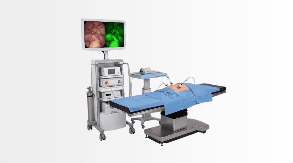
Quick answer
Pinpoint is a fluorescence imaging system that helps surgeons assess blood flow, tissue perfusion, and vascular supply in real time during an operation. At Acibadem in Turkey, it may be used in minimally invasive general surgery to support evaluation of tissue viability and surgical decision-making.
Pinpoint Fluorescence Imaging System
Pinpoint is an advanced fluorescence imaging system that helps surgeons view blood flow and tissue perfusion in real time during surgery. It is used as an additional visual guidance tool, particularly in minimally invasive laparoscopic procedures performed by General Surgery teams.
During an operation, a surgeon must often assess whether tissue has a healthy blood supply. Although this can be evaluated through clinical experience and direct observation, fluorescence imaging can provide an additional, dynamic view of blood flow. Pinpoint supports the surgical team by showing how blood reaches tissues and surgical connections at key points during the procedure.
For appropriate patients, this technology may support more informed intraoperative decisions, greater precision and careful planning of the surgical approach. It does not replace the judgement of an experienced surgeon. Instead, it provides useful real-time information that can complement the surgeon’s clinical assessment.
At Acibadem International, Pinpoint technology is used within a personalised treatment pathway. Your care plan is based on your diagnosis, imaging, previous medical history, planned surgery and overall health. Our multidisciplinary teams work together to determine whether fluorescence guidance may be helpful for your procedure.
What Is Pinpoint Fluorescence Imaging?
Pinpoint is a near-infrared fluorescence imaging system designed for use during surgery. It works with a fluorescent dye, most commonly indocyanine green (ICG), which is administered by the medical team when clinically appropriate. Once the dye circulates through the bloodstream, the imaging system can detect its fluorescence using specialised light and camera technology.
The surgeon can then view images showing blood flow to tissues in real time. This may help identify tissue perfusion, evaluate vascular supply and assess the blood flow of organs or surgical reconstructions.
The system can be used during open surgery or minimally invasive surgery, depending on the procedure and the equipment available at the treating hospital. In laparoscopic surgery, it can be integrated into the operative workflow while the surgeon is working through small incisions using a camera and specialised surgical instruments.
Pinpoint is not a diagnostic test performed before surgery. It is an intraoperative imaging tool used while you are in the operating theatre. The images support the surgical team at important stages of the operation, such as before creating or assessing a surgical connection between sections of bowel.
How Does Fluorescence Guidance Work?
Fluorescence imaging involves a carefully coordinated process led by the surgical, anaesthesia and nursing teams. If your surgeon considers Pinpoint suitable, the details will be discussed as part of your surgical consent and preparation.
1. Preoperative assessment
Before surgery, your specialist reviews your diagnosis, scans, laboratory results, medications, allergy history and any previous operations. This assessment helps determine the safest and most appropriate surgical plan. If fluorescence imaging may be useful, your team will explain its role in the operation and any individual considerations related to the dye or procedure.
2. Administration of the fluorescent dye
During the operation, the medical team may administer indocyanine green through an intravenous line. The timing and dosage are determined by the surgical and anaesthesia teams according to the clinical situation. The dye travels through the bloodstream and is then visualised by the imaging system.
3. Near-infrared imaging
Pinpoint uses near-infrared light and a dedicated camera to detect fluorescence. The system produces an image that can help the surgeon see patterns of blood flow that are not always visible under standard surgical lighting alone.
4. Surgical decision support
The surgeon interprets the imaging findings alongside all other clinical information. Depending on the procedure, this may support assessment of tissue viability, help guide the choice of surgical margins or help evaluate blood supply to an area before completing a reconstruction.
Every operation is different. The exact use of Pinpoint varies according to your condition, the anatomy involved, the planned technique and the professional judgement of your surgical team.
Clinical Applications
Pinpoint fluorescence imaging is most commonly associated with general surgical procedures where assessing tissue perfusion can be clinically valuable. It may be considered in minimally invasive and open surgical techniques when the surgeon needs additional real-time visual information about vascular supply or tissue blood flow.
| Clinical application | How fluorescence imaging may support the surgical team | Examples of conditions or procedures |
|---|---|---|
| Colorectal surgery | May help assess blood flow to sections of bowel before or after a surgical connection is created. | Colorectal cancer surgery, diverticular disease surgery, inflammatory bowel disease surgery and selected bowel resections. |
| Gastrointestinal surgery | May provide additional information about tissue perfusion during reconstruction or resection procedures. | Selected stomach, small bowel and complex abdominal operations. |
| Hepatobiliary surgery | May assist with visualisation of biliary anatomy or tissue characteristics in selected cases. | Gallbladder surgery, liver procedures and selected bile duct operations. |
| Emergency and complex abdominal surgery | May support assessment of tissue viability when blood supply is a particular concern. | Selected bowel obstruction, intestinal ischaemia and complex reoperative cases. |
| Minimally invasive surgery | Can be integrated with laparoscopic imaging to provide real-time fluorescence guidance through small-incision approaches. | Appropriate laparoscopic General Surgery procedures, depending on the patient and surgeon’s plan. |
Not every patient undergoing these procedures needs fluorescence imaging, and not every operation can be performed using minimally invasive techniques. Your surgeon will recommend the approach that best balances safety, treatment effectiveness and recovery needs.
Conditions Pinpoint May Help Surgeons Manage
Pinpoint does not treat a condition on its own. It is a technology that may support the surgical treatment of conditions requiring an operation. It can be particularly relevant when the surgeon needs to evaluate blood supply to tissue during a procedure.
Depending on your individual assessment, fluorescence imaging may be considered in surgery for colorectal cancer, bowel tumours, diverticular disease, inflammatory bowel disease, bowel narrowing, selected intestinal emergencies and some gallbladder, liver or bile duct conditions. It may also be useful in complex surgery where previous treatment, inflammation, altered anatomy or vascular considerations affect the surgical plan.
For cancer care, Pinpoint may be part of a broader surgical strategy developed by specialists in General Surgery, Medical Oncology, Radiation Oncology, Radiology, Pathology and other relevant disciplines. The aim is to provide coordinated care based on the nature and stage of the disease, your general health and your treatment goals.
Your treatment recommendation will always be individual. The availability and use of fluorescence guidance depend on the procedure, hospital resources, your clinical needs and the surgeon’s assessment during planning and surgery.
Benefits and Advantages for Patients
The primary purpose of Pinpoint is to support surgical precision by providing additional real-time visual information. For suitable cases, this may help the surgical team make more informed decisions during the operation.
When used as part of a well-planned minimally invasive procedure, advanced imaging technology may also contribute to the advantages often associated with laparoscopic surgery. These can include smaller incisions, less postoperative discomfort and a potentially faster return to everyday activities when compared with some open surgical approaches. However, recovery varies significantly between patients and procedures.
| Patient benefit at a glance | What it may mean for your care |
|---|---|
| Real-time blood flow assessment | The surgeon can evaluate tissue perfusion during the operation using additional visual information. |
| Support for surgical precision | Fluorescence guidance may help inform decisions about tissue viability and vascular supply at key stages of surgery. |
| Compatibility with minimally invasive techniques | In appropriate laparoscopic procedures, the system can support surgery performed through small incisions. |
| Individualised treatment planning | The technology is used only when it is clinically relevant to your diagnosis, procedure and overall surgical plan. |
| Coordinated specialist care | Surgeons, anaesthesia specialists, nurses and other clinical teams work together throughout your treatment journey. |
| Potentially smoother recovery pathway | When minimally invasive surgery is suitable, patients may experience benefits associated with smaller interventions, although outcomes vary. |
It is important to understand that no imaging system can guarantee a particular surgical result. The benefits of Pinpoint depend on the reason for surgery, the complexity of the procedure, tissue condition, previous treatments and many other personal factors. Your surgeon will explain what is realistic for your case.
What to Expect During a Procedure Using Pinpoint
Before you travel or begin treatment
International patients can share medical reports, imaging results, pathology reports and treatment summaries for preliminary review. Our international patient team helps coordinate the evaluation process and supports communication between you, your family and the relevant specialists.
Following review, you may be advised on the recommended specialty consultation, additional tests that may be needed and whether surgery is likely to be considered. If Pinpoint fluorescence imaging may be appropriate, this will be included in the broader discussion of your surgical plan.
Before surgery
After you arrive, you will meet the treating team for a detailed consultation and preoperative assessment. You may undergo blood tests, imaging or other examinations depending on your condition and the type of surgery planned. The anaesthesia team will also review your health history, medications, allergies and fasting instructions.
Your surgeon will explain the planned operation, the possible surgical approaches, expected hospital stay, recovery considerations and potential risks. You will have the opportunity to ask questions and discuss the role of fluorescence imaging in your care.
During surgery
You will receive anaesthesia appropriate to your operation. Once the procedure is underway, the surgeon may use Pinpoint at one or more stages to assess blood flow or visualise tissue. If ICG is used, it is administered under the supervision of the operating and anaesthesia teams.
The surgical team will not rely on a single image alone. Fluorescence findings are evaluated together with direct examination, imaging studies, laboratory results, anatomy and the surgeon’s experience. If the surgical plan needs to be adapted for safety or treatment reasons, the team will make decisions according to accepted clinical practice and your consented treatment plan.
After surgery
After the operation, you will be monitored in a recovery area and then transferred to an inpatient room if hospital admission is required. Your recovery plan may include pain management, early mobilisation, breathing exercises, nutrition guidance, wound care and prevention of complications such as blood clots or infection.
The length of recovery depends on the type and extent of surgery. Some patients are able to return to light daily activities relatively soon after minimally invasive procedures, while others need a longer recovery period. Your team will provide personalised discharge instructions and guidance about travelling home safely.
Safety Considerations
Pinpoint fluorescence imaging is used by trained surgical teams as part of a controlled operating theatre environment. Like all medical technologies, it is used only after considering the potential benefits and any risks for the individual patient.
Indocyanine green is generally used in clinical imaging applications, but it may not be suitable for everyone. Your doctor will ask about previous allergies, medication use, liver function, pregnancy status where relevant and other medical conditions. Any concerns regarding dye administration or the surgical procedure itself will be discussed with you before treatment.
Fluorescence imaging does not eliminate the general risks associated with surgery or anaesthesia. These may include bleeding, infection, pain, blood clots, injury to nearby structures, wound complications, reactions to medicines and the possibility that a minimally invasive procedure may need to be converted to an open operation when clinically necessary.
Your surgeon and anaesthesia specialist will explain the risks relevant to your particular procedure. You will be asked to provide informed consent only after you have had an opportunity to understand the recommended approach and ask questions.
Quality, Safety and Accreditation at Acibadem International
Acibadem International supports international patients seeking access to multidisciplinary care in JCI-accredited hospital environments in Türkiye. JCI accreditation reflects a structured commitment to recognised standards in patient safety, quality improvement, clinical processes and continuity of care.
For surgical patients, this includes coordinated preoperative assessment, operating theatre safety protocols, infection prevention measures, anaesthesia monitoring, medication management and postoperative follow-up. Technology such as Pinpoint is used within these wider clinical systems, not in isolation.
Our teams focus on clear communication, careful documentation and individual treatment planning. When surgery involves cancer care, complex gastrointestinal disease or a high-risk health condition, relevant specialists may review the case together to help ensure that recommendations are comprehensive and clinically appropriate.
Why Pinpoint Matters for International Patients
Travelling abroad for surgery requires confidence in both the medical plan and the coordination around it. International patients may arrive with questions about treatment options, language, travel timing, recovery away from home and follow-up after discharge. Our international patient services are designed to help make this process more manageable.
From the first review of your reports, the team can help coordinate specialist consultations, examinations, treatment scheduling and communication in your preferred language where available. You and your family can receive support in understanding the sequence of care, preparing for hospital admission and planning an appropriate period of recovery before travel.
If Pinpoint fluorescence imaging is recommended, your surgeon will explain why it may add value to your specific operation. The decision is never based on technology alone. It is based on whether its use is relevant to your diagnosis, surgical goals and safety needs.
For patients returning home after treatment, discharge planning is an important part of the care pathway. Your team will provide medical documentation, postoperative guidance and recommendations for follow-up. If further communication with your local physician is needed, relevant clinical records can be prepared to support continuity of care.
Frequently Asked Questions
Is Pinpoint fluorescence imaging safe?
Pinpoint is used routinely in surgical settings by trained specialists. The system is designed to provide additional imaging information during an operation. If fluorescent dye is planned, your team will assess whether it is appropriate for you and discuss relevant precautions, allergies and possible risks.
Will Pinpoint be used for every General Surgery procedure?
No. The use of fluorescence imaging depends on the procedure and your individual clinical needs. Your surgeon will decide whether it may provide meaningful support during your operation.
Does Pinpoint mean my surgery will definitely be minimally invasive?
Not necessarily. Pinpoint can be used in minimally invasive and open surgery. Whether laparoscopic surgery is appropriate depends on your condition, anatomy, previous operations, the complexity of the procedure and the surgeon’s assessment.
Will fluorescence imaging guarantee a faster recovery?
No technology can guarantee a particular recovery time or surgical outcome. When minimally invasive surgery is appropriate, some patients may benefit from smaller incisions and a shorter recovery compared with certain open procedures. Your expected recovery will be discussed based on your specific operation.
Will my insurance cover the procedure?
Insurance coverage depends on your policy, insurer and the recommended treatment plan. You can share your insurance information with our international patient team, who can help clarify the available options and explain financial arrangements before treatment begins.
How can I find out whether Pinpoint is suitable for me?
You can submit your medical reports, imaging and pathology results for a preliminary consultation request. A relevant specialist can review your case and advise whether surgery is indicated and whether fluorescence imaging may have a role in your treatment plan.
Take the Next Step
If you are considering General Surgery in Türkiye and would like to learn whether Pinpoint fluorescence imaging may be relevant to your procedure, our international patient team can help coordinate a specialist review.
Please share your available medical records, recent imaging reports, pathology findings and treatment history. Following clinical assessment, the appropriate specialist can explain your treatment options, the possible use of advanced imaging guidance and the next steps for planning your care.
Related Medical Units
Available at these Hospitals


















Related Treatments
Speak with our medical team
Find out how this technology could support your diagnosis or treatment.