Achalasia Treatment
Achalasia care focuses on confirming the esophageal motility disorder and relieving swallowing difficulty through endoscopic dilation, POEM, botulinum toxin injection, or surgical myotomy.
Quick answer
Achalasia is a disorder of the oesophagus in which the lower oesophageal sphincter fails to relax, so food and liquid struggle to reach the stomach. Treatment relieves that obstruction through pneumatic balloon dilation, peroral endoscopic myotomy (POEM), laparoscopic Heller myotomy or botulinum toxin injection. The right option depends on the achalasia subtype, the shape of the oesophagus, prior procedures and overall health.
Achalasia: Understanding the Condition Before Choosing Treatment
Achalasia is a disorder of oesophageal movement. The lower oesophageal sphincter, the muscular valve between your oesophagus and your stomach, fails to relax when you swallow, and the oesophagus itself loses its normal, coordinated squeezing action. Food and liquid become trapped above the valve instead of passing into the stomach. Treatment exists, it works by relieving the obstruction at that valve, and choosing between the treatment options is the central decision you will face after diagnosis.
The condition rarely announces itself suddenly. Most people describe a slow progression: occasional trouble with solid food, then difficulty with liquids, then regurgitation of undigested food, chest discomfort, coughing at night or unintended weight loss. Meals become slow and stressful. Some people quietly stop eating in company. Others start sleeping propped upright to reduce night-time regurgitation, or begin to worry that the symptoms point to cancer or heart disease. That fear is understandable, and it is one of the reasons a proper diagnostic work-up matters so much.
Achalasia does not usually resolve on its own. Modern care rests on three steps: confirming the diagnosis accurately, understanding the type and severity of the motility disorder, and selecting a treatment that relieves the obstruction at the lower oesophageal sphincter. This page walks through each of those steps, what achalasia is, what causes it, how it is diagnosed, what the four main treatments involve, and what recovery and long-term follow-up realistically look like.
What is achalasia esophagus?
Achalasia esophagus, the phrase many people type into a search engine, simply means achalasia of the oesophagus, the muscular tube that carries food from your throat to your stomach. There is no other kind: achalasia is by definition an oesophageal condition, so “achalasia”, “oesophageal achalasia” and “achalasia of the oesophagus” all describe the same disorder. In a healthy swallow, waves of muscle contraction, peristalsis, push food downward, and the lower oesophageal sphincter opens briefly to let it into the stomach, then closes to stop stomach contents flowing back. In achalasia both mechanisms fail: the wave weakens or disappears, and the valve stays tight. The result is a column of food and fluid held above a valve that will not open properly, which is what produces the sensation of food sticking behind the breastbone.
What is achalasia cardiospasm?
Achalasia cardiospasm is an older name for exactly the same condition. The “cardia” is the region where the oesophagus joins the stomach, and early physicians assumed the problem was an active spasm of that region, hence cardiospasm. Later research showed that the real problem is a failure to relax rather than a spasm: the nerve cells that normally instruct the sphincter to open are lost, so the muscle simply never receives the signal. The modern name, achalasia, comes from Greek roots meaning “does not relax”. You may still meet the phrase cardiospasm achalasia in older textbooks, translated records or medical documents from some countries. If your paperwork uses that term, it refers to the condition described on this page, and nothing about the diagnosis or treatment changes.
Achalasia, achlasia or acalasia, which spelling is right?
Achlasia and acalasia are common misspellings of achalasia; all three spellings lead people to the same condition, and the correct medical term is achalasia. This matters mainly when you search for information or read your own medical records. Reports written in other languages, or transcribed in a hurry, sometimes carry a variant spelling, and it is worth knowing that they all mean the same diagnosis.
 Dr. Tarek ArafatMDBoard Commentary
Acıbadem physicians have also reported their clinical experience with peroral endoscopic myotomy (POEM) in patients with achalasia. In a single-center series of 27 patients treated between 2019 and 2022, POEM achieved a 100% technical success rate, with a mean procedure time of approximately 72 minutes and a mean hospital stay of 2.7 days. Only one minor adverse event was reported. These findings support POEM as a minimally invasive treatment option when performed by an experienced therapeutic endoscopy team, although long-term clinical outcomes depend on achalasia subtype, disease severity and post-procedure reflux.
Achalasia Symptoms
Achalasia symptoms usually build over months or years, which is a large part of why the diagnosis is so often delayed. The exact pattern varies from person to person, but the core features all trace back to one mechanical fact: swallowed material is not reaching the stomach efficiently.
Difficulty swallowing (dysphagia)
The most common symptom is dysphagia, difficulty swallowing. In achalasia, dysphagia typically affects both solids and liquids, which helps distinguish it from a purely mechanical narrowing, where solids usually cause trouble first while liquids still pass. You may feel food stopping behind the breastbone, need large amounts of water to wash each mouthful down, or find yourself standing up, straightening your back or lifting your arms to help food pass. Many people adapt so gradually, softer foods, smaller portions, longer mealtimes, that they only realise how narrow their diet has become when a doctor asks them to describe a typical day’s eating.
Regurgitation and other symptoms
Regurgitation is the second hallmark. Undigested food or fluid comes back up, often when you bend over or lie down, and usually without nausea. This is not vomiting in the ordinary sense: the material never reached the stomach, so it returns unchanged, sometimes hours after a meal. Night-time regurgitation can wake you coughing or choking.
Other achalasia symptoms include chest pressure or pain, a heartburn-like burning that can mislead both patients and doctors toward a reflux diagnosis, chronic cough, hoarseness, bad breath from retained food, hiccups, and repeated chest infections caused by aspiration, retained material slipping into the airway. Weight loss is common, not because appetite disappears but because eating becomes slow, uncomfortable and unpredictable.
Why the diagnosis is often delayed
Early achalasia mimics more common problems. Burning and regurgitation look like acid reflux, so many patients spend months or years on reflux treatment that never addresses the true cause. Chest pain can trigger cardiac investigations that come back normal. Swallowing complaints are sometimes put down to stress or anxiety; genuinely psychological presentations, such as dissociative symptoms, do exist in medicine, but achalasia is a physical disorder that objective testing can confirm or exclude. When swallowing difficulty is persistent, progressive, or involves liquids as well as solids, specialised oesophageal testing is what settles the question.
What is the main cause of achalasia?
The main cause of achalasia is the loss of nerve cells in the wall of the oesophagus, specifically the ganglion cells of the myenteric plexus, the network that coordinates muscle contraction and tells the lower sphincter when to relax. When those cells degenerate, the sphincter stays contracted and the oesophageal body loses its rhythm. Why the nerve cells are lost is not fully understood. Current research points toward an autoimmune process, in which the body’s own immune system damages the nerves, possibly triggered in some people by an earlier viral infection; a small genetic contribution is suspected in rare cases. What is clear is what does not cause achalasia: it is not caused by diet, by stress, or by anything you did or failed to do. A separate condition, Chagas disease, an infection found mainly in parts of Latin America, can destroy the same nerve cells and produce an achalasia-like picture, which is one reason your travel and medical history form part of the assessment.
How Achalasia Is Diagnosed
Diagnosis begins with a detailed history and physical examination, but achalasia cannot be confirmed by symptoms alone. Three tests carry most of the weight, and each answers a different question.
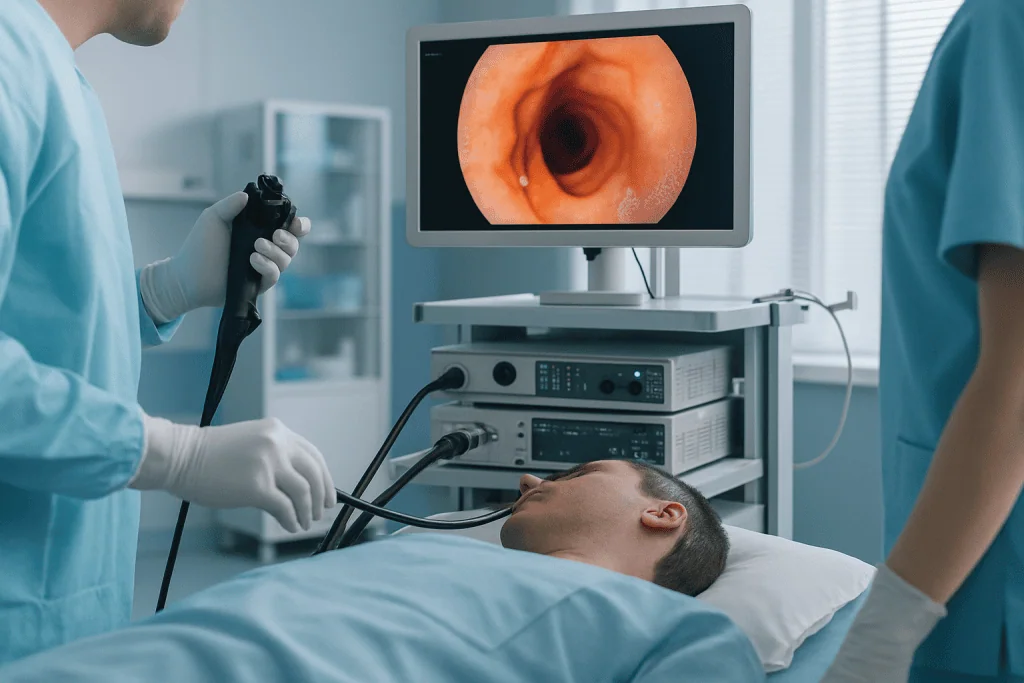
Upper endoscopy lets the physician look directly at the oesophagus and stomach. Its main job in suspected achalasia is to exclude cancer or a mechanical blockage, assess how much food or fluid is retained in the oesophagus, and check the lining for inflammation or other disease. Endoscopy alone often cannot prove achalasia, the findings can be subtle, especially early on, but it is essential for ruling out conditions that would change the plan entirely.
Barium swallow study shows how a swallowed contrast liquid moves through the oesophagus on X-ray images. In achalasia it may reveal a tight, tapered narrowing at the lower sphincter, often described as a “bird’s beak” appearance: a dilated oesophagus above it, and delayed emptying. A timed version of the study, the timed barium oesophagram, measures how much contrast remains after set intervals and is useful for comparing emptying before and after treatment.
High-resolution oesophageal manometry is usually the key diagnostic test. A thin pressure-sensing catheter is passed through the nose into the oesophagus, and you take a series of swallows while the system records pressure along the entire length of the tube. Manometry confirms that the lower sphincter fails to relax, documents the loss of normal peristalsis, and, critically, classifies the achalasia into one of three subtypes, which directly shapes the treatment recommendation.
Additional tests are used selectively: pH monitoring to evaluate acid reflux, cross-sectional imaging such as CT when another cause must be excluded, and repeat barium studies to track emptying over time. Not every patient needs every test; the sequence is tailored to the individual picture.
Ruling out pseudoachalasia
A vital part of the diagnostic pathway is excluding pseudoachalasia: a condition in which the symptoms and even the test findings resemble achalasia, but the real cause is something else, most importantly a tumour at the junction between the oesophagus and stomach. Certain features raise suspicion: rapid weight loss, a short history of symptoms, or an older age at onset. This is precisely why endoscopy, careful imaging when indicated, and expert review of the whole picture come before any procedure. Treatment should not proceed until the diagnosis is sufficiently clear, because treating pseudoachalasia as achalasia delays care for a condition that needs a completely different approach.
Types of Achalasia
High-resolution manometry divides achalasia into three subtypes. The distinction is practical rather than academic: it influences which treatment is most likely to help and how the procedure is planned.
Type I achalasia shows minimal pressurisation within the oesophagus and absent normal peristalsis. It often reflects longer-standing disease in which the oesophageal body has become weak and, in some cases, dilated.
Type II achalasia involves panoesophageal pressurisation, pressure building simultaneously along the whole oesophagus with each swallow. This subtype often responds well to therapies that relieve the obstruction at the lower sphincter.
Type III achalasia is the spastic form, with abnormal, premature contractions that may extend well above the sphincter. Because the problem involves a longer segment of muscle, type III may require a longer myotomy, and this is weighed carefully when choosing between POEM and surgical options.
Recurrent symptoms after previous treatment
Treatment is also considered for people whose symptoms persist or return after prior therapy. Some patients have had dilation, botulinum toxin injection or a myotomy years earlier and later develop renewed difficulty swallowing. Re-treatment is entirely possible, but it demands more detailed assessment: scar tissue, reflux, changes in the shape of the oesophagus and the anatomy left by the previous procedure all affect both the risk and the expected benefit of the next step. If this is your situation, the reports from earlier procedures, what was done, when, and with what result, become an important part of the evaluation.
Advanced achalasia and the dilated oesophagus
Longstanding achalasia can leave the oesophagus markedly enlarged or bent into a sigmoid shape. These cases need special planning. In some patients, relieving the sphincter still improves emptying and symptoms meaningfully. In others, the oesophageal body has lost so much function that opening the valve helps only partially, and more complex surgical considerations enter the discussion. Part of the specialist’s job is to distinguish honestly between achalasia that is likely to respond to sphincter-directed therapy and cases where expectations need to be set differently or alternative strategies considered. An honest conversation about what treatment can and cannot achieve in advanced disease is a mark of good care, not a lack of it.
Achalasia Treatment: The Main Options
Achalasia treatment is a group of procedures designed to help food and liquid pass from the oesophagus into the stomach more easily. Because the underlying problem is a lower oesophageal sphincter that will not relax, every effective treatment works the same way at its core: it reduces the pressure of that sphincter. This can be done endoscopically, surgically or with injection therapy.
There are four main options. Pneumatic dilation uses a special balloon, placed endoscopically, to stretch and partially disrupt the tight sphincter muscle. Peroral endoscopic myotomy (POEM) is an advanced endoscopic procedure in which the physician creates a tunnel within the oesophageal wall and cuts the tight muscle from the inside, no external incision at all. Laparoscopic Heller myotomy is minimally invasive surgery that divides the sphincter muscle through small abdominal incisions, usually combined with an anti-reflux procedure. Botulinum toxin injection temporarily relaxes the sphincter and suits selected patients who are not good candidates for the more definitive procedures.
Medication plays only a limited role. Some drugs can lower sphincter pressure for a short time, but they are generally less effective than procedural treatment and can cause side effects. For most people with significant symptoms, durable improvement requires an intervention that directly addresses the sphincter. Any decisions about medicines, including reflux medication after treatment, sit with the treating doctor.
The right treatment is not the same for every patient. Planning depends on the manometry subtype, your age and general health, prior procedures, the degree of oesophageal dilation, reflux risk, and whether other oesophageal conditions are present. A patient with early type II achalasia is approached differently from someone with a markedly dilated oesophagus, a previously failed dilation, or complex spastic symptoms. This is why the diagnostic pathway comes first and the procedure choice second, never the other way round.
Can achalasia be cured?
No. Achalasia cannot currently be cured, because the nerve cells that have been lost do not grow back. What treatment can do, and often does well, is relieve the obstruction so that food passes, symptoms ease, nutrition recovers and daily life becomes far more normal. It is more accurate to think of achalasia as a chronic condition that can be treated effectively than as a disease that is eliminated. Some patients need one procedure; others need a repeat intervention years later; almost everyone benefits from some long-term follow-up.
You may come across personal accounts online titled “how I cured my achalasia” describing diets, supplements or exercises. Read these cautiously. Symptoms can fluctuate, mild cases can be temporarily managed by adaptation, and misdiagnosed conditions can improve for unrelated reasons, but no diet or exercise reopens a sphincter whose nerve supply has been lost. Claims of a self-cure usually reflect one of those situations rather than a reversal of the disease itself.
How Achalasia Treatment Is Performed
Before the procedure
Treatment begins well before the procedure itself. The first step is a structured consultation covering your symptoms, prior testing, medical history, medications, allergies, nutritional status and anaesthetic risk. You will typically be asked how long the dysphagia has lasted, whether it involves solids, liquids or both, whether you regurgitate or aspirate, and what interventions you have already had. Existing records, endoscopy reports, manometry tracings, barium images, pathology results and operative notes, are reviewed as part of this assessment wherever they exist.
Preparation depends on the planned treatment. Because food and liquid can sit in the oesophagus for hours, you may need a liquid diet for a period beforehand and fasting instructions stricter than those used for routine endoscopy; in some cases the oesophagus is cleared endoscopically before the definitive treatment. Blood tests, an electrocardiogram, chest imaging and an anaesthesia assessment may be required. If you take blood thinners or manage heart, lung or metabolic conditions, the treating team reviews these and gives you specific instructions as part of the plan.
Pneumatic dilation
Pneumatic dilation is performed during upper endoscopy, usually with sedation or anaesthesia. The physician positions a balloon across the lower oesophageal sphincter and inflates it in a controlled way to stretch and partially disrupt the tight muscle; imaging guidance may confirm the balloon’s position. The procedure itself is typically brief, but you are monitored afterwards for pain, bleeding, fever or signs of perforation, which is the main risk the technique must respect. Some programmes use graded dilation, increasing balloon size in stages across separate sessions depending on your response and on safety considerations, rather than aiming for maximal stretch in a single sitting.
Botulinum toxin injection
Botulinum toxin injection is also performed endoscopically. Small amounts of the medication are injected directly into the sphincter muscle to reduce its contraction. It is the least invasive option and can be useful for frail patients, older patients with significant medical risk, or people who need temporary symptom control while a longer-term decision is made. Its main limitation is durability: the effect tends to fade over time, and repeated injections can cause tissue changes that make a later definitive procedure technically more demanding. That trade-off is discussed openly before choosing this route.
Peroral endoscopic myotomy (POEM)
POEM is an advanced endoscopic procedure performed under general anaesthesia. The endoscopist enters the oesophagus through the mouth, makes a small opening in the inner lining, and creates a tunnel within the wall of the oesophagus. Working through this tunnel, the tight muscle fibres of the lower sphincter, and, when needed, part of the oesophageal muscle above it, are carefully divided. The opening in the lining is then closed with endoscopic clips or another closure method. Because the length of the muscle cut can be tailored, POEM is particularly relevant for the spastic type III form, where the abnormal muscle extends higher than a standard surgical myotomy usually reaches. There are no external incisions.
Laparoscopic Heller myotomy
Laparoscopic Heller myotomy is performed through small abdominal incisions under general anaesthesia. The surgeon divides the tight sphincter muscle from the outside of the oesophagus, and the procedure is commonly combined with a partial fundoplication, wrapping part of the stomach around the lower oesophagus, to reduce the risk of reflux afterwards. Heller myotomy has a long track record and remains an important option, particularly when reflux control is a priority, when anatomy or previous treatment favours a surgical approach, or when the surgeon’s assessment points that way for the individual patient.
The technology behind the procedures
The tools used in achalasia care support both diagnostic accuracy and procedural precision. High-resolution manometry maps pressure along the whole oesophagus and classifies the disease. Endoscopy provides direct visualisation, biopsy when needed, and therapeutic access without external incisions. Barium imaging shows the shape of the oesophagus and how it empties over time. During procedures, high-definition endoscopic visualisation, imaging guidance, advanced closure tools and minimally invasive surgical instruments help physicians work accurately while limiting tissue trauma, and anaesthesia monitoring supports safety throughout sedation or general anaesthesia. None of this replaces judgement; it gives judgement better information to work with.
How long do the procedure and hospital stay take?
Duration varies with the treatment, your anatomy, prior interventions and complexity. Botulinum toxin injection and pneumatic dilation are generally the shorter procedures. POEM and laparoscopic Heller myotomy take longer, because both involve a controlled division of muscle followed by careful closure or reconstruction. Afterwards, you are observed in a recovery area, and the length of any hospital stay depends on the procedure and your clinical condition. Some patients need imaging or a swallow assessment before the team confirms it is safe to start drinking and eating again.
Eating again: diet after treatment
The return to eating is deliberately gradual. A typical sequence, adjusted to the procedure and your individual instructions, looks like this:
- Step 1, clear liquids: water and other clear fluids once the team confirms it is safe to begin oral intake.
- Step 2, full liquids: milk-based drinks, smooth soups and similar fluids over the following days.
- Step 3, soft foods: well-cooked, moist, easily chewed foods as healing progresses.
- Step 4: a broader diet: gradual expansion toward normal eating, at the pace your care team advises.
Throughout recovery, eating slowly, chewing thoroughly, taking small bites and drinking fluids with meals are commonly recommended. Acid-suppressing medication is often prescribed by the treating doctor, particularly after POEM or myotomy, because lowering the sphincter pressure raises the chance of reflux.
What foods should you not eat with achalasia?
There is no single forbidden-foods list that applies to everyone with achalasia, because tolerance varies widely from person to person. That said, certain textures cause trouble for many patients: dry or dense foods such as plain bread, tough meat and rice; sticky foods; and large, hurried mouthfuls of almost anything. Some people find very cold items uncomfortable, while others manage them well. How you eat often matters as much as what you eat, small bites, thorough chewing, sips of fluid between mouthfuls, eating upright and avoiding large meals close to bedtime all reduce the chance of food impaction and night-time regurgitation. The most useful approach is to build your own list of reliable and unreliable foods with guidance from your care team and, where available, a dietitian, and to revisit it after treatment, since many previously difficult foods become manageable once the obstruction is relieved.
Why Acting Early Matters
Early evaluation and appropriate treatment make a real difference in achalasia. Symptoms may fluctuate, but the underlying motility disorder persists, and delay carries specific consequences: progressive dilation of the oesophagus, worsening food retention, nutritional decline, and a greater chance of regurgitated material entering the airway. Many patients also spend long periods on treatment for presumed reflux while the true cause of their dysphagia goes unaddressed.
Aspiration is one of the more serious concerns, especially at night. Food, saliva or fluid held in the oesophagus can move upward during sleep and slip into the airway, causing coughing and choking episodes, bronchitis or pneumonia. Waking with coughing fits, needing to sleep propped up, and recurring chest infections are the features that usually prompt earlier specialist assessment.
Longstanding obstruction also changes the oesophagus itself. It can become enlarged, tortuous and progressively less able to empty. In advanced cases, opening the sphincter still helps, but the improvement may be more limited because the oesophageal body has lost much of its pumping and reservoir function. Earlier treatment gives the oesophagus a better chance of retaining useful function.
Finally, acting early is a matter of diagnostic safety. Symptoms resembling achalasia can, rarely, be caused by malignancy or other structural disease. Rapidly progressive swallowing difficulty, significant weight loss, older age at symptom onset or a short symptom history all argue for a prompt, thorough evaluation, both to confirm achalasia and to avoid delaying care for something else.
How long can you live with achalasia?
Achalasia itself is a chronic condition rather than a directly life-shortening one, and with proper diagnosis, treatment and follow-up, people live with it for decades. The risks that matter come mainly from its complications rather than the disorder itself: aspiration and chest infections, malnutrition when eating becomes too difficult, and the structural deterioration of a chronically obstructed oesophagus. Effective treatment addresses exactly these risks by restoring passage into the stomach. Long-term follow-up remains sensible even after a good result, because longstanding achalasia is associated with lasting changes in the oesophageal lining and structure that specialists prefer to keep under periodic review. The honest summary: the condition needs managing, not merely enduring, and managed achalasia is compatible with a long life.
Can you live a normal life with achalasia?
Many people live a substantially normal life with achalasia after effective treatment. Eating becomes easier, weight stabilises, sleep improves once night-time regurgitation settles, and social meals stop being a source of dread. Some adjustments often remain: eating more slowly than others at the table, being careful with certain textures, staying upright after meals, and taking reflux medication if the treating doctor prescribes it. People with advanced disease and a severely dilated oesophagus may see more partial improvement, and a minority need repeat procedures over the years. “Normal” is therefore a realistic goal for most, with the honest caveat that it is a managed normal, supported by sensible habits and periodic follow-up rather than achieved once and forgotten.
Potential Benefits of Achalasia Treatment
The expected benefits depend on the procedure, the achalasia subtype and your individual health, but every treatment shares the same aim: reduce the obstruction and improve daily function.
| Benefit | What It Means for You |
|---|---|
| Improved swallowing | Food and liquids may pass into the stomach more easily, reducing the sensation of blockage or pressure during meals. |
| Less regurgitation | Reducing oesophageal retention can decrease the return of undigested food or fluid, especially when lying down. |
| Better nutrition and weight stability | When eating becomes easier, you may be able to broaden your diet and regain weight lost to swallowing difficulty. |
| Reduced aspiration risk | Improved emptying may lower the chance of retained material entering the airway, though ongoing precautions may be needed in advanced disease. |
| Improved quality of life | Meals may become less stressful, with greater confidence in social eating, travel and daily routines. |
| More precise long-term planning | A confirmed diagnosis and documented treatment response guide follow-up, reflux management and decisions if symptoms ever return. |
Recovery Timeline After Achalasia Treatment
Recovery varies with the treatment type, the anaesthesia used and individual healing, but most patients follow a staged return to eating and normal activity.
| Time Period | What to Expect |
|---|---|
| Day 1 | You are monitored after the procedure. Some chest or throat discomfort, mild abdominal bloating or temporary nausea is common. Oral intake may be restricted until the team confirms it is safe to begin liquids. |
| First week | Diet is usually limited to liquids and soft foods, depending on the procedure. You eat slowly and follow the written instructions your team provides, including the specific warning signs, such as fever, severe pain, persistent vomiting or breathing difficulty, that they will ask you to watch for during healing. |
| First month | Most patients gradually expand their diet. Swallowing often improves early, but adaptation takes time. Reflux symptoms may appear during this period and are discussed at follow-up, since acid suppression or further testing may be arranged by the treating doctor. |
| Longer term | Follow-up focuses on symptom control, nutrition, reflux monitoring and whether any additional treatment is needed. Periodic endoscopy, pH testing or repeat imaging may be suggested based on symptoms and risk factors. |
Factors That Influence the Result
A good result begins with an accurate diagnosis. High-resolution manometry is central because it distinguishes achalasia from other motility disorders and identifies the subtype, and treatment tends to work best when the procedure matches the motility pattern, the oesophageal anatomy, the reflux risk and your overall condition. Someone with type III achalasia may need a different myotomy strategy from someone with type II disease; the test result is what makes that distinction visible before anyone operates.
Disease duration and severity matter too. Patients treated before the oesophagus becomes markedly dilated or tortuous tend to have more predictable improvement. In advanced achalasia, the sphincter can be successfully opened while the oesophageal body still empties slowly, which is why expectations should rest on imaging, manometry and clinical judgement rather than on averages or on another patient’s story.
Prior treatments shape the next step. Botulinum toxin injections, repeated dilations or a previous myotomy can create scarring or alter the anatomy. None of this necessarily prevents further care, but it changes procedural planning and risk. Previous endoscopy reports, manometry results, dilation details, operative notes and imaging all help the evaluating team understand exactly what has been done and what remains possible.
Physician experience is a real variable, not a marketing line. POEM demands precise work within the layers of the oesophageal wall. Pneumatic dilation must balance adequate disruption of the sphincter against the risk of perforation. Heller myotomy requires accurate muscle division and thoughtful reflux prevention. A multidisciplinary setting lets complex cases be examined from more than one specialist perspective, particularly valuable when symptoms have recurred or the diagnosis is not straightforward.
Reflux management drives much of long-term satisfaction. Any treatment that effectively lowers sphincter pressure can increase gastro-oesophageal reflux afterwards. Some patients notice typical heartburn or sour regurgitation; others have silent acid exposure detectable only by testing. Follow-up may therefore include acid-reducing medication prescribed by the treating doctor, lifestyle guidance, endoscopy or pH monitoring. Untreated acid exposure over many years can lead to changes in the oesophageal lining such as Barrett’s oesophagus, which is one reason surveillance endoscopy is sometimes recommended. Reflux is not a reason to avoid appropriate achalasia treatment, but it should be anticipated, looked for and managed rather than discovered by accident years later.
Finally, your own participation counts. Following the diet plan, taking prescribed medication as directed by your doctor, resisting the temptation to advance your diet too quickly, attending follow-up appointments and raising problems early all support safer healing.
Achalasia Care at Acibadem
Achalasia is a condition that rewards coordinated care, because good treatment is never just a single procedure. It requires diagnostic confirmation, a considered choice among several options, experienced procedural teams and structured follow-up. At Acibadem, patients with suspected or confirmed achalasia are assessed through a pathway that can involve gastroenterology, advanced endoscopy, gastrointestinal surgery, radiology, anaesthesiology, nutrition and other specialties as the individual case requires.
This collaborative model matters for achalasia precisely because the best treatment is individualised. Some patients are better suited to POEM, others to pneumatic dilation, laparoscopic Heller myotomy, botulinum toxin injection, or a staged approach, and the recommendation follows the medical findings rather than a one-size-fits-all preference. Complex cases, recurrent symptoms after previous treatment and uncertain diagnoses may be discussed in multidisciplinary settings so that more than one specialist perspective informs the plan.
The diagnostic and therapeutic infrastructure supports that process: high-resolution manometry to define the motility disorder, endoscopy for evaluation and incisionless treatment, barium imaging to show oesophageal shape and emptying, and minimally invasive surgical systems and advanced endoscopic instruments for controlled myotomy or dilation. Anaesthesia assessment, infection prevention, endoscopy safety protocols, medication review and discharge planning are built into the pathway rather than added afterwards.
Care also continues after swallowing improves. Long-term management may include reflux surveillance, nutritional recovery, symptom review, medication adjustment by the treating doctor and reassessment if dysphagia returns. Documentation is shared so that ongoing care can continue smoothly with the physicians who follow you afterwards.
Deciding on Treatment: What a Careful Review Involves
If you have been diagnosed with achalasia, or your symptoms suggest it, the most useful thing to understand is what a thorough review looks like, because that is what protects you from both undertreatment and the wrong procedure. A careful review draws on the full record: endoscopy reports, high-resolution manometry tracings, barium swallow images, details of any previous dilations or injections, operative notes and pathology results. With those in hand, a specialist can confirm the subtype, judge the state of the oesophageal body, weigh reflux risk and set out which of the four treatment options fits your situation, and why.
Useful questions to raise with any treating team include: which achalasia subtype the manometry shows; how dilated the oesophagus is and what that means for expected improvement; why the recommended procedure is preferred over the alternatives for your case; what the reflux plan is afterwards; and what the follow-up schedule looks like, including who monitors you once you are home. A team that answers these questions specifically, with your test results, not generalities, is giving you the basis for a sound decision.
Achalasia is demanding, but it is one of the better-understood oesophageal motility disorders, with several established treatments and a clear diagnostic pathway. Understood early, tested properly and treated by an experienced team, it is a condition most people learn to live with well, eating, travelling and getting on with life.
Preparation
- Evaluation usually includes upper endoscopy, esophageal manometry, and sometimes a barium swallow to confirm diagnosis and plan treatment. Patients are generally asked to fast before the procedure and may need medication adjustments, especially blood thinners. An anesthesia assessment is completed when endoscopic or surgical treatment is planned.
Aftercare
- Patients are monitored for swallowing comfort, chest discomfort, fever, or signs of reflux after treatment. A liquid diet is often started first, then gradually advanced as advised by the care team. Follow-up may include reflux management and repeat testing if symptoms persist.
Frequently Asked Questions
What affects the cost of achalasia treatment?
The final cost depends on diagnostic testing, the selected treatment, anaesthesia, hospital stay, specialist fees, medications, follow-up needs and whether travel or translation services are included. A personalised quote can be prepared after a medical review.
How can I get a personalised quote for achalasia care in Turkey?
You can request a free consultation by sharing your symptoms, previous endoscopy or manometry reports, imaging results and medical history. The clinical team can then advise which assessments are needed and what the proposed package includes.
Is POEM usually more expensive than dilation or botulinum toxin injection?
POEM often involves advanced endoscopic expertise, specialised equipment, anaesthesia and post-procedure monitoring, so its cost structure may differ from dilation or injection. The most appropriate option should be chosen by a specialist, not by cost alone.
Does a package include all achalasia tests and follow-up?
Package inclusions vary. Some include consultation, procedure, anaesthesia and hospital services, while additional diagnostic tests, extended stay, medications or later follow-up may be separate. Written confirmation of inclusions is recommended before travel.
Can previous test results reduce the cost or waiting time?
Recent and complete results may help the specialist plan care more efficiently, but some tests may need to be repeated to confirm the diagnosis or ensure safety. The medical team will decide what is acceptable after reviewing your documents.
Is achalasia treatment abroad safe for international patients?
Safety depends on correct diagnosis, specialist experience, hospital standards, anaesthesia assessment and clear follow-up planning. International patients should choose an accredited centre, ask what support is included and attend a free consultation for individual guidance.
Medically reviewed by the Acıbadem International Medical Board September 13, 2026
See our medical review board →
Update history
- PublishedJune 8, 2026
- Medical review approvedSeptember 13, 2026
- Board commentary addedAugust 26, 2026
- Last content updateSeptember 12, 2026
Trusted care for international patients
From the Acibadem Blog

How Is Achalasia Confirmed and Typed? Esophageal Manometry, Barium Swallow and Endoscopy
Achalasia is confirmed when high-resolution esophageal manometry shows that the lower esophageal sphincter fails to relax on swallowing and the esophageal body has lost…

POEM vs Heller Myotomy for Achalasia: Endoscopic or Laparoscopic, and the Reflux Trade-Off
POEM and laparoscopic Heller myotomy both cut the tight muscle ring at the bottom of the esophagus to relieve achalasia, and a randomized trial…