Optical Coherence Tomography in Neuro-Ophthalmology: What Eye Nerve Scans Can Reveal

OCT is a fast, noninvasive scan that shows tiny structural changes in the retina and optic nerve. Neuro-ophthalmologists use OCT to support diagnosis and follow-up in optic neuritis, glaucoma, papilledema, and other disorders.
Key Takeaways
- OCT is a fast, noninvasive scan that shows tiny structural changes in the retina and optic nerve.
- Neuro-ophthalmologists use OCT to support diagnosis and follow-up in optic neuritis, glaucoma, papilledema, and other disorders.
- The scan does not usually touch the eye and is often completed within minutes.
- OCT findings are interpreted together with symptoms, eye examination, visual field testing, and sometimes MRI or blood tests.
- OCT can help track whether swelling, thinning, or damage is stable, improving, or progressing over time.
Optical coherence tomography, often called OCT, is a painless imaging test that creates detailed cross-sectional pictures of the retina and optic nerve. In neuro-ophthalmology, it helps doctors detect, monitor, and better understand conditions that affect vision, the optic nerve, and the brain’s visual pathways.
Overview
Optical coherence tomography in neuro-ophthalmology is an advanced imaging method used to examine the retina and optic nerve in very fine detail. It uses light waves, not radiation, to produce cross-sectional images of the layers at the back of the eye. Because these structures are closely connected to the visual system and the brain, OCT can offer valuable clues about neurological and eye-related conditions.
In practical terms, OCT acts like a highly detailed “eye nerve scan.” It can measure the thickness of the retinal nerve fiber layer, the ganglion cell layer, and other retinal structures that may change when the optic nerve is inflamed, swollen, compressed, or damaged. These measurements help doctors identify patterns that may not be visible during a standard eye exam alone.
Neuro-ophthalmologists often use OCT alongside a complete history, visual acuity testing, color vision testing, pupil examination, and visual field tests. In some cases, additional evaluation such as MRI, blood tests, or lumbar puncture may be needed. OCT is helpful, but it is one part of a broader clinical picture rather than a stand-alone diagnosis.
What OCT Can Reveal About the Eye Nerve
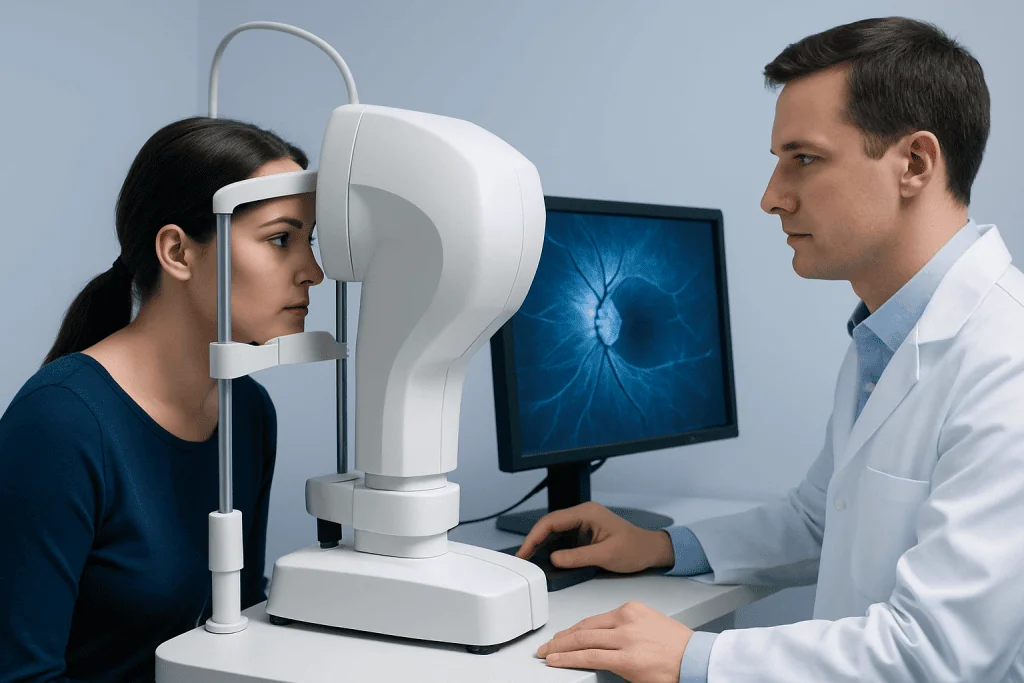
The optic nerve carries visual information from the eye to the brain. When it is affected by inflammation, pressure, poor blood flow, compression, or degeneration, subtle structural changes can occur in the retina and optic nerve head. OCT can detect some of these changes earlier and more precisely than a routine examination alone.
One of the main OCT measurements is the retinal nerve fiber layer, which contains the fibers of the optic nerve. Thinning in this layer may suggest prior optic nerve injury or loss of nerve tissue. In contrast, thickening or elevation around the optic disc may suggest swelling, such as papilledema or other causes of optic nerve head edema.
OCT can also assess the macular ganglion cell layer, which may show damage in disorders affecting the optic nerve or the visual pathway. This is particularly useful when symptoms are unclear or when doctors need to distinguish between true disease and findings that may look similar on examination.
- Optic nerve swelling
- Loss of nerve fiber thickness
- Retinal layer changes linked to neurological disease
- Differences between one eye and the other
- Structural changes over time during follow-up
Symptoms and Conditions That May Lead to OCT Testing
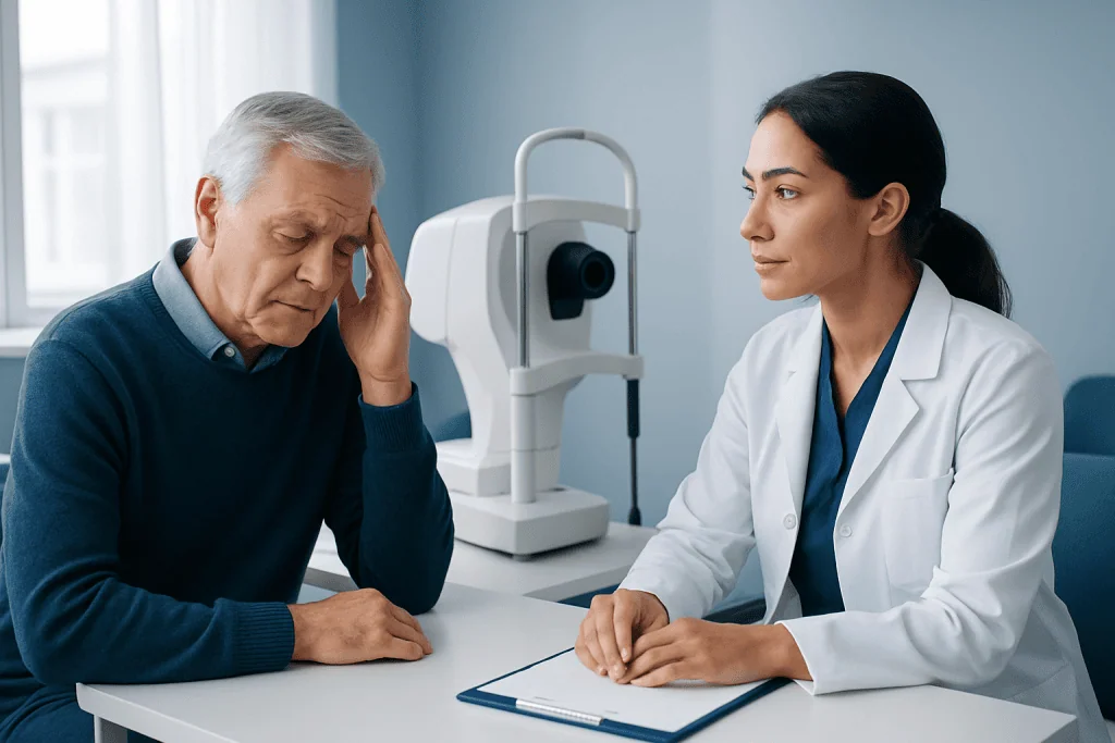
OCT may be recommended when a person has symptoms such as blurred vision, loss of color vision, eye pain with movement, double vision, blind spots, reduced side vision, or unexplained visual loss. It may also be used when the optic nerve looks abnormal during an eye exam, even if symptoms are mild or absent.
In neuro-ophthalmology, OCT is commonly used in the evaluation of optic neuritis, papilledema, ischemic optic neuropathy, compressive optic neuropathy, and some hereditary or toxic optic nerve disorders. It can also help doctors distinguish swelling from pseudo-swelling, such as crowded optic discs or optic disc drusen, which may require a different management approach.
Although glaucoma is usually managed within ophthalmology rather than neuro-ophthalmology, OCT can sometimes help when the diagnosis overlaps with neurological causes of optic nerve damage. The test may also contribute information in patients with multiple sclerosis, intracranial hypertension, or lesions affecting the visual pathway. For related conditions, doctors may also assess disorders such as optic neuritis or papilledema as part of the overall evaluation.
How the Test Is Performed and What to Expect
OCT is usually done in an outpatient clinic or eye center. During the test, the patient sits in front of a machine and places the chin and forehead on supports to keep the head steady. The person is then asked to look at a target while the machine scans the eye with light.
The test is painless and does not normally touch the eye. In many cases, no special preparation is needed. Some clinics may use dilating eye drops to widen the pupils for better images, but this depends on the type of scan and the doctor’s preference.
The scan itself is very quick, often taking only a few minutes for each eye. Good image quality depends on steady fixation and a clear view through the front of the eye. Cataracts, dry eyes, poor focus, or difficulty holding still can sometimes reduce image quality and make repeat scans necessary.
After the test, most people can return to their normal activities right away. If dilating drops were used, temporary light sensitivity or blurred near vision may occur for a few hours. The images are then reviewed by the eye specialist, who interprets them in the context of symptoms and examination findings.
How Doctors Interpret OCT Results
OCT images are not interpreted in isolation. A neuro-ophthalmologist looks at the scan pattern, compares one eye with the other, and reviews whether findings match the patient’s symptoms and examination. Thickness maps, color-coded summaries, and serial measurements can all be useful, but they must be read carefully.
For example, a swollen optic disc on examination may be due to true papilledema, optic neuritis, ischemia, or an anatomic variation. OCT can help show whether there is subretinal fluid, retinal nerve fiber layer thickening, or buried disc drusen features that support one diagnosis over another. Over time, repeat OCT scans may show whether swelling is improving or whether thinning has developed after injury.
At the same time, OCT has limitations. It does not directly measure vision quality, and a person can have significant symptoms even when the scan appears close to normal. Results may also be affected by age, high myopia, media opacity, motion artifact, or differences between machines and scan protocols.
Because of these factors, doctors often combine OCT with tests such as visual fields, fundus photography, and neuroimaging when needed. In selected cases, additional diagnostic tools like MRI imaging or visual field testing may provide important complementary information.
Role of OCT in Diagnosis, Monitoring, and Treatment Planning
OCT is especially valuable not only for diagnosis but also for follow-up. A single scan may show what structures look like at one point in time, while repeat scans can reveal whether swelling, thinning, or atrophy is developing. This makes OCT helpful in conditions where treatment decisions depend on whether disease is active, stable, or progressing.
For example, after optic neuritis, swelling may initially be present, but later scans may reveal thinning of the retinal nerve fiber layer or ganglion cell layer. In papilledema, OCT can help track whether optic disc swelling is improving with treatment. In compressive optic neuropathy, it may show the extent of structural damage and support discussions about visual prognosis.
OCT can also help avoid unnecessary concern when findings are stable and consistent with benign anatomic variation. In other cases, it may prompt more urgent investigation if the pattern suggests active optic nerve disease. Treatment itself depends on the underlying cause and may include observation, medications, management of intracranial pressure, or referral for other therapies.
Depending on the diagnosis, a doctor may coordinate care with neurology, ophthalmology, radiology, or neurosurgery. When structural abnormalities or pressure-related causes are suspected, further care may involve options such as neurology evaluation or brain and pituitary tumor surgery if a compressive lesion is identified and surgery is appropriate.
When to Seek Medical Attention
A person should arrange medical evaluation if there is unexplained blurred vision, sudden loss of vision, reduced color vision, new double vision, eye pain with movement, or persistent blind spots. These symptoms do not always mean a serious condition is present, but they do deserve prompt professional assessment.
Urgent attention is especially important if visual symptoms develop suddenly, affect one or both eyes quickly, or occur together with severe headache, nausea, weakness, imbalance, or other neurological symptoms. In these situations, doctors may need to rule out problems involving the optic nerve, the brain, or raised intracranial pressure.
People who already have a diagnosed neuro-ophthalmic condition should attend follow-up visits as advised, even if they feel stable. Serial OCT scans can help detect change before it becomes obvious in day-to-day vision. Near the end of the care pathway, patients may find reassurance in centers where multidisciplinary specialists work together; Acibadem International’s JCI-accredited hospitals diagnose and treat neuro-ophthalmic conditions for international patients.
Frequently asked questions
Is OCT painful or invasive?
No. OCT is a noninvasive and painless scan that uses light to create detailed images of the retina and optic nerve. The test usually does not touch the eye, and most people tolerate it very easily.
How long does an OCT eye nerve scan take?
The scan itself is usually completed within a few minutes for each eye. The full visit may take longer if other tests are needed or if the pupils are dilated beforehand.
Can OCT diagnose optic neuritis by itself?
Not by itself. OCT can show structural changes that support the diagnosis and help with follow-up, but doctors also rely on symptoms, examination findings, and sometimes MRI or laboratory tests.
What is the difference between OCT and MRI?
OCT looks at the retina and optic nerve head in very fine detail from inside the eye. MRI shows deeper structures such as the optic nerves behind the eye, the brain, and surrounding tissues, so the two tests often answer different questions.
Why might a doctor repeat OCT scans over time?
Repeat scans help show whether a condition is stable, improving, or getting worse. This is useful for monitoring optic nerve swelling, nerve fiber loss, and response to treatment.
Can OCT detect all causes of vision loss?
No. OCT is very useful for many retinal and optic nerve problems, but it cannot identify every cause of visual symptoms. Some people may need additional testing because the source of vision loss can be neurological, vascular, inflammatory, or elsewhere in the eye.
References
- American Academy of Ophthalmology
- North American Neuro-Ophthalmology Society
- National Eye Institute
- American Society of Retina Specialists
- Royal College of Ophthalmologists
This article is for general information only and is not a substitute for professional medical advice. Please consult a qualified doctor about your individual situation.
Explore treatments in Turkey — costs, top hospitals & a free quote
JCI-accredited · board-certified surgeons · reply within 24h
Add us as a Preferred Source to see more of our trusted health content across Google Search, AI Overviews and Discover.

More from the Health Library



Related Specialists

Prof. Dr. Mehmet Veli Karaaltın
Aesthetic Plastic & Reconstructive Surgery
Assoc. Prof. Dr. Onur Dülgeroğlu
General Surgery
Assoc. Prof. Dr. Müjdat Kara
Endocrinology
Kenan Kesgin
Physical Medicine & Rehabilitation