Vena Cava: An Evidence-Based Guide for Patients

The body has two venae cavae: the superior vena cava in the chest and the inferior vena cava in the abdomen. Their main job is to return blood from the body to the right side of the heart.
Key Takeaways
- The body has two venae cavae: the superior vena cava in the chest and the inferior vena cava in the abdomen.
- Their main job is to return blood from the body to the right side of the heart.
- Problems affecting the vena cava may include compression, narrowing, congenital differences, or blood clots.
- Symptoms depend on which vein is involved and may include swelling, shortness of breath, visible chest veins, or leg swelling.
- Diagnosis usually involves imaging such as ultrasound, CT, MRI, or echocardiography, depending on the suspected cause.
- Treatment focuses on the underlying problem and may involve medications, minimally invasive procedures, or surgery.
The vena cava is not a disease but the name for the body’s two largest veins: the superior vena cava and the inferior vena cava. They carry oxygen-poor blood back to the heart, and when they become compressed, blocked, or affected by a clot, symptoms can range from mild swelling to more urgent circulation problems.
What the vena cava is and why it matters
The vena cava refers to the two largest veins in the body. The superior vena cava carries blood from the head, neck, arms, and upper chest back to the heart. The inferior vena cava returns blood from the abdomen, pelvis, and legs. Both empty into the right atrium, the chamber of the heart that receives oxygen-poor blood before it is sent to the lungs.
In everyday health, most people never notice the vena cava working. However, because these veins handle a large volume of blood, any blockage, compression, narrowing, or clot can affect circulation noticeably. Problems involving the vena cava may be temporary or chronic, and their seriousness depends on the cause, the speed of onset, and how much blood flow is reduced.
This topic can be confusing because “vena cava” may refer to normal anatomy, a symptom pattern, or a specific medical problem such as superior vena cava syndrome or inferior vena cava thrombosis. A patient-friendly way to understand it is this: the vena cava is a major return route to the heart, and when that route is disrupted, swelling and pressure often develop upstream from the blockage.
The two main venae cavae and how they work
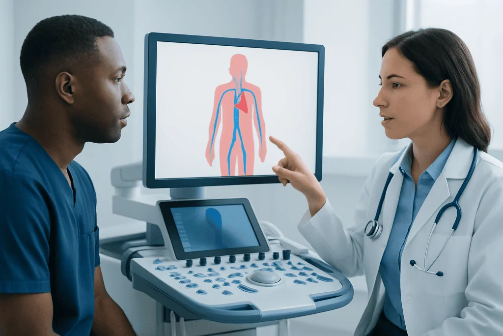
The superior vena cava (SVC) is a short, large vein in the upper chest. It forms from veins that drain the head, neck, arms, and upper torso. Because it sits close to the lungs, lymph nodes, and other structures in the chest, it can sometimes be compressed from the outside by a mass or enlarged tissue. This is why symptoms affecting the face, neck, and upper chest may point to an SVC problem.
The inferior vena cava (IVC) is the larger of the two and runs through the abdomen. It collects blood from the lower body, including the legs, pelvis, kidneys, and liver, before reaching the heart. The IVC can be affected by clots, external pressure, pregnancy-related changes, or rarely congenital differences in how the vein developed before birth.
Veins rely on low pressure and one-way blood flow to return blood efficiently. When that flow is interrupted, the body may partly compensate by using smaller nearby veins, called collateral veins. These alternate pathways can help, especially if the problem develops slowly, but they may not fully prevent symptoms if the obstruction is significant.
Common vena cava problems and symptoms

Symptoms depend on which vena cava is involved. If the superior vena cava is narrowed or blocked, a person may notice swelling of the face, neck, or arms, a feeling of fullness in the head, visible veins on the chest, cough, or shortness of breath. Symptoms may become more noticeable when bending forward or lying down. This pattern is often described as superior vena cava syndrome.
If the inferior vena cava is affected, symptoms may include swelling in one or both legs, pelvic discomfort, abdominal fullness, or enlarged abdominal veins. Some people develop symptoms related to a deep vein clot in the leg, while others may have more general signs of poor venous return such as heaviness and tightness in the lower body.
Not every vena cava problem causes obvious symptoms at first. Some are found during imaging for another reason. When symptoms do occur, they often reflect venous congestion rather than pain alone. This means swelling, pressure, and visible veins are often more informative than sharp discomfort.
- Upper-body swelling may suggest superior vena cava involvement.
- Lower-body swelling may suggest inferior vena cava involvement.
- Sudden symptoms deserve faster evaluation than slowly developing ones.
- Breathing difficulty, chest symptoms, or neurologic symptoms should be assessed urgently.
Causes and risk factors
Vena cava problems can happen for several reasons. One major cause is external compression, where a nearby structure presses on the vein. In the chest, this may happen because of enlarged lymph nodes, inflammation, or a tumor. In the abdomen, compression may relate to pregnancy, abdominal masses, or less commonly scarring and anatomic abnormalities.
Another important cause is thrombosis, which means a blood clot forms inside the vein. Clots may arise in the vena cava itself or extend from another vein, especially from the legs. Risk factors for clotting include prolonged immobility, major surgery, cancer, inherited clotting conditions, central venous catheters, dehydration, and a history of deep vein thrombosis.
Some patients have congenital variations of the inferior vena cava that are present from birth. These do not always cause symptoms, but they may increase the risk of clotting in certain situations. Less often, inflammation, scar tissue, or previous procedures involving large veins can contribute to narrowing or obstruction.
Because the vena cava is connected closely with heart and vascular function, doctors also assess for related problems such as reduced heart function, other vein disorders, or conditions that increase pressure in the circulation. The exact cause matters because treatment is directed at the source of the blockage or impaired flow.
How doctors diagnose vena cava conditions
Diagnosis begins with a medical history and physical examination. Doctors ask when symptoms started, whether they came on suddenly or gradually, and whether there are risk factors for clots, chest disease, abdominal masses, or recent hospital care. The pattern of swelling can provide useful clues, especially when it affects the face and arms or, alternatively, the legs and abdomen.
Imaging is usually needed to confirm the diagnosis and locate the problem. Depending on the suspected condition, this may include Doppler ultrasound, CT scan, MRI, venography, or echocardiography. Ultrasound is often used to assess veins in the legs and may help if an IVC problem is suspected indirectly. CT or MRI can show whether the vena cava is compressed, narrowed, or blocked and can also reveal surrounding structures.
Blood tests may be used to look for clotting tendency, inflammation, kidney function, or signs of an underlying illness. In selected cases, doctors investigate for cancer, inherited clotting disorders, or other vascular problems. If symptoms suggest a broader circulatory issue, a patient may also be evaluated by specialists in cardiology care or vascular surgery.
Diagnosis is not just about naming the problem. It also helps determine urgency. A slowly developing partial blockage may be monitored and treated in a planned way, while a sudden clot or severe upper-body swelling with breathing symptoms may require more immediate treatment.
Treatment options and what they aim to do
Treatment for a vena cava problem depends on the cause, location, severity, and the patient’s overall health. If a blood clot is present, treatment often involves anticoagulant medicines to reduce further clotting and help the body gradually break the clot down. In selected cases, doctors may consider catheter-based procedures to remove or dissolve the clot, especially when symptoms are severe or blood flow is markedly reduced.
If the vein is compressed from the outside, treatment focuses on relieving that pressure. This may involve treating inflammation, addressing a nearby mass, or using a stent to hold the vein open. In some cases, a minimally invasive approach performed by specialists in interventional radiology can improve blood flow without open surgery.
For patients with superior vena cava syndrome, doctors first determine whether the problem is caused by clotting, compression, or a chest condition that needs its own treatment. If a clot is involved, anticoagulation may be used; if a mass is involved, treatment targets the underlying disease. Supportive measures such as elevating the head of the bed or easing fluid overload may help symptoms while the main cause is being treated.
Some patients need longer-term follow-up, especially if they have a clotting disorder, recurrent thrombosis, or a chronic narrowing of the vein. Rarely, surgery is considered when less invasive options are not suitable. In multidisciplinary centers such as Acibadem International, JCI-accredited hospitals and specialist teams evaluate these cases using imaging, vascular medicine, and procedural expertise tailored to the underlying cause.
Prevention and self-care
Not all vena cava problems can be prevented, but some steps can reduce risk, especially for clot-related conditions. Staying mobile during long travel or recovery, following medical advice after surgery, staying well hydrated, and managing chronic health conditions can all support healthy circulation. People with a past history of clotting should discuss prevention plans with their doctor before major procedures, pregnancy, or extended immobility.
Patients who have already had a venous clot may be advised to continue follow-up care, take prescribed medication exactly as directed, and watch for new swelling or breathing symptoms. Compression stockings may be recommended in some lower-limb venous conditions, although they are not suitable for every patient. A doctor can explain whether they are helpful in an individual case.
Self-care should not replace medical evaluation when symptoms suggest vena cava obstruction. Home measures may improve comfort, but they do not treat the underlying cause. New facial swelling, one-sided or sudden leg swelling, or visible enlarged veins should be assessed rather than self-diagnosed.
When to seek medical care
Medical review is appropriate if swelling of the face, neck, arms, abdomen, or legs develops without a clear reason, especially if it is persistent or worsening. A doctor should also assess unexplained shortness of breath, visible enlarged veins over the chest or abdomen, or symptoms that begin after surgery, prolonged travel, or use of a central venous catheter.
Urgent care is important if symptoms appear suddenly, become severe, or are accompanied by chest pain, fainting, confusion, marked breathing difficulty, or signs of a possible pulmonary embolism. These symptoms do not always mean a vena cava problem, but they do need prompt medical attention.
Patients already diagnosed with a clotting disorder, cancer, or a previous major vein problem should seek advice early if new swelling appears. Timely assessment can help doctors identify whether the issue is related to the vena cava, another large vein, or a different heart, lung, or kidney condition.
Frequently asked questions
What is the vena cava in simple terms?
The vena cava is the name for the body’s two largest veins. They bring oxygen-poor blood back to the heart so it can be pumped to the lungs and then out to the body again.
What is the difference between the superior and inferior vena cava?
The superior vena cava drains blood from the upper body, including the head, neck, and arms. The inferior vena cava drains blood from the lower body, including the abdomen, pelvis, and legs.
Can a vena cava problem cause swelling?
Yes. Because the vena cava returns blood to the heart, a blockage or narrowing can cause blood to back up, leading to swelling above or below the affected area. The location of swelling often helps doctors identify which vena cava may be involved.
Is vena cava syndrome the same as a blood clot?
Not always. Vena cava syndrome describes symptoms caused by reduced flow through the vein, and that can happen because of a clot, outside compression, or narrowing. A blood clot is one possible cause, but not the only one.
How is a vena cava problem diagnosed?
Doctors usually combine a physical exam with imaging tests. Depending on the situation, these may include ultrasound, CT, MRI, venography, or echocardiography to show where blood flow is impaired and why.
Are vena cava conditions treatable?
Many are treatable, but the best approach depends on the cause. Treatment may include medication for clotting, procedures to open the vein, or treatment of a nearby condition that is pressing on it.
References
- National Heart, Lung, and Blood Institute
- American Heart Association
- Society for Vascular Surgery
- Merck Manual Professional Edition
- Radiological Society of North America
This article is for general information only and is not a substitute for professional medical advice. Please consult a qualified doctor about your individual situation.
Explore treatments in Turkey — costs, top hospitals & a free quote
JCI-accredited · board-certified surgeons · reply within 24h
Add us as a Preferred Source to see more of our trusted health content across Google Search, AI Overviews and Discover.