Eye Anatomy — Explained by Medical Evidence, Not Myths
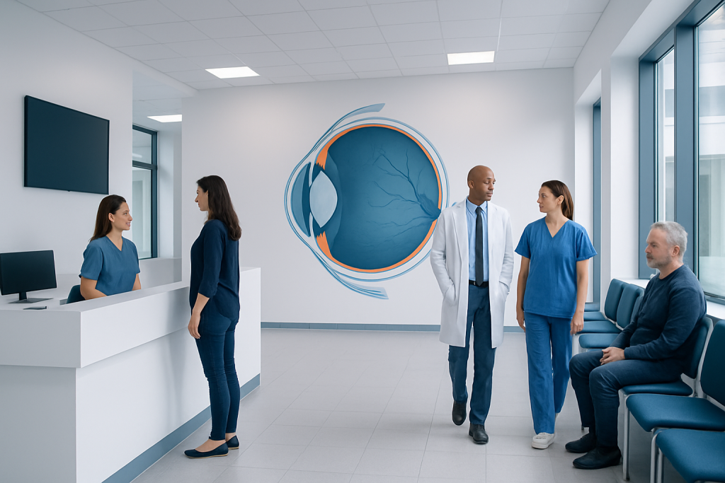
Eye anatomy includes the outer protective layers, the focusing system, and the light-sensing retina. The cornea and lens bend light so it can focus clearly on the retina.
Key Takeaways
- Eye anatomy includes the outer protective layers, the focusing system, and the light-sensing retina.
- The cornea and lens bend light so it can focus clearly on the retina.
- The retina converts light into nerve signals, and the optic nerve carries those signals to the brain.
- Symptoms such as sudden vision loss, severe eye pain, flashes, or a curtain-like shadow need urgent medical care.
- Regular eye examinations help detect problems early, even before symptoms appear.
Eye anatomy describes the structures that collect light, focus images, and send visual information to the brain. Understanding the basic parts of the eye can make symptoms, eye exams, and common conditions easier to understand.
Overview: what eye anatomy means
Eye anatomy is the study of the parts of the eye and how they work together to produce vision. In simple terms, the eye acts like a highly specialized optical system: it lets light in, focuses that light, converts it into electrical signals, and sends those signals to the brain, where vision is interpreted.
Medical explanations of eye anatomy are most useful when they connect structure to function. Rather than treating the eye as a list of parts to memorize, it helps to understand how each structure supports a specific job such as protection, focusing, fluid balance, or signal transmission. This approach can also make common eye conditions easier to understand.
The eye does not work alone. Eyelids, tear glands, eye muscles, blood vessels, and the brain all support normal sight. When one part is affected by injury, aging, infection, or disease, symptoms may include blurred vision, dryness, double vision, pain, or changes in visual field.
The main parts of the eye and what they do
The outermost visible structures include the eyelids, eyelashes, conjunctiva, and sclera. The eyelids protect the eye from injury and spread tears across the surface with each blink. The conjunctiva is a thin, clear membrane covering the white of the eye and inner eyelids, while the sclera is the tough white outer coat that helps maintain the eye’s shape.
At the front of the eye is the cornea, a clear dome that begins the focusing process by bending incoming light. Behind it is the anterior chamber, which contains aqueous humor, a clear fluid that nourishes internal tissues and helps maintain pressure. The iris gives the eye its color, and the pupil is the central opening that adjusts how much light enters the eye.
Behind the pupil sits the lens, which changes shape to fine-tune focus, especially for near vision. The ciliary body and its muscles help the lens adjust. Farther back, the vitreous humor, a gel-like substance, fills most of the inside of the eye and helps it keep its round form.
The innermost layer is the retina, which senses light. At the center of the retina is the macula, responsible for sharp central vision needed for reading and recognizing faces. The optic nerve then carries visual information from the retina to the brain. Surrounding and nourishing many of these structures is the choroid, a vascular layer rich in blood supply.
How the eye works to create vision

Vision begins when light enters the eye through the cornea. The cornea provides much of the eye’s focusing power. Light then passes through the aqueous humor and pupil, where the iris adjusts the pupil size depending on the brightness of the environment.
The lens further refines focus by changing shape, a process called accommodation. This is particularly important when shifting attention from distant objects to near ones. Once light reaches the retina, specialized photoreceptor cells called rods and cones convert it into electrical signals. Rods are more sensitive in dim light, while cones support color vision and fine detail.
Those signals travel through retinal nerve cells to the optic nerve, which carries them to visual processing centers in the brain. The brain combines information from both eyes to support depth perception, spatial awareness, and coordinated movement.
Clear vision depends on precise alignment of many structures. If the cornea or lens does not focus light accurately, refractive errors such as nearsightedness, farsightedness, or astigmatism may occur. If the retina, optic nerve, or brain pathways are affected, the problem is not simply one of focus but of signal detection or processing.
Important supporting structures often overlooked
Many people think only of the eyeball itself, but normal vision also depends on surrounding support systems. The tear film is one of the most important. It coats the eye’s surface, keeps it smooth for clear vision, protects against infection, and reduces irritation. Problems with tear production or quality can lead to burning, grittiness, and fluctuating vision.
Six extraocular muscles move each eye in different directions. These muscles allow tracking, reading, and coordinated binocular vision. If they do not work together properly, symptoms such as double vision or eye strain can occur. Eye movement problems may be due to local muscle issues, nerve problems, or neurologic conditions.
Blood vessels are also essential to eye anatomy. The retina and optic nerve require a continuous blood supply. Reduced circulation can damage delicate tissues and affect vision. Intraocular pressure is another key factor: pressure inside the eye must remain within a healthy range to support structure without harming the optic nerve.
Because the eye is closely connected to the nervous system and circulation, a general health problem can affect vision. Diabetes, hypertension, autoimmune disease, infection, and aging-related changes may all alter the function of normal eye anatomy over time.
Common conditions linked to eye anatomy
Understanding eye anatomy helps explain why certain diseases cause specific symptoms. For example, a problem in the cornea may cause pain, light sensitivity, or blurred vision because the cornea has many nerve endings and plays a major role in focusing. A condition affecting the lens, such as cataract, can gradually cloud vision because light no longer passes through a clear structure.
Diseases of the retina and optic nerve may be less painful but more serious for sight. In retinal detachment, the retina pulls away from underlying supportive tissue, which can cause flashes, floaters, or a curtain-like shadow. In glaucoma, damage to the optic nerve is often related to eye pressure and may slowly reduce peripheral vision.
Changes in the macula can affect central vision, making reading or face recognition difficult. Conditions involving the conjunctiva or eyelids may lead to redness, irritation, discharge, or swelling. The same symptom, such as blurry vision, can come from very different structures, which is why a professional eye examination is important.
When treatment is needed, it depends on which part of the eye is affected. This may include medications, optical correction, laser procedures, or surgery such as cataract surgery or retinal surgery when medically appropriate.
How doctors examine eye anatomy
Eye examinations assess both structure and function. A clinician usually begins by asking about symptoms, medical history, medications, and vision changes. Visual acuity testing checks how clearly a person sees at distance or near. Refraction may be performed to determine whether glasses or contact lenses can improve focus.
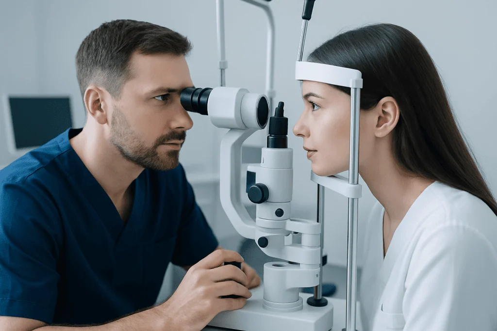
The front of the eye is often examined with a slit lamp, a specialized microscope that provides a detailed view of the eyelids, conjunctiva, cornea, iris, and lens. Eye pressure may be measured to help screen for glaucoma. Doctors may also check pupil reactions, eye movements, and visual fields to assess how different parts of the eye and nervous system are functioning.
To examine the retina and optic nerve, the pupils may be dilated with eye drops. This allows a wider view into the back of the eye. In some cases, imaging tests such as optical coherence tomography or retinal photography are used to look more closely at retinal layers or the optic nerve head.
If symptoms suggest a more complex problem, additional testing may be needed. This can include ultrasound, fluorescein angiography, corneal mapping, or neurologic imaging. In centers with multidisciplinary eye services, treatments such as glaucoma treatment may be planned based on detailed structural and functional findings.
Protecting eye health through prevention and self-care
Good eye care starts with daily habits that support the structures of the eye. Wearing sunglasses with UV protection may help protect the lens and retina from cumulative sunlight exposure. Protective eyewear is important during sports, home repairs, and work activities that could cause eye injury.
Screen use can increase eye strain and dryness, especially when blinking decreases. Taking regular breaks, adjusting screen distance, and using appropriate lighting can help. People who wear contact lenses should follow cleaning and replacement instructions carefully, because poor lens hygiene can increase the risk of corneal infection.
General health also matters. Managing diabetes, blood pressure, and cholesterol supports blood flow to the eyes. Not smoking, eating a balanced diet rich in vegetables and omega-3 sources, and attending regular checkups all contribute to long-term eye health.
Routine eye examinations remain one of the most effective preventive steps. Some eye diseases progress silently at first, particularly glaucoma and diabetic retinal disease. Early detection can preserve vision and broaden treatment options.
When to seek medical care
Many minor eye symptoms improve with rest or simple care, but some changes should not be ignored. A prompt medical assessment is important for sudden vision loss, severe eye pain, a chemical splash, significant trauma, new double vision, or rapid swelling and redness. Flashes of light, a sudden increase in floaters, or a dark curtain over part of the vision can signal a retinal emergency.
Medical review is also recommended for persistent blurred vision, ongoing dryness or irritation, recurring headaches with visual symptoms, or any eye symptom that worsens instead of improving. Children should be assessed if they squint frequently, tilt the head to see, rub the eyes often, or seem to have wandering eye movements.
People with diabetes, a family history of glaucoma, prior eye surgery, autoimmune disease, or long-term steroid use may need more regular follow-up because of higher risk for certain eye problems. If there is uncertainty about a symptom, speaking with a qualified eye specialist is the safest approach.
For patients seeking specialist evaluation, Acibadem International’s multidisciplinary eye teams in JCI-accredited hospitals diagnose and treat a wide range of eye conditions for international patients, including medical and surgical care such as cornea treatment when clinically indicated.
Frequently asked questions
What are the main parts of the eye?
The main parts of the eye include the cornea, iris, pupil, lens, retina, macula, optic nerve, sclera, and the fluids that fill the front and back of the eye. Each structure has a different role in protecting the eye, focusing light, or sending visual information to the brain.
Which part of the eye is most important for vision?
Vision depends on several parts working together rather than one single structure. The cornea and lens focus light, the retina detects it, and the optic nerve carries signals to the brain. Damage to any of these can affect sight.
What is the difference between the retina and the optic nerve?
The retina is the light-sensitive tissue at the back of the eye that converts light into electrical signals. The optic nerve is the pathway that carries those signals from the retina to the brain. Both are essential for normal vision.
Why does the pupil change size?
The pupil changes size to control how much light enters the eye. In bright light it becomes smaller to reduce light entry, and in dim light it becomes larger to allow more light in. This adjustment helps the eye function across different lighting conditions.
Can eye anatomy change with age?
Yes. The lens becomes less flexible over time, which can make near vision more difficult. Aging can also increase the risk of cataracts, glaucoma, macular changes, and dry eye, so routine eye examinations become more important.
How do doctors check if the inside of the eye is healthy?
Doctors use vision testing, slit-lamp examination, pressure measurement, and often dilated retinal examination to assess internal eye health. In some cases, imaging such as retinal scans or photographs provides more detailed information about the retina and optic nerve.
This article is for general information only and is not a substitute for professional medical advice. Please consult a qualified doctor about your individual situation.
Explore treatments in Turkey — costs, top hospitals & a free quote
JCI-accredited · board-certified surgeons · reply within 24h
Add us as a Preferred Source to see more of our trusted health content across Google Search, AI Overviews and Discover.

More from the Health Library

Synovial: What Patients Need to Know

Jugular: What Patients Need to Know

Nerve Block: An Evidence-Based Guide for Patients
Sucralfate — Explained by Medical Evidence, Not Myths

Intellectual Disability: What Patients Need to Know