

Cardiac Sarcoidosis
Cardiac sarcoidosis is an inflammatory condition where sarcoidosis affects the heart, potentially causing rhythm problems, heart failure, or conduction block. Care focuses on diagnosis, inflammation control, and arrhythmia prevention.
Quick answer
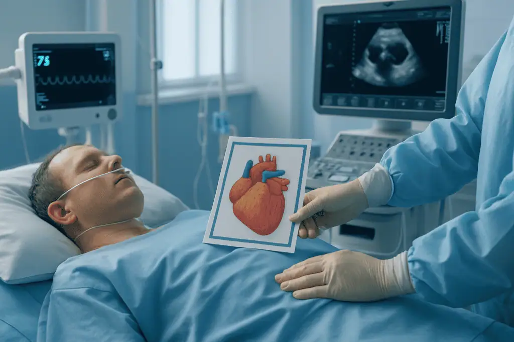
Cardiac sarcoidosis is inflammation in the heart caused by sarcoidosis, and treatment aims to control inflammation, protect heart function, and reduce the risk of rhythm and conduction problems. At Acibadem in Turkey, evaluation may include cardiac imaging and rhythm assessment, with care tailored through medicines and, when needed, devices or other cardiac procedures.
When Sarcoidosis Affects the Heart, the Decisions Can Feel Urgent and Complex
Cardiac sarcoidosis can be difficult to understand because it often begins quietly. A person may already know they have sarcoidosis in the lungs, lymph nodes, skin, or eyes, and then learn that the heart may also be involved. Others have no previous diagnosis of sarcoidosis and first seek medical care because of palpitations, fainting, shortness of breath, chest discomfort, or an abnormal electrocardiogram. For many patients and families, the concern is immediate: Is my heart rhythm safe? Is my heart muscle weakening? Do I need medication, a device, or long-term monitoring?
These are appropriate concerns. Cardiac sarcoidosis is an inflammatory condition in which clusters of immune cells, called granulomas, develop in the heart tissue. Inflammation may affect the heart’s electrical system, the heart muscle, or both. This can lead to rhythm disturbances, conduction block, reduced pumping strength, heart failure symptoms, or, in some cases, serious ventricular arrhythmias.
The encouraging aspect is that cardiac sarcoidosis can often be managed effectively when it is recognized early and treated in a coordinated way. Care usually focuses on three priorities: confirming the diagnosis as accurately as possible, controlling inflammation, and reducing the risk of dangerous heart rhythms. Because the condition can involve both inflammatory disease and advanced cardiology, patients benefit from a structured evaluation by specialists who understand the full spectrum of sarcoidosis and its cardiac complications.
For international patients considering care abroad, the process should feel clear, organized, and clinically rigorous. At Acibadem, evaluation and treatment planning for cardiac sarcoidosis may involve cardiology, electrophysiology, heart failure, pulmonology, rheumatology, nuclear medicine, radiology, and intensive care teams when needed. The goal is to define the patient’s individual risk, choose treatment based on evidence and international standards, and provide a plan that can be continued safely after returning home.
What Cardiac Sarcoidosis Treatment Is
Cardiac sarcoidosis treatment is not a single procedure. It is a comprehensive care pathway designed to diagnose heart involvement, suppress active inflammation, protect the heart’s electrical system, prevent arrhythmia-related complications, and preserve heart function. Treatment is personalized because cardiac sarcoidosis can behave differently from one patient to another.
In some patients, the main problem is inflammation causing swelling and irritation of the heart tissue. In others, previous inflammation has left scar tissue that disrupts electrical signals. Some patients have mild imaging abnormalities with few symptoms, while others present with heart block, ventricular tachycardia, or heart failure. The treatment plan depends on which of these patterns is present and how active the disease appears to be.
The main elements of care may include anti-inflammatory medication, rhythm monitoring, heart failure therapy, implantable devices, catheter-based procedures for certain arrhythmias, and long-term follow-up imaging. Corticosteroids are often used as first-line therapy to reduce inflammation. In selected cases, additional immune-modulating medications may be added to help control disease activity, reduce steroid exposure, or treat recurrent inflammation. These medications require careful monitoring because they can affect the immune system, liver, kidneys, blood counts, blood sugar, bone health, and infection risk.
When the heart’s electrical system is affected, treatment may involve a pacemaker or an implantable cardioverter-defibrillator. A pacemaker can support a slow heartbeat caused by conduction block. An implantable defibrillator can detect and treat certain dangerous ventricular rhythms. Some patients with recurrent arrhythmias may also be evaluated for catheter ablation, especially when rhythm problems persist despite medication or device therapy. If heart pumping function is reduced, guideline-based heart failure treatment is used to support the heart and reduce symptoms.
The purpose of cardiac sarcoidosis care is therefore broader than reducing inflammation alone. It aims to lower risk, improve symptoms where possible, protect against rhythm emergencies, and create a long-term surveillance plan that matches the patient’s disease activity and lifestyle.
Who May Need Evaluation and Treatment for Cardiac Sarcoidosis
Patients may need evaluation for cardiac sarcoidosis if they have known sarcoidosis and develop cardiac symptoms, or if heart findings suggest an inflammatory cause. Because cardiac involvement can occur without obvious symptoms, some patients with sarcoidosis are referred for screening even when they feel well, particularly if tests suggest electrical abnormalities or reduced heart function.
Common symptoms that may lead to evaluation include palpitations, skipped beats, unexplained dizziness, fainting, near-fainting episodes, shortness of breath, reduced exercise tolerance, swelling in the legs, chest discomfort, or unusual fatigue. Some patients notice that they cannot climb stairs or exercise as they previously could. Others experience sudden episodes of rapid heartbeat. In more advanced cases, symptoms may resemble heart failure, with breathlessness while lying flat, waking at night short of breath, weight gain from fluid retention, or persistent tiredness.
Cardiac sarcoidosis may also be suspected after abnormal test results. An electrocardiogram may show conduction delays, atrioventricular block, bundle branch block, or arrhythmias. Ambulatory rhythm monitoring may reveal frequent premature ventricular contractions, nonsustained ventricular tachycardia, pauses, or intermittent heart block. Echocardiography may show reduced ejection fraction, regional wall motion abnormalities, ventricular enlargement, or valve-related changes caused by heart remodeling.
Diagnosis usually requires a combination of clinical assessment, imaging, rhythm evaluation, and evidence of sarcoidosis in the body. Cardiac magnetic resonance imaging can identify scar and inflammation-related changes in the heart muscle. Positron emission tomography can help show active inflammation and may be useful for monitoring response to treatment. Echocardiography provides information about pumping function, heart chamber size, valve function, and pressures. Blood tests may assess inflammation, organ function, heart strain markers, and medication safety. In some patients, biopsy is considered, though heart biopsy may miss the affected area because sarcoidosis can be patchy. When tissue confirmation is needed, biopsy from lymph nodes, lung, skin, or another involved site may be more practical.
The diagnosis can be challenging because cardiac sarcoidosis may resemble other heart conditions, including viral myocarditis, inherited cardiomyopathies, ischemic heart disease, arrhythmogenic cardiomyopathy, or idiopathic conduction disease. For this reason, patients often benefit from review by a multidisciplinary specialist board, where imaging, rhythm data, symptoms, and systemic sarcoidosis findings are considered together.
Conditions and Indications Addressed by Cardiac Sarcoidosis Care
Cardiac sarcoidosis care addresses both the inflammatory disease process and the complications that may develop in the heart. A patient may need treatment because of active myocardial inflammation, scarring from previous inflammation, or the electrical and mechanical consequences of either.
One major indication is conduction system disease. Sarcoid inflammation can affect the pathways that carry electrical impulses through the heart, leading to atrioventricular block or other conduction abnormalities. A patient may feel lightheaded, faint, or have an abnormally slow pulse. In some people, conduction block appears suddenly and requires urgent evaluation.
Another important indication is ventricular arrhythmia. Inflammation and scar tissue can create unstable electrical circuits, leading to ventricular tachycardia or other potentially serious rhythms. These rhythms may cause palpitations, dizziness, fainting, or cardiac arrest. Risk assessment for ventricular arrhythmia is a central part of cardiac sarcoidosis management.
Cardiac sarcoidosis can also cause cardiomyopathy, meaning disease of the heart muscle. The heart may pump less effectively, and the ejection fraction may decline. Patients may develop symptoms of heart failure, such as breathlessness, fluid retention, and fatigue. In these cases, treatment often combines inflammation control with heart failure medication and close follow-up.
Some patients have extracardiac sarcoidosis and abnormal cardiac imaging without major symptoms. Even in this situation, evaluation is important because imaging findings may indicate inflammation or scarring that carries future risk. The treatment decision depends on disease activity, rhythm findings, heart function, and the patient’s overall health.
Care may also address recurrent sarcoidosis activity despite previous treatment, medication intolerance, device-related decisions, pregnancy planning in selected patients, athletic or occupational restrictions, and coordination with physicians in the patient’s home country. Because cardiac sarcoidosis is often a chronic condition, the indication for care may not be limited to diagnosis; it may include long-term monitoring and adjustment of therapy over time.
How Cardiac Sarcoidosis Treatment Is Performed
The care pathway begins with a detailed medical review. Physicians assess symptoms, previous sarcoidosis history, medications, family history of heart disease or sudden cardiac death, prior imaging, rhythm studies, biopsy results, and any episodes of fainting or emergency treatment. For international patients, reviewing records before travel can help determine which tests are needed and whether urgent care is advisable.
The first step is usually risk-focused cardiac testing. An electrocardiogram checks baseline electrical conduction. Ambulatory monitoring, such as a Holter monitor or longer rhythm recorder, can detect intermittent arrhythmias that may not appear during a clinic visit. Echocardiography evaluates heart structure and pumping function. Cardiac magnetic resonance imaging provides detailed tissue characterization and can identify areas of scarring or inflammation-related injury. Nuclear imaging, including positron emission tomography when appropriate, can help determine whether inflammation is active. Blood tests are used to evaluate heart strain, organ function, metabolic status, inflammation, and medication safety.
If the diagnosis is uncertain, the team may consider additional testing to confirm sarcoidosis elsewhere in the body. Chest imaging may identify lymph node or lung involvement. Pulmonary function tests may be performed when lung disease is suspected. Biopsy may be recommended if tissue confirmation has not already been obtained. The safest and most informative biopsy site is chosen individually.
Once the diagnostic picture is clear, specialists define whether the patient has active inflammation, scar-related disease, conduction disease, ventricular arrhythmia risk, reduced heart function, or a combination of these. This distinction matters because anti-inflammatory therapy may reduce active inflammation, but scar-related rhythm risk may persist even when inflammation improves.
Medical treatment often begins with corticosteroids when active cardiac inflammation is likely and there are no contraindications. The dose and duration are individualized. Physicians may gradually taper the dose while monitoring symptoms, imaging findings, rhythm data, and side effects. In some patients, steroid-sparing immune-modulating medications are added. These may be considered when inflammation is persistent, relapse occurs during tapering, long-term steroid toxicity is a concern, or multi-organ sarcoidosis requires broader control. Medication planning includes infection screening, vaccination review when appropriate, blood test monitoring, and counseling about side effects.
If heart failure is present, guideline-based heart failure therapy may be started or optimized. This can include medications that reduce strain on the heart, improve fluid balance, support pumping function, and lower the risk of hospitalization. Lifestyle guidance may include sodium management, fluid recommendations, exercise advice, weight monitoring, and recognition of worsening symptoms. These recommendations are tailored to the patient’s heart function and overall condition.
When conduction block is significant, a pacemaker may be needed. If the patient has risk factors for dangerous ventricular arrhythmias, an implantable cardioverter-defibrillator may be recommended instead of, or in addition to, pacing capability. The decision is based on rhythm history, ejection fraction, imaging evidence of scar, syncope history, and guideline criteria. Device implantation is performed in a specialized cardiac setting under sterile conditions, usually with imaging guidance. Leads are positioned in the heart through veins, and the generator is placed beneath the skin, commonly in the upper chest. After implantation, the device is programmed and tested, and the patient receives instructions for wound care, movement restrictions, and follow-up checks.
For patients with recurrent ventricular tachycardia or difficult-to-control arrhythmias, electrophysiology evaluation may be recommended. Catheter ablation can be considered in selected cases. During ablation, thin catheters are guided through blood vessels into the heart. Mapping systems help identify abnormal electrical circuits, and targeted energy is delivered to interrupt arrhythmia pathways. In cardiac sarcoidosis, arrhythmia circuits may be complex because scar can be patchy and located in different layers of the heart muscle. Ablation may reduce arrhythmia burden, but ongoing medical therapy and device protection may still be necessary.
Typical duration varies according to the care plan. Diagnostic evaluation may take several days, especially when advanced imaging, rhythm monitoring, and multidisciplinary review are needed. Medication-based treatment begins as soon as the team is confident about the diagnosis and risk profile. Device implantation may require a short hospital stay, while catheter ablation can require more intensive monitoring depending on arrhythmia complexity and the patient’s heart function. Patients with severe heart failure, active arrhythmias, or new heart block may need inpatient care until stable.
Recovery depends on which interventions are performed. After diagnostic testing alone, patients can usually resume normal activities quickly. After device implantation, patients typically avoid heavy lifting and certain arm movements for a period while the pocket and leads heal. After catheter ablation, patients are monitored for vascular access site healing, rhythm stability, and medication adjustment. For those beginning immunosuppressive therapy, “recovery” means not only feeling better but also demonstrating controlled inflammation, stable rhythm findings, and protected heart function over time.
Why Acting Early Matters
Early evaluation matters because cardiac sarcoidosis can progress silently. Inflammation may damage the heart’s electrical system before symptoms become obvious. A person may have intermittent conduction block or short runs of ventricular tachycardia that are not felt but still carry risk. Identifying these changes early allows physicians to treat inflammation, monitor rhythm, and consider protective therapies before a serious event occurs.
Delay can lead to preventable complications. Untreated inflammation may contribute to scarring, and scar tissue can create long-term arrhythmia risk even after inflammation improves. Progressive heart muscle involvement may reduce ejection fraction and lead to heart failure symptoms. Intermittent heart block can cause fainting or injury. Ventricular arrhythmias may be life-threatening, particularly when they occur without warning.
Early care also helps avoid both undertreatment and overtreatment. Not every patient with sarcoidosis needs aggressive cardiac therapy, but every patient with suspected cardiac involvement deserves accurate risk stratification. Modern imaging, rhythm monitoring, and specialist review help determine whether inflammation is active, whether scar is present, and whether device therapy or medication is appropriate. This careful approach is especially important for international patients who need a plan that is safe, understandable, and practical to continue after they return home.
Potential Benefits of Treatment
When cardiac sarcoidosis care is carefully planned, treatment may offer several important benefits depending on the patient’s disease pattern and severity.
| Benefit | What It Means for You |
|---|---|
| Better control of inflammation | Medication may reduce active cardiac inflammation and help limit further injury when disease is active. |
| Lower arrhythmia risk through monitoring and protection | Rhythm testing, medication, device therapy, or ablation may help detect and manage dangerous heart rhythms. |
| Support for heart function | Heart failure therapy can help reduce symptoms, improve fluid balance, and support daily activity in patients with reduced pumping function. |
| Clearer diagnosis and risk stratification | Advanced imaging and specialist review help distinguish active inflammation, scar, conduction disease, and other possible causes. |
| A long-term management plan | Follow-up imaging, rhythm surveillance, medication monitoring, and home-country coordination help maintain continuity of care. |
Recovery and Follow-Up Timeline
Recovery varies according to whether care involves medication only, device implantation, ablation, or treatment for heart failure, but many patients follow a structured monitoring pathway similar to the one below.
| Time Period | What Patients Can Expect |
|---|---|
| Day 1 | Initial assessment may include ECG, blood tests, imaging review, symptom evaluation, and rhythm risk assessment. Urgent rhythm or heart failure issues are addressed promptly. |
| First Week | Advanced imaging, rhythm monitoring, medication planning, and multidisciplinary review may be completed. If a device or procedure is needed, preparation and recovery instructions are provided. |
| First Month | Patients starting immunosuppressive therapy are monitored for response and side effects. Device wounds, rhythm data, and heart failure symptoms are reviewed when relevant. |
| First 3 to 6 Months | Medication doses may be adjusted. Repeat imaging or rhythm monitoring may be recommended to assess inflammation control and arrhythmia burden. |
| Longer Term | Ongoing follow-up focuses on relapse detection, device checks if applicable, heart function, medication safety, and coordination with local physicians. |
Factors That Influence Outcomes
Outcomes in cardiac sarcoidosis depend on several clinical factors. One of the most important is whether the disease is detected before significant scarring or heart muscle damage has occurred. Active inflammation may respond to immunosuppressive treatment, while established scar may remain a source of arrhythmia risk. This is why imaging that distinguishes inflammation from scar is so valuable.
Heart pumping function at diagnosis is another major factor. Patients with preserved ejection fraction and limited rhythm abnormalities may have a different risk profile than those with reduced ejection fraction, ventricular tachycardia, or advanced heart failure symptoms. Improvement is possible in some patients when inflammation is controlled and heart failure therapy is optimized, but the degree of recovery varies.
The type and burden of arrhythmia also influence the care plan. A patient with occasional palpitations and minimal rhythm findings may need monitoring and medication adjustment. A patient with sustained ventricular tachycardia, fainting, or extensive scar may require more intensive protection, including an implantable defibrillator and electrophysiology care.
Response to medication is also important. Some patients respond well to corticosteroids and can taper gradually under supervision. Others have recurrent inflammation, side effects, or multi-organ disease that requires additional immune-modulating therapy. Medication adherence and laboratory monitoring are essential because treatment often continues for months or longer and must be balanced against side effects.
Other health conditions can affect outcomes. Diabetes, kidney disease, lung sarcoidosis, pulmonary hypertension, coronary artery disease, obesity, sleep apnea, and previous infections may influence medication choices, procedural risk, and recovery. A complete evaluation helps the team avoid a narrow focus on the heart alone when other systems may affect the safest treatment path.
Continuity of follow-up is especially important. Cardiac sarcoidosis can relapse, remain stable, or evolve over time. Patients benefit from a written plan that explains medication schedules, warning symptoms, device follow-up if applicable, timing of repeat imaging, and which test results should prompt urgent medical attention. For international patients, this plan should be understandable to both the patient and the physicians who will continue care at home.
Why International Patients Choose Acibadem for Cardiac Sarcoidosis Care
International patients with suspected or confirmed cardiac sarcoidosis often seek care where advanced cardiology and inflammatory disease expertise can be coordinated in one setting. The condition may require detailed imaging, rhythm interpretation, immunosuppressive treatment decisions, and device or ablation expertise. Fragmented evaluation can leave patients uncertain about whether they have active inflammation, permanent scar, or an immediate rhythm risk. A coordinated approach helps clarify these questions.
Acibadem Hospitals provide care within JCI-accredited hospital environments, with diagnostic and treatment pathways designed around international evidence-based practice. For cardiac sarcoidosis, patients may be evaluated by cardiologists, electrophysiologists, heart failure specialists, radiologists, nuclear medicine physicians, pulmonologists, rheumatologists, and other specialists depending on the pattern of disease. Complex cases may be discussed in multidisciplinary boards so that imaging, rhythm findings, medication options, and procedural decisions are reviewed together.
Technology plays an important role, but its value lies in how it is applied. Cardiac magnetic resonance imaging can show tissue changes that are not visible on routine tests. Nuclear imaging can help identify active inflammation and guide treatment response assessment. Echocardiography provides practical information about heart function and pressures. Long-term rhythm monitoring helps detect intermittent arrhythmias. Electrophysiology laboratories support device implantation and selected catheter ablation procedures. Intensive care and advanced cardiac monitoring are available when patients present with unstable rhythms, severe conduction block, or decompensated heart failure.
Experienced physicians are central to interpreting these findings correctly. Cardiac sarcoidosis is not diagnosed from one test alone in many cases. The diagnosis often depends on pattern recognition: where inflammation appears, how rhythm abnormalities behave, whether sarcoidosis is proven elsewhere in the body, and whether other heart diseases have been excluded. This is particularly important for patients who have received different opinions or incomplete answers in the past.
Personalized treatment planning is also essential. A patient with active inflammation and preserved heart function may need a different approach than a patient with scar-related ventricular tachycardia or advanced heart failure. Some patients need medication and close follow-up; others require device therapy, ablation evaluation, or inpatient stabilization. Treatment decisions are explained in terms of expected benefit, uncertainty, alternatives, and follow-up requirements.
For patients traveling from abroad, Acibadem International supports the practical aspects of care in more than 20 languages. Assistance may include appointment coordination, medical record collection, translation support, hospital admission guidance, travel-related communication, and coordination between departments. This support is particularly valuable for cardiac sarcoidosis because patients may need multiple tests and specialist visits within a defined travel window.
International patients also need a plan for what happens after they leave Turkey. The care team can provide medical summaries, imaging reports, medication plans, device information when applicable, and follow-up recommendations that can be shared with physicians at home. This continuity helps patients avoid uncertainty after returning and supports safer long-term management.
A Careful, Coordinated Path Forward
Cardiac sarcoidosis can be frightening because it affects the heart in ways that may be unpredictable. Yet a clear diagnosis and a thoughtful treatment plan can change the experience from uncertainty to structured management. The most important steps are to identify whether inflammation is active, understand the rhythm risk, assess heart function, and choose therapies that match the patient’s individual situation.
If you have sarcoidosis and new heart symptoms, an abnormal ECG, unexplained fainting, reduced ejection fraction, or concerns about cardiac involvement, a specialist evaluation is appropriate. If you have already received a diagnosis and would like a second opinion, reviewing your imaging, rhythm studies, biopsy results, and current medications can help clarify whether your treatment plan is complete and whether additional protection or monitoring is needed.
Acibadem’s multidisciplinary teams can help international patients navigate diagnosis, inflammation control, arrhythmia prevention, and long-term follow-up planning in a coordinated hospital setting. To learn more, you may request a consultation or second opinion and share your current medical records for review.
This information is general and is not a substitute for professional medical advice. Diagnosis and treatment decisions should be made with a qualified physician who can evaluate your individual medical condition.
Preparation
- Patients typically undergo cardiac evaluation, blood tests, ECG, echocardiography, cardiac MRI, or PET-CT when indicated. Bring previous sarcoidosis records, imaging, biopsy results, medication lists, and device reports if available. Your cardiologist may review steroid or immunosuppressive treatment options and assess arrhythmia risk.
Aftercare
- Follow-up is important to monitor inflammation, heart rhythm, and heart function. Take prescribed medications exactly as directed and report palpitations, fainting, chest pain, or worsening shortness of breath promptly. Some patients may need long-term rhythm monitoring or device therapy such as a pacemaker or ICD.
Turkey vs UK, Germany & USA
Cardiac sarcoidosis care can involve advanced imaging, immunosuppressive treatment, rhythm monitoring and, in selected patients, device or ablation procedures. Costs and patient experience vary by country, hospital pathway, specialist team and the complexity of heart involvement.
The comparison below focuses on practical factors that may influence the overall cost and experience of receiving cardiac sarcoidosis care in different healthcare systems.
| Factor | Turkey | UK | Germany | USA |
|---|---|---|---|---|
| Care pathway | Private international patient pathway with coordinated cardiology, electrophysiology, imaging and rheumatology input where needed. | Public and private pathways; access may depend on referral route, urgency and local specialist availability. | Structured specialist care with strong hospital-based diagnostics; access depends on insurance or self-pay pathway. | Highly specialized care available, often with detailed insurance authorization or self-pay arrangements. |
| Main price drivers | Advanced imaging, laboratory work-up, hospital stay, medications, device implantation, ablation and follow-up planning. | Public care may reduce direct patient payment for eligible residents; private care varies by consultant, hospital and tests. | Costs vary by hospital category, diagnostic complexity, inpatient care and whether electrophysiology procedures are needed. | Costs are often itemized and can vary widely by hospital, physician fees, imaging, procedures, devices and insurance coverage. |
| Hospital and specialist factors | Choice of JCI-accredited hospitals, experienced cardiology and electrophysiology teams, and international patient coordination may affect package scope. | Costs and timing may differ between public hospitals, private hospitals and specialist cardiac centers. | University hospitals and specialist cardiac centers may offer broad diagnostics and multidisciplinary review. | Major academic and private centers may offer extensive subspecialty care, with separate billing for different services. |
| Waiting times | Private scheduling can often be coordinated around travel plans, subject to medical urgency and appointment availability. | Public pathways may involve waiting lists; private appointments may be faster depending on capacity. | Timing depends on specialist availability, insurance pathway and need for advanced imaging or procedures. | Timing may be influenced by insurance approval, center availability and coordination between specialists. |
| Travel and language logistics | International patient services may assist with appointment scheduling, translation, airport transfers and local coordination. | English language care is straightforward; international patients may need private coordination and travel planning. | Interpreter support may be needed for non-German speakers; international offices are available in many large centers. | English language care is standard; travel, accommodation and insurance administration may require separate planning. |
| Typical package elements | Packages may include consultation, diagnostic planning, translation support, selected tests, hospital coordination and procedure arrangements when applicable. | Private packages may include consultation and selected tests, while additional imaging or procedures may be billed separately. | Packages or estimates may separate consultation, imaging, inpatient care, procedures and medications. | Estimates often separate hospital, physician, imaging, device, anesthesia and facility-related charges. |
What affects your final cost
- Whether the focus is diagnosis, inflammation control, rhythm protection, heart failure care or a combination of these.
- The need for cardiac MRI, PET imaging, rhythm monitoring, echocardiography, blood tests or biopsy.
- Whether immunosuppressive therapy, hospital admission, intensive monitoring or medication adjustment is required.
- Whether a pacemaker, implantable defibrillator or catheter ablation is recommended by the specialist team.
- The hospital category, physician experience, accreditation status and multidisciplinary team involvement.
- Travel, accommodation, interpreter services, follow-up visits and remote review arrangements.
Compare your options
Cardiac sarcoidosis treatment is individualized according to inflammation activity, rhythm risk, heart function and symptoms. Suitability for any option is decided by a specialist after clinical assessment and appropriate testing.
| Option | What it is | Typical use | Key considerations |
|---|---|---|---|
| Diagnostic work-up | Assessment with specialist consultation, ECG, echocardiography, cardiac MRI, PET imaging, rhythm monitoring and laboratory tests as needed. | Used to confirm heart involvement, evaluate inflammation and assess risk of rhythm problems or heart failure. | Costs depend on the number and type of tests, imaging availability and whether inpatient monitoring is needed. |
| Anti-inflammatory treatment | Medication-based treatment, commonly involving corticosteroids and sometimes additional immunosuppressive medicines. | Used when active inflammation is suspected or confirmed and treatment is expected to reduce cardiac damage risk. | Requires specialist monitoring for response, side effects, dose changes and interaction with other heart medications. |
| Heart rhythm monitoring | Use of ambulatory monitors, device checks or hospital-based observation to detect abnormal rhythms or conduction block. | Used when patients have palpitations, fainting, conduction abnormalities or imaging findings that raise concern. | The scope of monitoring influences cost and may determine whether a device or electrophysiology procedure is needed. |
| Pacemaker or implantable defibrillator | A cardiac device implanted to treat slow heart rhythms or protect against dangerous rhythm disturbances. | Considered for conduction block, high-risk rhythm findings or reduced heart function depending on specialist evaluation. | Device type, hospital stay, anesthesia, follow-up programming and future checks affect the overall care plan. |
| Catheter ablation | A minimally invasive electrophysiology procedure that targets selected abnormal rhythm circuits. | May be considered for recurrent or difficult-to-control arrhythmias despite medication or device therapy. | Suitability depends on rhythm type, inflammation activity, heart function and procedural risk assessment. |
| Heart failure management | Medication, lifestyle guidance, imaging follow-up and advanced cardiac care when heart pumping function is affected. | Used for patients with reduced heart function, fluid retention, breathlessness or exercise limitation. | Cost depends on medication needs, monitoring frequency, hospitalization and whether advanced heart failure evaluation is required. |
Trusted care for international patients
General information only — not medical or financial advice. Final costs depend on the factors above and your individual case; request a free, personalised quote.
Doctors Performing This Treatment

Prof. Dr. Ahmet Akyol
Cardiology
Prof. Dr. Ahmet Karabulut
Cardiology
Prof. Dr. Ahmet Kaya Bilge
Cardiology
Prof. Dr. Ahmet Oytun Baykan
Cardiology
Prof. Dr. Aleks Değirmencioğlu
Cardiology
Prof. Dr. Ali Aydinlar
Cardiology
Prof. Dr. Alpay Turan Sezgin
Cardiology
Prof. Dr. Alper Özkan
Cardiology
Prof. Dr. Barış Kılıçaslan
Cardiology
Prof. Dr. Bekir Sıtki Cebeci
Cardiology
Prof. Dr. Burak Pamukçu
Cardiology
Prof. Dr. Cahide Soydaş Çınar
Cardiology
Prof. Dr. Duhan Fatih Bayrak
Cardiology
Prof. Dr. Elif Eroğlu Büyüköner
Cardiology
Prof. Dr. Ender Semiz
Cardiology
Prof. Dr. Ercüment Yılmaz
Cardiology
Prof. Dr. Ergün Seyfeli
Cardiology
Prof. Dr. Ertuğrul Zencirci
Cardiology
Prof. Dr. Ethem Kumbay
Cardiology
Prof. Dr. Gültekin Karakuş
Cardiology
Prof. Dr. Haldun Akgöz
Cardiology
Prof. Dr. Mert İlker Hayıroğlu
Cardiology
Prof. Dr. Metin Gürsürer
Cardiology
Prof. Dr. Murat Turfan
CardiologyAvailable at These Hospitals












Guides for This Treatment
Frequently Asked Questions
What affects the cost of cardiac sarcoidosis care?
The main factors are the diagnostic tests required, the level of inflammation and rhythm risk, medication planning, possible hospital admission, and whether procedures such as device implantation or ablation are needed.
How can I get a personalised quote?
You can request a free consultation by sharing your medical reports, imaging results, ECG records, medication list and symptoms. The specialist team can then recommend a care plan and provide a personalised estimate.
Is cardiac sarcoidosis treated by one doctor or a team?
Care is often multidisciplinary. Depending on your condition, cardiology, electrophysiology, cardiac imaging, rheumatology, pulmonology and heart failure specialists may be involved.
Does a package usually include all tests and treatment?
Packages vary by patient need. A package may include consultation, selected tests, translation support and hospital coordination, while additional imaging, medications, devices or procedures may be quoted separately.
Will I need to travel again for follow-up?
Some follow-up can often be planned remotely, especially for report review and medication guidance. In-person follow-up may be needed for imaging, rhythm assessment, device checks or changes in symptoms.