Skin Cancer Pictures: Diagnosis, Outlook, and Modern Treatment Approaches
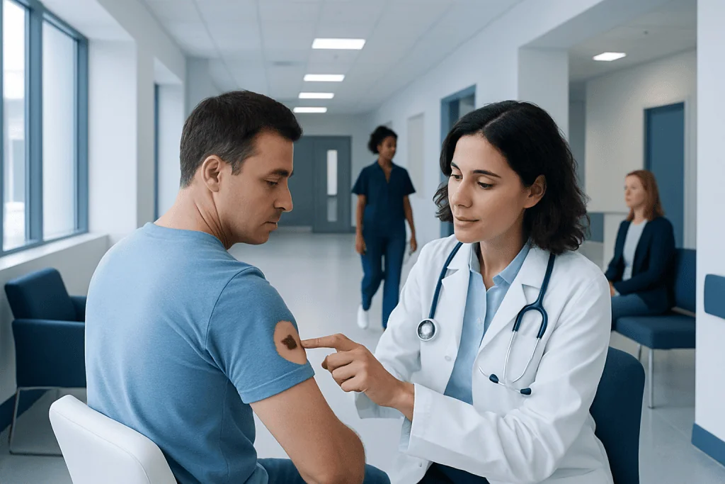
Skin cancer pictures are educational tools, not a substitute for medical diagnosis. Basal cell carcinoma, squamous cell carcinoma, and melanoma can look different from one person to another.
Key Takeaways
- Skin cancer pictures are educational tools, not a substitute for medical diagnosis.
- Basal cell carcinoma, squamous cell carcinoma, and melanoma can look different from one person to another.
- Warning signs include a new lesion, a sore that does not heal, bleeding, itching, or change in size, shape, or color.
- Doctors diagnose skin cancer with a skin exam and often a biopsy.
- Early diagnosis usually allows more treatment options and a better outlook.
Skin cancer pictures can be useful for learning what suspicious spots may look like, but images alone cannot confirm a diagnosis. The safest approach is to compare changing skin findings with trusted examples, then have any concerning lesion examined by a qualified clinician.
Overview: What Skin Cancer Pictures Can and Cannot Show
Skin cancer pictures can help people recognize patterns that deserve attention, such as an unusual mole, a pearly bump, a scaly patch, or a sore that keeps returning. They are most useful as a starting point for awareness. A photograph may suggest that a lesion looks suspicious, but it cannot determine the exact type of skin cancer or rule out a harmless condition.
Appearance varies widely by skin tone, body location, age, sun exposure history, and the type of cancer involved. The same diagnosis may look different in different people. Some skin cancers are dark, but many are pink, red, skin-colored, or even nearly invisible unless viewed closely. This is one reason why relying on online images alone can be misleading.
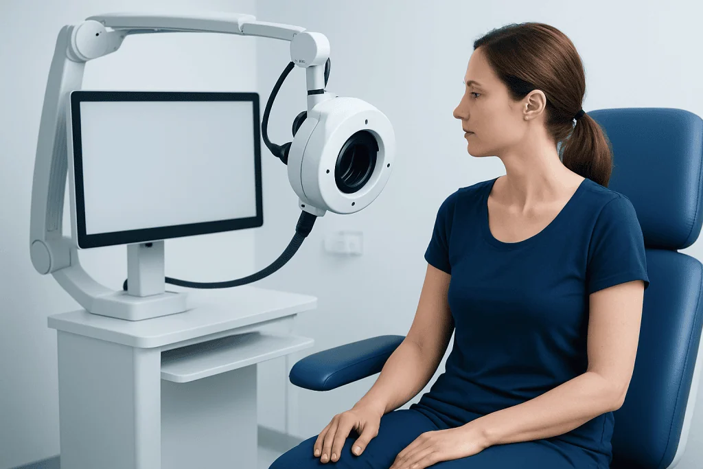
In practice, clinicians use more than appearance. They consider the lesion’s history, the pattern of change over time, the patient’s risk factors, and often a magnified examination called dermoscopy. If needed, a biopsy is performed to confirm the diagnosis. People concerned about a suspicious lesion may also wish to learn about skin cancer more broadly as they prepare for a clinical visit.
How Skin Cancer May Look in Pictures
There is no single “classic” image of skin cancer. Some lesions are obvious, while others are subtle. Looking at many examples can be more helpful than focusing on one picture, because this shows the range of possible appearances. It is also important to compare any image with changes seen on one’s own skin over time.
Common visual patterns include:
- Asymmetry: one half of a mole or spot does not match the other.
- Border irregularity: edges look blurred, notched, ragged, or uneven.
- Color variation: more than one shade may be present, such as tan, brown, black, red, white, or blue.
- Diameter or growth: a lesion becomes larger or looks different from nearby moles.
- Evolving change: any spot that changes in appearance, sensation, or behavior.
Photos may also show warning signs that are not limited to moles. A pink bump that looks shiny, a rough patch that crusts, or a lesion that bleeds with minimal friction may all warrant assessment. In people with darker skin tones, suspicious lesions may appear on the palms, soles, nails, or mucosal areas, not only on sun-exposed skin.
Because common benign conditions can resemble cancer in pictures, self-checks should be viewed as screening rather than diagnosis. Rashes, warts, seborrheic keratoses, cysts, and inflammatory conditions can all imitate more serious disease.
Main Types of Skin Cancer and Their Typical Appearance
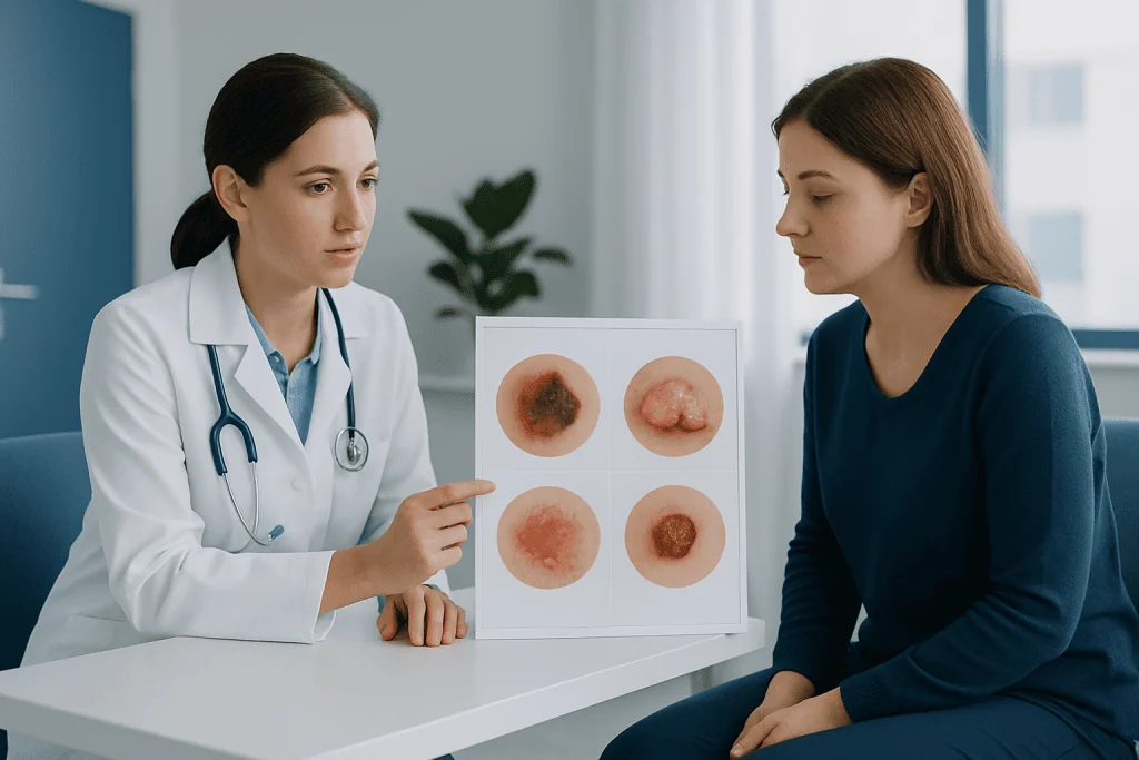
Basal cell carcinoma is the most common type. In pictures, it may appear as a shiny or pearly bump, a pink patch, a sore that heals and returns, or a scar-like flat area. Tiny visible blood vessels may run across the surface. Although it often grows slowly, it should still be treated because it can damage nearby tissue over time.
Squamous cell carcinoma may look like a rough, scaly patch, a firm red bump, a crusted growth, or a sore that does not heal. It often develops on sun-exposed areas such as the face, ears, scalp, neck, forearms, and hands, but it can occur elsewhere too. Some lesions are tender, bleed, or become thickened.
Melanoma is less common but more likely to spread if not detected early. In pictures, melanoma may appear as a new dark spot, a changing mole, or a lesion with multiple colors and irregular borders. Not all melanomas are dark brown or black. Some are pink or skin-colored, which can delay recognition. People looking for more focused information may also read about melanoma.
There are also less common forms of skin cancer. These include Merkel cell carcinoma and rare tumors of sweat glands or other skin structures. Their appearance can overlap with benign lesions, which is another reason medical assessment is important when a spot seems unusual or persistently changes.
Risk Factors and Why Some Lesions Are Easier to Miss
Several factors increase skin cancer risk. The most important is ultraviolet exposure from sunlight or tanning beds. Repeated sunburns, especially earlier in life, can add to risk over time. Fair skin, light eyes, light hair, a tendency to burn easily, and a high number of moles are also well-known risk factors, but skin cancer can occur in any skin tone.
Family history, a personal history of skin cancer, immunosuppression, certain inherited conditions, and increasing age may also raise risk. For some people, workplace or environmental exposures play a role. However, a person does not need to have every risk factor to develop a suspicious lesion, so new skin changes still deserve attention even in those who consider themselves low risk.
Some cancers are missed because they do not match common expectations seen in pictures online. For example, melanoma may develop under a nail, on the sole of the foot, or in an area not regularly examined. Basal or squamous cell cancers may be mistaken for eczema, a pimple, a scratch, or a nonhealing insect bite. Hair-bearing areas, the back, the scalp, and the ears can also be difficult to check without help.
How Doctors Diagnose Skin Cancer
Diagnosis starts with a clinical history and full skin examination. The clinician asks when the lesion first appeared, whether it has changed, and whether there has been bleeding, pain, itching, or repeated crusting. A dermatoscope may be used to magnify details that are not visible to the naked eye. This can improve the assessment of pigmented and nonpigmented lesions.
If a lesion looks suspicious, the next step is often a biopsy. This means removing all or part of the spot so it can be examined under a microscope. The biopsy is the only way to confirm the diagnosis and identify the specific cancer type. If cancer is found, the pathology report helps guide the next stage of care.
For some cancers, especially melanoma or larger aggressive tumors, doctors may recommend additional tests to understand depth, spread, or lymph node involvement. Imaging is not required for every patient, but it can be appropriate in selected cases. People undergoing evaluation may receive a skin biopsy as part of the diagnostic process.
It can be helpful for patients to bring photos showing how the lesion looked weeks or months earlier. A timeline of change, along with information about personal and family history, can make the clinical assessment more accurate and efficient.
Modern Treatment Approaches and Outlook
Treatment depends on the type of skin cancer, its size and depth, where it is located, and whether it has spread. Many early skin cancers are treated successfully with a minor procedure. Common approaches include surgical removal, curettage and cautery in selected cases, cryotherapy for some precancerous or superficial lesions, and topical therapies in carefully chosen situations.
When tissue preservation is important, such as on the face, ears, or around the eyes, a doctor may recommend Mohs surgery. This technique removes the cancer in stages while checking the edges under the microscope, helping save as much healthy tissue as possible. Standard excision remains highly effective for many lesions as well.
More advanced disease may require a combination of treatments. Depending on the cancer type, options can include radiation therapy, immunotherapy, targeted therapy, or wider surgery. Melanoma treatment can differ from the management of basal or squamous cell carcinoma, especially if there is concern about spread.
The outlook is often very good when skin cancer is found early. Small basal and squamous cell cancers are frequently cured with local treatment. Melanoma outcomes also improve significantly with prompt diagnosis and treatment. Near the end of the care pathway, some patients seek multidisciplinary support; Acibadem International’s specialists in JCI-accredited hospitals diagnose and treat skin cancers for international patients when coordinated care is needed.
Self-Checks, Prevention, and Safer Use of Skin Cancer Pictures
Skin cancer pictures are most useful when combined with regular self-checks. A practical method is to examine the skin once a month in good lighting, using a mirror for hard-to-see areas. People can look for new spots, changes in existing moles, nonhealing sores, or lesions that bleed or itch. Taking well-lit photos may help track change over time, but any concerning lesion should still be assessed by a clinician.
Sun protection reduces risk. Helpful measures include using broad-spectrum sunscreen, seeking shade during strong midday sun, wearing protective clothing and a hat, and avoiding tanning beds. These habits matter for all skin tones. Prevention does not eliminate all risk, but it can lower cumulative UV damage.
People with many moles, prior skin cancer, a strong family history, or immunosuppression may benefit from regular professional skin checks. Some patients with suspicious pigmented lesions may be evaluated by specialists with experience in dermatology and skin oncology. Consistent follow-up is especially important after treatment, because new skin cancers can develop later.
When to Seek Medical Care
Medical attention is advisable if a skin lesion is new, changing, bleeding, crusting, painful, itchy, or not healing after several weeks. A spot that looks different from a person’s other moles, sometimes called the “ugly duckling,” also deserves evaluation. Prompt review is especially important for lesions on the face, scalp, ears, lips, nails, palms, soles, or genital area.
Urgent assessment may be needed if a lesion is rapidly growing, repeatedly bleeding, becoming ulcerated, or associated with a swollen nearby lymph node. People with a history of skin cancer or weakened immunity should have a lower threshold for seeking care. Even when a lesion turns out to be benign, professional reassurance can be valuable and can prevent delays if treatment is needed.
Frequently asked questions
Can skin cancer pictures diagnose a suspicious mole or spot?
No. Skin cancer pictures can help a person recognize warning signs, but they cannot confirm or exclude cancer. Diagnosis requires a clinician’s examination and often a biopsy.
What does melanoma usually look like in pictures?
Melanoma often appears as a changing mole or a new spot with asymmetry, irregular borders, multiple colors, or growth over time. However, some melanomas are pink or skin-colored, so they do not always look dark or classic.
Are all skin cancers dark brown or black?
No. Many skin cancers are pink, red, pearly, scaly, or skin-colored rather than dark. This is one reason they can be mistaken for harmless conditions such as eczema, a pimple, or a sore.
What is the difference between basal cell, squamous cell, and melanoma?
Basal cell carcinoma often grows slowly and may look like a pearly bump or a sore that comes back. Squamous cell carcinoma is more likely to look rough, crusted, or thickened. Melanoma commonly develops from pigment-producing cells and is more likely to spread if not treated early.
When should someone see a doctor about a skin lesion?
A doctor should assess any lesion that is new, changing, bleeding, crusting, painful, itchy, or not healing. A spot that looks unlike a person’s other moles or changes quickly also needs prompt evaluation.
How is skin cancer confirmed?
The diagnosis is confirmed with a biopsy, which allows the tissue to be examined under a microscope. The pathology result identifies the cancer type and helps guide the most appropriate treatment.
References
- American Academy of Dermatology
- National Cancer Institute
- American Cancer Society
- Skin Cancer Foundation
- World Health Organization
This article is for general information only and is not a substitute for professional medical advice. Please consult a qualified doctor about your individual situation.
Skin Cancer in Turkey — costs, top hospitals & a free quote
JCI-accredited · board-certified surgeons · reply within 24h
Add us as a Preferred Source to see more of our trusted health content across Google Search, AI Overviews and Discover.