Pupil Size Chart: An Evidence-Based Guide for Patients

A pupil size chart is a guide, not a diagnosis on its own. Normal pupils usually measure about 2 to 8 mm, depending on lighting and other factors.
Key Takeaways
- A pupil size chart is a guide, not a diagnosis on its own.
- Normal pupils usually measure about 2 to 8 mm, depending on lighting and other factors.
- Pupils should generally be round and react equally to light, although small natural differences can occur.
- Sudden unequal pupils, eye pain, vision loss, or a severe headache need prompt medical assessment.
- Eye doctors assess pupil size together with vision, eye movements, and overall neurological findings.
A pupil size chart helps patients understand what eye doctors mean when they describe pupils in millimeters. In most healthy adults, pupil size usually falls between about 2 and 8 mm and changes naturally with light, focus, emotions, age, and some medicines.
What a pupil size chart shows
A pupil size chart is a simple reference that shows the diameter of the dark center of the eye in millimeters. It helps explain whether a pupil appears small, average, or large under certain conditions, especially in bright light versus dim light. For most adults, normal pupil size is usually somewhere between about 2 mm and 8 mm.
The most important point is that pupil size is not fixed. Pupils constantly adjust to control how much light enters the eye, and they can also change with age, emotional state, focusing on near objects, and the use of certain medicines. Because of this, a pupil measurement only makes sense when it is interpreted in context.
A chart can be useful for understanding a doctor’s notes, but it cannot confirm whether something is healthy or abnormal by itself. Eye specialists look not only at the number in millimeters, but also at whether both pupils are equal, whether they are round, and how they respond to light and near focus.
Normal pupil ranges by lighting conditions
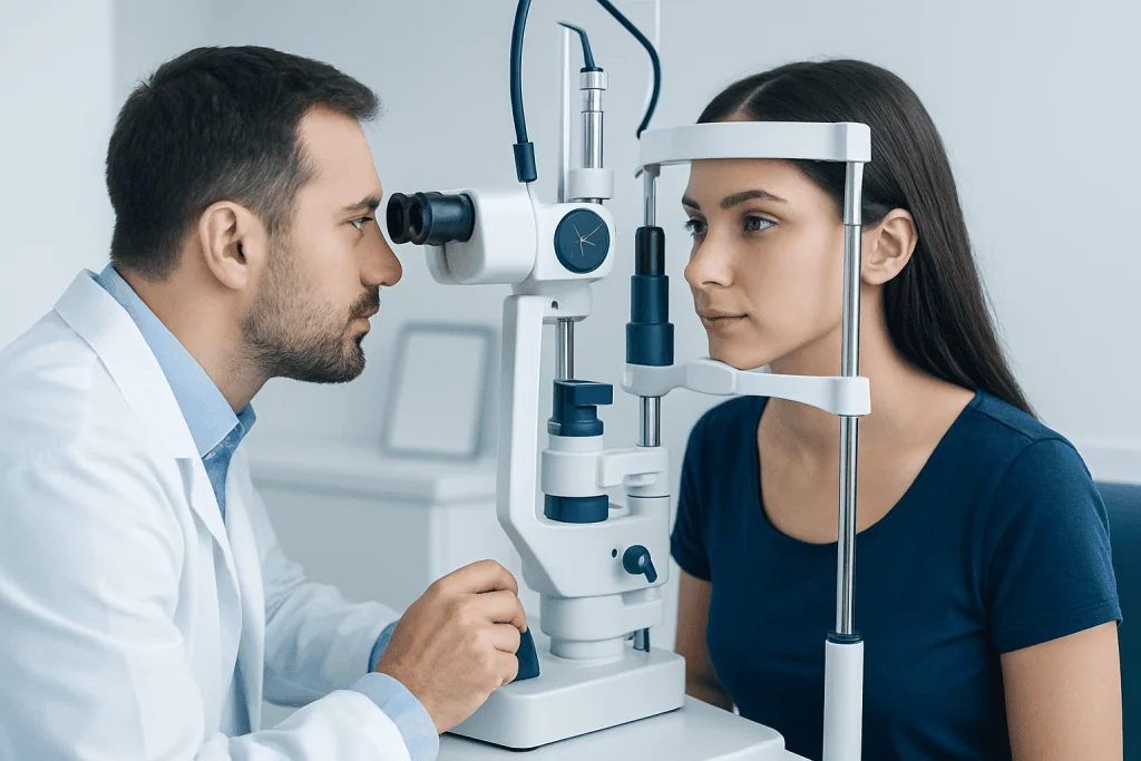
In bright light, pupils normally become smaller to protect the retina and improve visual clarity. In dim light, they enlarge to allow more light into the eye. This is why the same person may have noticeably different pupil sizes at different times of day or in different rooms.
A practical patient-friendly pupil size chart looks like this:
- Bright light: often around 2 to 4 mm
- Indoor or moderate light: often around 3 to 5 mm
- Dim light or darkness: often around 4 to 8 mm
These are general ranges rather than strict cutoffs. Children and younger adults may have larger pupils than older adults, while some healthy people naturally have pupils at the smaller or larger end of the usual range. Doctors also compare the two eyes rather than looking at a single measurement alone.
A small difference between pupils can be normal. Mild physiologic anisocoria, meaning naturally unequal pupils, is fairly common and often harmless when the difference is small and stable over time, and when vision and eye movements are normal.
Why pupil size changes

The pupil changes size because muscles in the iris respond to signals from the nervous system. Bright light causes constriction, while darkness causes dilation. This automatic adjustment is called the pupillary light reflex and is a normal part of healthy eye and brain function.
Pupil size can also change for non-dangerous reasons. Looking at something up close, strong emotions, fatigue, and some everyday medications can all affect the pupils. Eye drops used during an eye exam are another common reason for temporary dilation.
Age matters as well. As people get older, pupils often become smaller and may react a bit more slowly. This does not always mean disease. However, when a change is sudden, one-sided, or accompanied by symptoms such as double vision, drooping eyelid, pain, or blurred vision, a medical assessment is important.
Doctors may also consider whether a pupil problem is related to an eye condition, a nerve problem, trauma, or medication exposure. If other signs suggest disease, they may evaluate for conditions that affect the optic nerve or surrounding structures, including glaucoma and other eye disorders.
When a pupil size chart may suggest a problem
A chart becomes clinically useful when pupil size is interpreted alongside symptoms and exam findings. Very small pupils may occur with certain medications, inflammation, or neurological causes. Very large pupils may be seen after dilating drops, with some drug effects, after eye injury, or in certain nerve disorders.
Unequal pupils, called anisocoria, are not always dangerous, but the pattern matters. A larger difference in bright light may suggest that the bigger pupil is not constricting properly. A larger difference in darkness may suggest that the smaller pupil is not dilating properly. Eye doctors use this clue to decide what to examine next.
Doctors also look at shape and reactivity. A healthy pupil is usually round and should react to light. An irregularly shaped pupil, a fixed pupil that does not respond, or a pupil change after trauma may need urgent attention. Problems affecting the retina, optic nerve, or brain pathways can also alter the pupil response.
Sometimes pupil changes occur together with eye pain, halos around lights, nausea, and blurred vision, which can raise concern for urgent eye pressure problems such as glaucoma. In other cases, a person may have decreased vision from disorders such as cataract, although cataracts typically affect lens clarity rather than pupil size itself.
How doctors measure and assess pupils
During an exam, a clinician measures pupil size in millimeters under specific lighting conditions. They may use a handheld pupil gauge, a penlight, or digital devices. The measurement is usually recorded for each eye separately in both light and dark settings.
The doctor also tests how each pupil responds to direct light and whether both pupils react together. This helps assess the pathways between the eye and the brain. They may then check the near response by asking the patient to shift focus from a distant object to a closer one.
A full eye examination often includes vision testing, eye movement assessment, and inspection of the front and back of the eye. If needed, the doctor may recommend a more detailed eye exam or further imaging and neurological evaluation, depending on the pattern of findings.
When pupil findings are linked to retinal or optic nerve concerns, specialized tests may be useful. In some cases, patients may need retina treatment or other care directed at the underlying cause rather than the pupil itself.
Common causes of abnormal pupil size
Abnormal pupil size can result from many different causes, ranging from harmless to urgent. Common benign causes include prescribed eye drops, some cold or motion-sickness medications, mild natural anisocoria, and temporary effects after an ophthalmology visit. Contact with certain substances or accidental exposure to medicated patches or sprays may also change the pupils.
Eye-related causes include inflammation inside the eye, trauma, prior surgery, and pressure-related emergencies. Neurological causes may include problems involving the nerves that control the iris, migraine-related changes, or, more rarely, serious disorders affecting the brain or blood vessels. Because the same sign can have several explanations, self-diagnosis is not reliable.
A clinician may ask about recent injuries, new medications, headaches, facial sweating changes, double vision, eyelid drooping, and light sensitivity. This history helps narrow the cause. If an abnormal pupil is painful or associated with sudden visual change, fast assessment is especially important.
Treatment depends on the reason for the finding. Some cases require only observation, while others need medication, urgent pressure-lowering care, neurological assessment, or surgery. If a structural eye condition is present, care may involve services such as glaucoma treatment or cataract surgery when clinically appropriate.
What patients can do at home
Patients can use a pupil size chart for general understanding, but it should not replace a professional exam. Home checks are less reliable because phone flash, room brightness, camera settings, and the angle of viewing can all make pupils appear different. A single photo can be misleading.
It can still help to observe patterns. For example, a patient may note whether one pupil always seems slightly larger, whether the difference is new, and whether symptoms such as headache, eye pain, drooping eyelid, nausea, or double vision are present. Writing down recent medication changes or accidental chemical exposures is also useful.
General eye health habits may reduce avoidable problems. These include using protective eyewear during sports or hazardous work, attending routine eye examinations, following medication instructions carefully, and not using another person’s eye drops. Patients with diabetes, high blood pressure, or known eye disease should keep regular follow-up appointments.
Near the end of the care pathway, some patients may benefit from multidisciplinary assessment, especially when symptoms overlap between eye health and neurology. Acibadem International’s multidisciplinary specialists and JCI-accredited hospitals diagnose and treat eye and neurological conditions for international patients when further evaluation is needed.
When to seek medical care
Prompt medical care is recommended if a pupil change is sudden, follows an eye or head injury, or is accompanied by severe headache, eye pain, vomiting, blurred vision, double vision, fainting, confusion, weakness, or a drooping eyelid. These features can point to an urgent eye or neurological problem.
A same-day eye assessment is also sensible if one pupil becomes fixed, very large, or very small and does not react normally to light. New unequal pupils in a person with vision changes should not be ignored, even if there is no pain. Children with unusual pupil findings should be assessed by a qualified clinician.
Less urgent but still important reasons to book an appointment include persistent light sensitivity, recurrent episodes of uneven pupils, or uncertainty about whether a medication is affecting the eyes. If symptoms are worsening or do not improve, professional advice is the safest next step.
Frequently asked questions
What is a normal pupil size on a pupil size chart?
In many healthy adults, pupils usually range from about 2 mm to 8 mm depending on lighting. They are typically smaller in bright light and larger in dim light. Doctors interpret this range together with the pupil’s shape and reaction to light.
Is it normal for one pupil to be slightly bigger than the other?
A small stable difference can be normal and is often called physiologic anisocoria. It is more reassuring when there are no other symptoms and the difference has not suddenly changed. A new or clearly increasing difference should be checked by a clinician.
Can medications change pupil size?
Yes. Prescription eye drops, some cold medicines, motion-sickness medicines, and other drugs can make pupils larger or smaller. Patients should tell their doctor about all medicines and recent eye drops if they notice a change.
Does a large pupil always mean a serious problem?
No. A large pupil can occur normally in dim light or after dilating eye drops. However, if one pupil is suddenly much larger and there is eye pain, drooping eyelid, double vision, or headache, urgent medical assessment is important.
Can a phone photo accurately measure pupil size?
Not reliably. Camera flash, screen brightness, filters, and room lighting can change pupil appearance. A clinical measurement in controlled lighting is much more accurate.
What kind of doctor checks abnormal pupil size?
An ophthalmologist or optometrist often performs the first evaluation, especially if the concern seems eye-related. If there are neurological symptoms, a neurologist or emergency physician may also be involved. The exact specialist depends on the overall pattern of symptoms.
References
- American Academy of Ophthalmology
- National Eye Institute
- Merck Manual Professional Edition
- MedlinePlus
- Royal College of Ophthalmologists
This article is for general information only and is not a substitute for professional medical advice. Please consult a qualified doctor about your individual situation.
Explore treatments in Turkey — costs, top hospitals & a free quote
JCI-accredited · board-certified surgeons · reply within 24h
Add us as a Preferred Source to see more of our trusted health content across Google Search, AI Overviews and Discover.

More from the Health Library

Manic: An Evidence-Based Guide for Patients

Septum — Explained by Medical Evidence, Not Myths

Babymoon: An Evidence-Based Guide for Patients
Lithotripsy procedure: Symptoms, Causes, and Treatment — Complete Patient Guide

Neuropsychological Testing — Explained by Medical Evidence, Not Myths