

Anisocoria Treatment: How It Works, Results and What to Expect

Anisocoria means the pupils are unequal in size, and it may be harmless or a sign of an underlying eye, nerve or brain condition. Treatment targets the cause, such as inflammation, medication effects, injury, glaucoma or a nerve disorder.
Key Takeaways
- Anisocoria means the pupils are unequal in size, and it may be harmless or a sign of an underlying eye, nerve or brain condition.
- Treatment targets the cause, such as inflammation, medication effects, injury, glaucoma or a nerve disorder.
- A sudden pupil change with severe headache, eyelid drooping, double vision, weakness or confusion requires emergency medical care.
- Some forms resolve when the trigger is removed or the underlying condition improves; others can remain stable without affecting vision.
- An eye examination and, when needed, neurological tests or imaging help determine the safest next steps.
Anisocoria treatment is guided by the reason the pupils are different sizes, not by pupil size alone. Many people have a harmless, stable difference that needs monitoring only, while new or changing anisocoria may require prompt eye and neurological assessment.
Overview: how anisocoria treatment works
Anisocoria treatment works by identifying and managing the cause of unequal pupil size. A small pupil-size difference that has been present for years, remains the same in light and darkness, and causes no other symptoms is often physiologic anisocoria. This common variation usually does not require treatment.
When anisocoria is new, becomes more noticeable, follows an eye or head injury, or occurs with visual or neurological symptoms, clinicians first assess whether an urgent condition may be present. Treatment may involve stopping or changing an offending medicine under medical guidance, treating an eye disorder, managing inflammation or infection, or addressing a neurological cause. The outlook depends more on the underlying diagnosis than on the amount of pupil difference.
Because pupil control involves the eye, nerves and brain, assessment may involve ophthalmology, neuro-ophthalmology, neurology, emergency medicine or other specialists. The goal is to protect vision, relieve associated symptoms and avoid missing conditions that need timely care.
What is the most common cause of anisocoria?
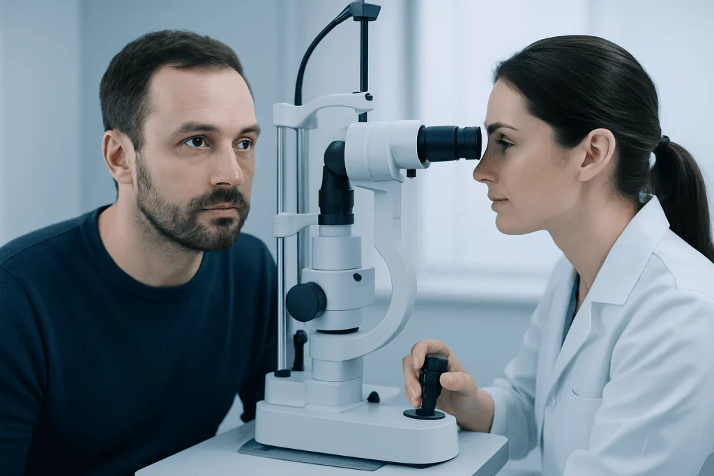
The most common cause of anisocoria overall is physiologic anisocoria: a normal, harmless difference in pupil size. In this situation, the difference is usually small, stable over time and present in otherwise healthy people without eye pain, drooping of the eyelid, double vision or vision loss.
Other frequent causes include exposure to medicines or chemicals that affect pupil muscles, including certain eye drops, inhaled medicines, skin patches and plant substances. A pupil may also change size after eye surgery, direct eye trauma, inflammation inside the eye, or conditions that interfere with the nerves controlling the iris.
Clinicians also consider Horner syndrome, third cranial nerve palsy, acute angle-closure glaucoma and, less commonly, brain or blood-vessel conditions. These possibilities cannot be distinguished reliably by appearance alone, which is why a new or unexplained pupil difference should be assessed by a qualified clinician.
Does anisocoria ever go away?

Anisocoria can go away when its cause is temporary or treatable. For example, unequal pupils related to accidental medicine exposure may improve after the substance clears, while pupil changes from eye inflammation may lessen as the inflammation is treated. The timing varies according to the trigger and should be discussed with the treating clinician.
Physiologic anisocoria commonly persists throughout life but is not harmful and does not need correction. Pupil differences caused by previous trauma, surgery or some nerve conditions may also remain, even after the underlying problem has been addressed.
It is important not to try to make pupils look equal with non-prescribed eye drops. These products can worsen certain eye conditions, obscure important examination findings or cause side effects. A clinician can explain whether observation, treatment or follow-up is appropriate.
How do you fix anisocoria?
There is no single procedure that fixes all anisocoria. The appropriate approach begins with a detailed history and examination, then treats the identified cause. If the difference is physiologic and stable, reassurance and observation may be all that is needed. If a medication or chemical exposure is responsible, avoiding the trigger and reviewing medicines with a clinician may allow the pupil to return toward its usual size.
For eye-related causes, treatment may include prescription drops or other therapies for inflammation, pressure problems or infection. Eye injury may require urgent ophthalmic care. When a nerve or neurological condition is suspected, management focuses on the specific diagnosis and may involve neurological evaluation, imaging and treatment by the relevant specialty team.
In selected cases where an iris injury causes a persistent pupil abnormality and troublesome glare, an ophthalmologist may discuss reconstructive options or tinted lenses. These are not routine treatments for anisocoria and are considered only after the eye is stable and the cause has been fully evaluated.
- Observation: appropriate for confirmed physiologic anisocoria without concerning symptoms.
- Cause-specific medical care: may address inflammation, pressure elevation, infection or medication effects.
- Specialist treatment: is needed when findings suggest a nerve, brain or blood-vessel cause.
- Vision-support measures: such as light-sensitive lenses, may help selected people with glare after iris injury.
Assessment, candidacy and step-by-step care
Anyone with new anisocoria, a worsening difference, eye symptoms or neurological symptoms is a candidate for medical assessment. The evaluation is particularly important after head, face or eye trauma, or when the pupil difference has no clear explanation. People with a known stable difference may still mention it during routine eye examinations.
The first step is a history. A clinician may ask when the difference began, whether it changes in bright or dim light, whether there has been injury, whether there are headaches or pain, and which prescription medicines, inhalers, patches, eye drops or supplements are used. Old photographs can sometimes help show whether unequal pupils were present previously.
The examination usually includes vision testing, pupil measurements in light and darkness, eyelid movement, eye movements and a neurological assessment. An eye specialist may examine the front and back of the eye and measure eye pressure. Depending on the findings, tests may include carefully selected pupil drops, blood tests or imaging such as CT or MRI.
Once the cause is clear, the clinician explains the treatment plan and follow-up schedule. Emergency treatment pathways are used when examination findings suggest a time-sensitive eye, nerve or brain condition. For international patients, Acibadem International’s multidisciplinary specialists and JCI-accredited hospitals can assess and treat conditions involving the eyes and nervous system.
Do people with anisocoria see normally?
Many people with anisocoria see normally, especially when the difference is physiologic and the eyes are otherwise healthy. Pupil size helps regulate the amount of light entering the eye, but a small stable difference alone does not necessarily reduce visual acuity or require treatment.
Vision may be affected when anisocoria is caused by an eye disorder or when the pupils no longer respond normally to light. Some people experience glare, light sensitivity, blurred vision, difficulty focusing or reduced comfort in bright settings. Double vision, new loss of vision or severe eye pain is not expected with simple physiologic anisocoria and needs urgent evaluation.
Any visual symptoms should be described clearly during assessment, including whether they began suddenly and whether they affect one or both eyes. Treating the underlying condition may improve visual comfort, but the expected result depends on the diagnosis and any pre-existing eye damage.
Benefits, risks and recovery expectations
The main benefit of evaluation and cause-directed treatment is that it clarifies whether anisocoria is a harmless variation or a sign of a condition requiring care. When treatment is needed, it aims to preserve vision, manage symptoms and address the underlying eye, nerve or systemic problem. Many people with benign anisocoria need no intervention and can continue normal daily activities.
Recovery timelines vary widely. Changes related to temporary medicine exposure may improve over hours to days, depending on the substance. Recovery from eye inflammation, injury or surgery may take longer and requires follow-up. Neurological causes have different timelines based on the diagnosis and response to treatment.
Risks depend on the treatment itself. Prescription drops can cause temporary stinging, blurred vision or light sensitivity, and some treatments are not suitable for every person. Procedures or surgery, when indicated for an underlying eye condition, carry their own risks and should be discussed with an ophthalmologist. Follow-up is important because a changing pupil pattern can provide useful information about recovery or a need to adjust care.
When to seek medical care
Seek emergency medical care immediately for a sudden new pupil-size difference accompanied by a severe or unusual headache, eye pain, red eye, reduced vision, double vision, a drooping eyelid, weakness, numbness, difficulty speaking, confusion, fainting or neck pain. These symptoms may indicate an urgent eye or neurological condition and should not be monitored at home.
Prompt, non-emergency medical assessment is also advisable for new anisocoria without other symptoms, a pupil difference after injury, persistent light sensitivity, or a change noticed after starting a medicine or using an eye product. Bringing a list of medicines and recent photographs, if available, can help the clinician assess the change.
People with confirmed physiologic anisocoria should seek review if the pattern changes or new symptoms develop. Regular eye examinations remain useful for general eye health, particularly for people with eye disease, diabetes, a history of eye injury or prior eye surgery.
Frequently asked questions
Is anisocoria always serious?
No. A small, longstanding and stable difference in pupil size is often physiologic anisocoria, a harmless normal variation. However, a new or changing pupil difference should be assessed, particularly if it occurs with pain, headache, eyelid drooping, double vision or vision changes.
Can stress cause anisocoria?
Stress can influence normal pupil size through the body’s autonomic nervous system, but it should not be assumed to explain a new persistent pupil difference. A clinician should assess unexplained anisocoria to rule out eye, medication-related or neurological causes.
Can eye drops cause one pupil to become larger?
Yes. Certain prescription or non-prescription eye products can alter pupil size, especially if they enter only one eye. Some inhaled medicines, skin patches and plant exposures can also affect a pupil, so it is important to tell the clinician about all recent exposures.
How long does medication-related anisocoria last?
The duration depends on the medicine, amount of exposure and the individual response. It may improve as the substance wears off, but a person should not wait for it to resolve if there are concerning symptoms or if the cause is uncertain.
Will anisocoria affect driving?
A small, stable pupil difference without vision symptoms generally does not prevent driving. If anisocoria causes blurred vision, glare, double vision, light sensitivity or an underlying condition that affects vision or alertness, driving should be avoided until a clinician advises it is safe.
Which doctor treats anisocoria?
An ophthalmologist or neuro-ophthalmologist commonly evaluates anisocoria, often alongside a neurologist when nerve or brain involvement is possible. Emergency clinicians may assess sudden anisocoria with concerning symptoms and arrange urgent specialist care.
References
- American Academy of Ophthalmology
- National Eye Institute
- Merck Manual Professional Edition
- Mayo Clinic
- Cleveland Clinic
This article is for general information only and is not a substitute for professional medical advice. Please consult a qualified doctor about your individual situation.
Explore treatments in Turkey — costs, top hospitals & a free quote
JCI-accredited · board-certified surgeons · reply within 24h
Add us as a Preferred Source to see more of our trusted health content across Google Search, AI Overviews and Discover.

More from the Health Library


Related Specialists

Ody. Nazlı Can Üstün
Audiology
Assoc. Prof. Dr. Deniz Atasoy
General Surgery
Prof. Dr. Evrim Kadriye Tezcanlı
Radiation Oncology
Dr. Tural Khalilov
Orthopedic Surgery & Traumatology