

Quick answer
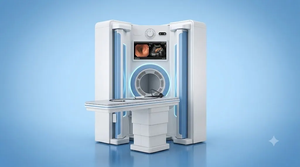
Endoscopic ultrasound (EUS), or endoultrasonography, combines a flexible endoscope with ultrasound imaging to examine the digestive tract and nearby organs in detail. At Acibadem in Turkey, EUS is used to assess areas including the oesophagus, stomach, pancreas, bile ducts and lymph nodes, and can guide tissue sampling when needed.
EUS (Endoultrasonography): Detailed Imaging of the Digestive System
Endoscopic ultrasound, also known as EUS or endoultrasonography, is an advanced imaging procedure that combines endoscopy with ultrasound. It allows Gastroenterology specialists to view the wall of the digestive tract and nearby structures in exceptional detail.
Unlike an ultrasound performed through the skin, EUS places a small ultrasound probe at the tip of a flexible endoscope. By bringing the ultrasound source close to the area being examined, specialists can obtain high-resolution images of organs and tissues that may be difficult to assess with standard imaging alone.
EUS can help evaluate the oesophagus, stomach, duodenum, pancreas, bile ducts, gallbladder, liver and nearby lymph nodes. When clinically appropriate, it can also guide the collection of tissue or fluid samples for laboratory analysis.
At Acibadem International, EUS supports precise diagnosis, careful treatment planning and coordinated care for patients travelling from abroad. Our teams use advanced technology alongside specialist experience, clear communication and an individual approach to every case.
What Is EUS?
EUS is a diagnostic procedure that combines two familiar medical techniques:
- Endoscopy, which uses a thin, flexible tube with a camera to view the inside of the upper digestive tract.
- Ultrasound, which uses sound waves to produce images of organs, soft tissues and blood vessels without using ionising radiation.
During EUS, the endoscope is usually passed through the mouth and gently advanced into the oesophagus, stomach and first part of the small intestine. The ultrasound transducer at its tip produces detailed images from inside the digestive tract.
This close-up perspective can help specialists identify changes in tissue layers, measure lesions accurately and assess the relationship between a finding and nearby organs or lymph nodes. It may be particularly valuable when doctors need more information after symptoms, blood tests, CT, MRI, abdominal ultrasound or another endoscopic examination.
EUS is not a replacement for every imaging test. Instead, it is used as part of a personalised diagnostic pathway. Your specialist will consider your symptoms, medical history, previous reports and the clinical question that needs to be answered.
How EUS Works
The EUS endoscope contains a miniature ultrasound device. This device sends sound waves into nearby tissues and receives returning signals. A computer converts these signals into real-time images that the specialist can review during the procedure.
Because the ultrasound probe is positioned inside the digestive tract, it can be very close to organs such as the pancreas and bile ducts. This can provide detailed images of structures that sit behind the stomach or duodenum and may be less clearly seen with an external ultrasound examination.
Depending on the purpose of the procedure, the specialist may use EUS to:
- Examine the layers of the oesophagus, stomach or intestinal wall.
- Assess a mass, cyst, narrowing or other abnormality.
- Evaluate the pancreas, pancreatic duct, bile ducts, gallbladder or surrounding tissues.
- Review lymph nodes near the digestive tract.
- Help determine the local extent of a known condition.
- Guide a fine-needle tissue or fluid sample when this is medically indicated.
If sampling is planned, the specialist may pass a very thin needle through the endoscope under ultrasound guidance. The ultrasound image helps the team target the selected area with precision. Samples are then assessed by pathology specialists, and results are discussed with you as part of your wider care plan.
Clinical Applications
EUS is commonly used in Gastroenterology and may contribute to the diagnosis, staging and management of a range of digestive and hepatobiliary conditions. Whether EUS is suitable depends on your individual symptoms, imaging findings and medical assessment.
| Clinical application | How EUS may help | Areas commonly assessed |
|---|---|---|
| Pancreatic conditions | Provides detailed assessment of pancreatic tissue, cystic lesions, masses and nearby ducts. It may also support targeted tissue or fluid sampling when appropriate. | Pancreas, pancreatic duct, nearby blood vessels and lymph nodes |
| Bile duct and gallbladder concerns | May help investigate suspected stones, duct narrowing, blockages or abnormalities identified on other scans. | Bile ducts, gallbladder, liver-adjacent structures and pancreas |
| Upper digestive tract lesions | Helps assess changes in the wall of the oesophagus, stomach or duodenum, including the depth and characteristics of selected lesions. | Oesophagus, stomach and duodenum |
| Lymph node assessment | Allows close evaluation of lymph nodes near the digestive tract and may guide sampling if clinically required. | Mediastinal, abdominal and peri-pancreatic lymph nodes |
| Digestive system cancer staging | Can contribute to staging by assessing local tissue involvement and nearby lymph nodes, supporting multidisciplinary treatment planning. | Oesophagus, stomach, pancreas, bile ducts and surrounding tissues |
| Unexplained symptoms or abnormal findings | May provide additional information when symptoms, laboratory results or previous imaging require closer investigation. | Upper digestive tract and nearby abdominal organs |
EUS findings are interpreted alongside other tests. In some cases, your specialist may recommend additional imaging, endoscopy, laboratory testing, pathology review or consultation with another specialty. This integrated approach supports accurate decisions rather than relying on a single test alone.
Conditions EUS May Help Evaluate
Endoscopic ultrasound may be considered for patients with a wide range of clinical concerns. These can include persistent upper abdominal pain, unexplained weight loss, jaundice, abnormal liver tests, pancreatic cysts, suspected bile duct stones, swallowing difficulties or findings identified during a previous scan or endoscopy.
It may also be used to investigate or monitor selected conditions affecting the upper digestive system, including:
- Pancreatic masses and pancreatic cysts.
- Chronic pancreatitis and selected pancreatic duct changes.
- Suspected bile duct stones or bile duct obstruction.
- Gallbladder and biliary system abnormalities.
- Submucosal lesions beneath the surface lining of the stomach or oesophagus.
- Oesophageal, stomach, pancreatic or biliary cancers, where local staging information is needed.
- Enlarged or abnormal-appearing lymph nodes.
- Selected fluid collections or cystic structures near the pancreas.
Not every symptom requires EUS, and not every EUS examination leads to tissue sampling. Your care team will recommend the most appropriate diagnostic approach based on your individual circumstances.
Benefits and Advantages for Patients
EUS can provide information that is difficult to obtain through standard endoscopy or imaging alone. Its value lies in the combination of direct endoscopic viewing and high-resolution ultrasound imaging from close to the target area.
For many patients, this means a clearer understanding of what is happening and a more focused next step. If a finding needs closer evaluation, EUS may help reduce uncertainty and support a personalised treatment plan.
| Patient benefit | What it can mean for your care |
|---|---|
| Detailed, close-range imaging | Specialists can assess the digestive tract wall and nearby organs with high-resolution ultrasound images. |
| No ionising radiation | Ultrasound uses sound waves rather than X-rays, which may be an important consideration in an individual diagnostic plan. |
| More precise assessment | Images can help identify the size, location and relationship of a finding to surrounding tissues. |
| Guided tissue sampling when needed | If clinically appropriate, EUS can help guide sampling from a selected lesion, cyst, lymph node or fluid collection. |
| Support for treatment planning | Results may assist Gastroenterology, Surgery, Oncology, Radiology and Pathology teams in coordinating next steps. |
| Usually performed without surgical incisions | EUS is performed with a flexible endoscope and does not usually require open surgery or skin incisions. |
Although EUS is often described as minimally invasive, it remains a medical procedure. The benefits, limitations and possible risks should always be discussed in relation to your own health condition.
What to Expect During an EUS Procedure
Your experience begins before you travel or arrive at the hospital. Acibadem International patient coordinators can help collect your available medical reports, imaging results and pathology information for preliminary review. Where possible, sharing these documents in advance helps the specialist understand your case and plan the next steps efficiently.
Before the procedure
A Gastroenterology specialist reviews your medical history, current symptoms, medicines, allergies and previous test results. You may need to stop eating and drinking for a period before the examination so that the stomach and upper digestive tract can be assessed safely and clearly.
It is important to tell your team about blood-thinning medicines, diabetes medications, heart or lung conditions, previous reactions to sedation or anaesthesia, pregnancy and any other relevant health concerns. Your doctor will provide individual instructions about medication adjustments and preparation.
For international patients, language support and coordination are available to help ensure that instructions are understood before the procedure. Your coordinator can also assist with appointment planning, document preparation and communication between you, your family and the clinical team.
During the procedure
EUS is usually performed in an endoscopy unit by an experienced Gastroenterology team. Sedation or anaesthesia may be used depending on the planned procedure, your clinical needs and the advice of the anaesthesia team.
Once you are comfortable, the specialist gently passes the flexible endoscope through the mouth into the upper digestive tract. The procedure does not usually affect breathing. The camera and ultrasound probe allow the specialist to examine the relevant structures in real time.
If tissue sampling is needed, the team may use ultrasound guidance to collect a sample. You may not be aware of the sampling itself because of sedation, but the clinical team will explain this possibility before the procedure and obtain the appropriate consent.
After the procedure
After EUS, you will rest in a recovery area while the effects of sedation wear off. You may experience temporary throat discomfort, bloating or mild abdominal discomfort. These symptoms are usually short-lived, but your team will advise you about what is expected and when to seek medical attention.
If sedation or anaesthesia has been used, you should not drive or make important decisions immediately afterward. International patients should arrange for a responsible adult or appropriate support person to accompany them, especially if they are returning to accommodation after same-day care.
In some cases, the specialist may share initial imaging findings shortly after the examination. If a sample has been taken, pathology analysis may require additional time. Your coordinator and doctor will explain when and how results will be discussed, including whether further appointments or treatment planning are recommended.
Safety, Quality and Accreditation
EUS is routinely performed by trained specialists in appropriately equipped endoscopy settings. Patient safety depends on careful preparation, appropriate sedation planning, infection prevention measures, experienced clinical teams and clear follow-up instructions.
As a JCI-accredited healthcare provider, Acibadem International follows structured quality and patient safety standards across the patient journey. This includes attention to identity verification, informed consent, medication review, safe sedation and anaesthesia practices, infection control and coordinated communication among clinical teams.
Like all endoscopic procedures, EUS has potential risks. These may vary depending on whether the examination is purely diagnostic or includes tissue sampling or another intervention. Possible risks can include reactions to sedation, bleeding, infection, perforation or inflammation, although serious complications are uncommon. Your specialist will explain the relevant risks, expected benefits and alternatives before the procedure.
Your care team will also tell you which symptoms require urgent medical advice after you leave the hospital. These may include severe or worsening abdominal pain, fever, difficulty breathing, vomiting blood, black stools, persistent vomiting or other unexpected symptoms.
Why EUS Matters for International Patients
Travelling for digestive health assessment can feel demanding, particularly when you are managing symptoms, uncertainty or a recent diagnosis. International patients often need more than access to advanced technology: they need clear information, timely coordination and confidence that their care will be connected from arrival through follow-up.
At Acibadem International, the patient journey is designed to support this process. Our international patient teams can help coordinate specialist appointments, pre-procedure assessments, translation support, medical documentation and communication regarding your treatment plan.
If you have already completed scans, endoscopy or laboratory tests in your home country, bringing these records can help avoid unnecessary repetition where clinically appropriate. Your specialist will review the quality and relevance of previous studies and recommend only the examinations needed to clarify your condition or guide care.
For patients who require multidisciplinary input, EUS findings can be shared with relevant specialists such as Gastroenterologists, Hepatobiliary Surgeons, Oncologists, Radiologists and Pathologists. This coordinated review can be especially important when planning treatment for pancreatic, biliary, oesophageal or stomach conditions.
Before you travel, your coordinator can help you understand the likely sequence of appointments and the expected recovery period after the procedure. If pathology review or additional consultations are needed, the team can support practical planning around your stay and follow-up arrangements.
Frequently Asked Questions
Is EUS safe?
EUS is widely used in modern Gastroenterology and is performed by trained specialists using established safety protocols. As with any medical procedure, there are possible risks, particularly if tissue sampling is performed or sedation is required. Your doctor will explain the considerations that apply to your individual case.
Is EUS painful?
Patients are commonly given sedation or anaesthesia support to help them remain comfortable. You may have mild throat irritation, bloating or temporary discomfort after the procedure. Your team will provide advice tailored to the sedation method and the type of EUS performed.
Will I need a biopsy during EUS?
Not always. Tissue or fluid sampling is only performed when the specialist believes it is clinically useful and appropriate. If sampling may be needed, this will be discussed with you before the procedure.
How long will I need to stay in Türkiye?
The appropriate length of stay depends on your diagnosis, whether sedation or sampling is planned, and whether you need further consultations or treatment. Your international patient coordinator can help organise a practical schedule after your specialist’s assessment.
Will my insurance cover EUS?
Coverage depends on your insurance policy and the medical reason for the procedure. You can share your insurance information with our team, who can help clarify the available process and explain financial arrangements before treatment.
How do I get started?
You can submit a consultation request and share your available reports, imaging studies and pathology results. A specialist can review your case and advise whether EUS may be suitable as part of your diagnostic or treatment plan.
Personalised Digestive Health Assessment
EUS can offer detailed insight into the digestive tract and nearby organs, helping specialists make more informed decisions at an early stage. Whether you need further investigation after an abnormal scan, evaluation of pancreatic or biliary symptoms, or support with treatment planning, your care pathway should be based on a thorough individual assessment.
Acibadem International combines advanced endoscopic imaging, experienced Gastroenterology teams, JCI-accredited quality standards and international patient coordination to support your care before, during and after EUS. Contact our international patient team to share your medical information and learn whether endoscopic ultrasound may be appropriate for you.
Related Medical Units
Available at these Hospitals


















Related Treatments
Speak with our medical team
Find out how this technology could support your diagnosis or treatment.