

Thyroid Ultrasound: Nodules, Goiter, and When Biopsy Is Needed
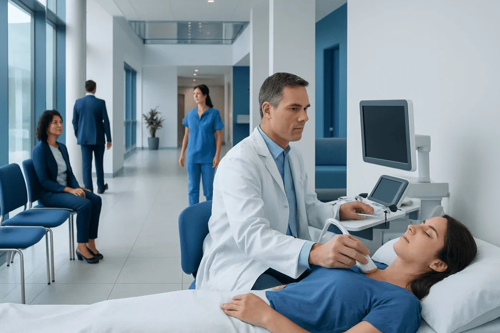
Thyroid ultrasound uses sound waves, not radiation, to create detailed images of the thyroid gland. Most thyroid nodules are benign, but ultrasound features help classify which nodules need closer evaluation.
Key Takeaways
- Thyroid ultrasound uses sound waves, not radiation, to create detailed images of the thyroid gland.
- Most thyroid nodules are benign, but ultrasound features help classify which nodules need closer evaluation.
- Goiter means an enlarged thyroid and may be diffuse or caused by one or more nodules.
- Fine needle aspiration biopsy is usually recommended based on a combination of nodule size and ultrasound risk features.
- Ultrasound findings should always be interpreted together with symptoms, physical examination, thyroid blood tests, and medical history.
Thyroid ultrasound is a safe, painless imaging test used to evaluate thyroid nodules, goiter, and nearby neck structures. It helps doctors decide whether a nodule can be monitored or should be sampled with a fine needle biopsy.
Overview
Thyroid ultrasound is an imaging test that uses high-frequency sound waves to show the thyroid gland in detail. The thyroid is a small, butterfly-shaped gland at the front of the neck that produces hormones involved in metabolism, heart rate, body temperature, and energy balance. Ultrasound can show the gland’s size, shape, texture, blood flow, and whether there are nodules or cysts.
This test is commonly requested when a doctor feels a lump in the neck, when a nodule is seen on another scan, or when blood tests suggest thyroid disease. It is also used to monitor known thyroid nodules over time and to guide fine needle aspiration biopsy when a tissue sample is needed. Because it does not use ionizing radiation, it is considered safe for adults, children, and pregnant patients when clinically indicated.
A thyroid ultrasound does not diagnose cancer by itself. Instead, it identifies imaging features that help estimate risk and guide next steps. Many nodules can be safely observed, while others require biopsy or specialist assessment. The goal is to investigate carefully without performing unnecessary procedures.
What Thyroid Ultrasound Can Show
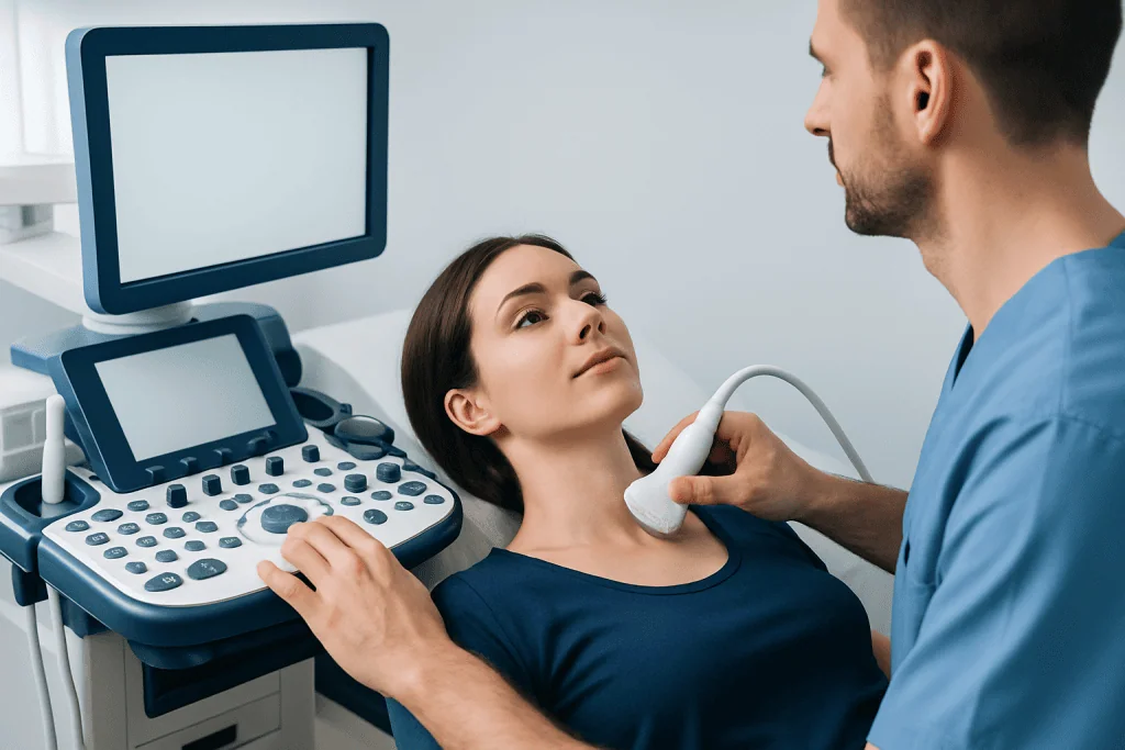
During a thyroid ultrasound, the radiologist or trained clinician evaluates both thyroid lobes and the isthmus, the thin bridge of tissue connecting them. The report usually includes gland measurements, the presence or absence of nodules, and descriptions of any important nodule features. It may also comment on lymph nodes in the neck, especially if a nodule has suspicious characteristics.
Ultrasound can distinguish between solid nodules, fluid-filled cysts, and mixed nodules that contain both solid and cystic parts. It can also assess whether a nodule is very dark compared with normal thyroid tissue, has irregular borders, contains tiny bright spots called microcalcifications, or is taller than it is wide. These details are important because some features are associated with higher suspicion, while others are typically reassuring.
Common reasons for thyroid ultrasound include:
- A lump, swelling, or fullness in the front of the neck
- A thyroid nodule found during physical examination
- An incidental thyroid finding on CT, MRI, or carotid ultrasound
- Monitoring a known thyroid nodule or multinodular goiter
- Guidance for fine needle aspiration biopsy
- Assessment after thyroid cancer treatment, when appropriate
Thyroid Nodules and Goiter
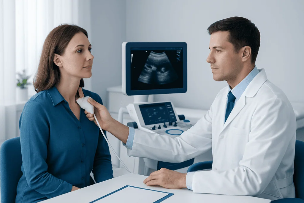
A thyroid nodule is a distinct lump or area within the thyroid gland. Nodules may be single or multiple, and they may be solid, cystic, or mixed. Many do not cause symptoms and are discovered incidentally. Some nodules produce thyroid hormone independently, which may lead to an overactive thyroid, while others are nonfunctioning and only visible on imaging.
Goiter means enlargement of the thyroid gland. It may be diffuse, where the whole gland is enlarged, or nodular, where enlargement is related to one or more nodules. Causes can include iodine imbalance, autoimmune thyroid disease such as Hashimoto’s thyroiditis or Graves’ disease, inflammation, and benign multinodular change. In some people, goiter may cause neck fullness, visible swelling, or pressure symptoms.
Ultrasound helps describe whether the thyroid enlargement is uniform or nodular, whether there are areas that need targeted evaluation, and whether nearby structures appear compressed. If a goiter extends below the breastbone into the chest, ultrasound may have limitations, and another imaging test may be needed. Blood tests such as thyroid-stimulating hormone, free thyroid hormones, and thyroid antibodies are often used alongside imaging to understand thyroid function.
How the Test Is Performed
Thyroid ultrasound is usually quick and does not require special preparation. The patient lies on an examination table with the neck gently extended. A clear gel is placed on the skin, and a handheld probe called a transducer is moved over the front and sides of the neck. The gel may feel cool, and the probe may create light pressure, but the test is generally painless.
The person performing the scan may ask the patient to turn the head slightly or swallow. Images are captured in different planes to measure the gland and any nodules accurately. Doppler ultrasound may be used to assess blood flow, although blood flow alone is not enough to determine whether a nodule is benign or suspicious.
After the examination, the gel is wiped off and normal activities can usually be resumed immediately. If a biopsy is planned on the same day, the care team may provide additional instructions. Patients taking blood-thinning medication or those with bleeding disorders should tell their doctor before any needle procedure, as individualized guidance may be needed.
Understanding Ultrasound Results
Thyroid ultrasound reports often use structured risk systems, such as ACR TI-RADS, EU-TIRADS, or guidance based on American Thyroid Association recommendations. These systems consider ultrasound features and nodule size to help determine whether observation or fine needle aspiration is appropriate. The exact wording and scoring may vary by country, hospital, and specialist preference.
Features that are generally more reassuring include a purely cystic appearance, spongiform pattern, smooth margins, and very small size without suspicious findings. Features that may raise concern include irregular or lobulated margins, marked hypoechogenicity, microcalcifications, a taller-than-wide shape, and evidence that a nodule extends beyond the thyroid capsule. Suspicious-looking lymph nodes may also change management.
It is important not to interpret one feature in isolation. For example, a large nodule may be benign, and a small nodule may still need attention if it has high-risk features. A normal thyroid blood test does not rule out a nodule, and an abnormal blood test does not automatically mean cancer. The most helpful interpretation comes from combining the ultrasound report with symptoms, examination findings, laboratory results, age, family history, and previous imaging.
When Biopsy Is Needed
The most common biopsy for a thyroid nodule is fine needle aspiration, often called FNA. In this procedure, a very thin needle is inserted into the nodule, usually under ultrasound guidance, to collect cells for microscopic examination. Local anesthesia may be used, and the procedure is typically performed as an outpatient test. Most people can return to routine activities soon afterward, following their clinician’s advice.
Biopsy is not recommended for every thyroid nodule. Doctors usually consider both the size of the nodule and its ultrasound risk pattern. Larger nodules with suspicious features are more likely to be sampled, while very small nodules may be monitored unless there are concerning lymph nodes, high-risk history, or other specific reasons. Nodules that appear purely cystic often do not need biopsy unless they cause symptoms or recur after drainage.
FNA results are commonly reported using a standardized cytology system, such as the Bethesda System for Reporting Thyroid Cytopathology. Results may be benign, nondiagnostic, indeterminate, suspicious, or malignant. A benign result often leads to ultrasound follow-up rather than surgery. Indeterminate results may require repeat biopsy, molecular testing where available, or surgical consultation depending on the overall risk profile.
Treatment Options and Follow-Up
Treatment depends on the cause of the thyroid finding. Many benign nodules require no immediate treatment and are followed with periodic ultrasound. Follow-up intervals vary according to the nodule’s size, appearance, growth pattern, and biopsy result. If a nodule remains stable and low risk, the interval between scans may become longer.
If a nodule produces excess thyroid hormone, additional testing such as a thyroid uptake scan may be considered, depending on the patient’s situation and local practice. Treatment options for overactive nodules can include medication, radioactive iodine, or surgery. For large benign nodules or goiter causing swallowing difficulty, breathing symptoms, voice changes, or cosmetic concern, treatment may include surgery or selected minimally invasive procedures in appropriate centers.
If biopsy suggests thyroid cancer or a high risk of cancer, referral to an endocrinologist, endocrine surgeon, or head and neck specialist is usually recommended. Treatment may involve surgery and, in selected cases, additional therapies or long-term surveillance. Many thyroid conditions are highly manageable, and decisions are best made through shared discussion of risks, benefits, and patient preferences.
Prevention, Self-Care, and When to See a Doctor
Not all thyroid nodules or goiters can be prevented. However, supporting overall thyroid health includes getting appropriate dietary iodine, avoiding unnecessary radiation exposure, and attending follow-up appointments when a nodule is being monitored. Patients should not start iodine supplements, thyroid hormone, or herbal thyroid products without medical advice, because these may be harmful in some thyroid conditions.
A person should see a doctor if they notice a new lump in the neck, persistent neck swelling, difficulty swallowing, breathing discomfort when lying down, unexplained hoarseness, or symptoms of thyroid hormone imbalance such as palpitations, heat intolerance, unusual fatigue, weight changes, or tremor. Urgent evaluation is appropriate for rapidly increasing neck swelling or significant breathing difficulty.
International patients who need coordinated assessment can be evaluated by multidisciplinary specialists in JCI-accredited Acibadem International hospitals, where thyroid ultrasound, laboratory testing, biopsy, endocrinology care, and surgical consultation can be arranged when clinically appropriate. Regardless of location, the safest approach is to discuss ultrasound findings with a qualified doctor who can personalize recommendations.
Frequently asked questions
Is thyroid ultrasound painful?
Thyroid ultrasound is usually painless. The probe may press gently on the neck, and the gel may feel cool, but there are no needles or radiation during the scan itself. Most people can return to normal activities immediately afterward.
Can ultrasound tell if a thyroid nodule is cancer?
Ultrasound can identify features that make a nodule more or less suspicious, but it cannot confirm cancer on its own. If a nodule meets criteria for sampling, fine needle aspiration biopsy may be recommended. The biopsy result is interpreted together with the ultrasound and clinical findings.
Do all thyroid nodules need biopsy?
No. Many thyroid nodules are small, benign-appearing, or cystic and can be monitored rather than biopsied. Doctors decide based on nodule size, ultrasound pattern, symptoms, thyroid function, and personal risk factors.
What does goiter mean on an ultrasound report?
Goiter means that the thyroid gland is enlarged. It may be evenly enlarged or enlarged because of multiple nodules. Blood tests and clinical evaluation help determine whether the goiter is related to autoimmune thyroid disease, iodine balance, hormone overproduction, or another cause.
How often should thyroid nodules be followed with ultrasound?
Follow-up timing depends on the nodule’s appearance, size, growth, and biopsy result if one was performed. Some nodules need repeat imaging within months, while others may be checked less often. The treating doctor should provide an individualized follow-up plan.
Is fine needle aspiration biopsy safe?
Fine needle aspiration is a commonly performed outpatient procedure and is generally well tolerated. Mild soreness or bruising can occur, and serious complications are uncommon. Patients should tell their doctor about blood thinners, bleeding disorders, allergies, or previous neck surgery before the procedure.
References
- American Thyroid Association
- American College of Radiology
- European Thyroid Association
- National Institute for Health and Care Excellence
- Endocrine Society
This article is for general information only and is not a substitute for professional medical advice. Please consult a qualified doctor about your individual situation.
Ultrasound in Turkey — costs, top hospitals & a free quote
JCI-accredited · board-certified surgeons · reply within 24h
Add us as a Preferred Source to see more of our trusted health content across Google Search, AI Overviews and Discover.
Check your numbers in seconds
BMI, calories, due date, blood pressure and 30+ more clinical calculators — free, instant, doctor-reviewed ranges.

Check-up & Preventive Medicine
Comprehensive health screenings designed for early detection and prevention.
3 specialists in this unitMore from the Health Library



Related Specialists

Prof. Dr. Ercan Karaarslan
Radiology
Prof. Dr. Bora Peynircioğlu
Interventional Radiology
Prof. Dr. Koray Güven
Interventional Radiology
Assoc. Prof. Dr. Deniz Can Aliş
Radiology