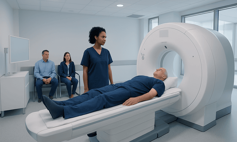
Diagnostic Ultrasound vs CT Scan: Which Test Answers What?

Ultrasound uses sound waves and does not expose the patient to ionizing radiation. CT scans use X-rays to create detailed cross-sectional images and are often useful in emergencies.
Key Takeaways
- Ultrasound uses sound waves and does not expose the patient to ionizing radiation.
- CT scans use X-rays to create detailed cross-sectional images and are often useful in emergencies.
- The best test depends on the body area, the suspected condition, and how quickly results are needed.
- Ultrasound is commonly used in pregnancy, soft tissue evaluation, and blood flow studies.
- CT is often better for trauma, lung conditions, complex abdominal problems, and many bone injuries.
Diagnostic ultrasound and CT scans are both valuable imaging tests, but they answer different clinical questions. Ultrasound is often preferred for real-time imaging without radiation, while CT is typically chosen for fast, detailed views of internal organs, bones, lungs, and urgent conditions.
Overview: how ultrasound and CT differ
Diagnostic ultrasound and computed tomography (CT) are imaging tools that help doctors look inside the body without surgery. Although both can be used to investigate pain, swelling, injury, or disease, they work in very different ways and are suited to different medical questions.
Ultrasound uses high-frequency sound waves to create images in real time. A handheld device called a transducer is moved over the skin, usually with gel, and images appear immediately on a screen. Because it does not use ionizing radiation, ultrasound is widely used for pregnancy, abdominal organs, muscles, tendons, thyroid, and blood vessels.
CT uses X-rays and computer processing to create detailed cross-sectional images, sometimes called slices. These images can show bones, organs, blood vessels, lungs, and internal bleeding with high detail. CT is especially useful when doctors need a broad overview quickly, such as after trauma or when a serious internal problem is suspected.
Rather than one test being better than the other in all situations, each has strengths and limitations. The most appropriate choice depends on the symptoms, the body part being examined, the need for speed, and whether the clinician needs to assess soft tissue, blood flow, bone, air-filled structures, or possible internal injury.
What questions can diagnostic ultrasound answer?
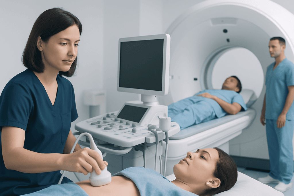
Ultrasound is often the first imaging test when doctors need a quick, safe look at soft tissues and fluid-filled structures. It is commonly used to examine the liver, gallbladder, kidneys, bladder, uterus, ovaries, testes, thyroid, breast, and superficial lumps. In many of these cases, ultrasound can show whether a structure looks normal, enlarged, inflamed, blocked, or fluid-filled.
Because ultrasound provides real-time imaging, it is especially useful for watching movement. This includes seeing how tendons move, how the heart pumps on echocardiography, and how blood flows through arteries and veins with Doppler ultrasound. It can help evaluate suspected blood clots, varicose veins, narrowed vessels, and blood supply to organs.
Ultrasound is also commonly used in pregnancy to assess fetal growth and position. In the abdomen, it is often chosen first for suspected gallstones, pelvic pain, or urinary obstruction. If more specialized evaluation is needed, a doctor may recommend additional imaging or a focused test such as ultrasound imaging.
However, ultrasound has limits. Image quality can be reduced by bowel gas, obesity, or structures located deep in the body. It is also less useful for looking inside the lungs or for providing the same level of overview that CT can offer in complex emergencies.
What questions can a CT scan answer?

CT scans are often chosen when doctors need detailed images quickly or when the area of concern is difficult to evaluate with ultrasound. CT is particularly valuable for the head, chest, abdomen, pelvis, spine, and bones. It can identify fractures, internal bleeding, organ injury, kidney stones, appendicitis, bowel problems, lung disease, and many types of tumors or complications.
In emergency medicine, CT is frequently used because it is fast and widely informative. After a fall, traffic accident, or sudden severe symptom, CT can help doctors rapidly assess whether there is bleeding, a fracture, obstruction, perforation, or other urgent problem. It is also central to evaluating some neurological emergencies and complex chest symptoms.
CT can be performed with or without contrast material. Contrast may be given by mouth, intravenously, or sometimes both, depending on what needs to be seen. This can make blood vessels, inflammation, tumors, or organ abnormalities easier to detect. In some cases, doctors may use CT scanning as the most efficient way to answer a time-sensitive question.
Like any imaging test, CT has limitations. It uses ionizing radiation, and some patients cannot safely receive contrast because of allergy history, kidney concerns, or other medical factors. For certain soft tissue or vascular questions, other tests such as MRI, ultrasound, or specialized imaging may be more informative.
Common situations where one test is preferred over the other
Doctors often choose ultrasound first when the concern involves the gallbladder, pelvis, pregnancy, thyroid, scrotum, or superficial soft tissues. It is also a common first test for children and younger patients when avoiding radiation is especially desirable. For suspected deep vein thrombosis, vascular narrowing, or some heart conditions, ultrasound offers important information about blood flow in real time.
CT is often preferred when the symptoms suggest trauma, severe abdominal pain of unclear cause, lung disease, complex fractures, kidney stones, or complications involving the bowel. It is also commonly used when ultrasound findings are unclear or incomplete, or when a wider internal survey is needed. In this way, the two tests frequently complement each other rather than compete.
For example, suspected gallstones may be assessed first with ultrasound, while suspected appendicitis may be evaluated by ultrasound in some patients but by CT in others, depending on age, body type, pregnancy status, and how clear the initial findings are. A doctor may also recommend CT if ultrasound raises a concern that requires more detailed mapping.
In neurological or stroke-related emergencies, CT is often one of the first imaging tests because it can quickly detect bleeding and other urgent causes. Related imaging pathways may overlap with advanced techniques used in MRI or in the assessment of conditions such as stroke, depending on the clinical picture.
Safety, radiation, contrast, and comfort
One of the biggest differences between the two tests is radiation. Ultrasound does not use ionizing radiation, which is why it is widely used in pregnancy and often favored when repeated imaging may be needed. CT does use radiation, but the amount varies depending on the body area and the scan protocol. Doctors generally request CT only when the expected benefit clearly outweighs the risk.
Contrast material is another important point. Standard ultrasound usually does not require contrast, although some specialized studies may use it in selected settings. CT often involves contrast to improve image detail. Before contrast is given, the medical team may ask about allergies, kidney disease, diabetes, medications, and previous reactions to imaging dye.
From a comfort perspective, ultrasound is usually simple and quiet. It is done while the transducer moves across the skin, and it may involve mild pressure on a tender area. CT is also painless, but the patient lies still on a table that moves through the scanner. If contrast is used, some people feel a temporary warm sensation or metallic taste.
Patients should tell the imaging team about pregnancy, implanted devices, kidney problems, breathing difficulty, claustrophobia, or any past reaction to contrast. Sharing this information helps the team choose the safest and most useful test and prepare the patient properly.
How doctors decide which imaging test to order
The choice between diagnostic ultrasound and CT scan is based on the clinical question, not only on the body part. Doctors consider the symptoms, physical examination, age, pregnancy status, medical history, urgency, and what condition is most likely. They also think about which test is most likely to change treatment decisions quickly and accurately.
For example, a person with sudden chest symptoms may need CT if a serious lung or vascular problem is suspected, while a person with a neck lump may start with ultrasound. Someone with abdominal pain may have ultrasound first if gallstones or pelvic causes are likely, but CT may be preferred if the pain is severe, widespread, or associated with possible bowel or kidney problems.
Sometimes one imaging test leads to another. Ultrasound may detect an abnormal mass but not fully define it, leading to CT or MRI for a clearer map. In other cases, CT identifies a finding that is then followed with targeted ultrasound, biopsy guidance, or a vascular study. This stepwise approach helps avoid unnecessary testing while improving diagnostic accuracy.
When care is complex, imaging decisions may involve radiologists, emergency physicians, surgeons, internists, or organ-specific specialists. Near the end of the diagnostic pathway, patients may also be referred to centers such as Acibadem International, where multidisciplinary specialists in JCI-accredited hospitals evaluate and treat international patients using coordinated imaging and clinical care.
Preparing for the test and understanding results
Preparation depends on the type of scan and the area being examined. Some ultrasounds require fasting for several hours, especially when the gallbladder or upper abdomen is being assessed. Pelvic ultrasound may require a full bladder. Other ultrasound exams need little or no preparation.
CT preparation varies more. Some scans require fasting, contrast drinking, blood tests, or temporary changes to medications, while others do not. The imaging team will explain what is needed in advance. Following instructions carefully helps improve image quality and reduce delays.
Results are usually interpreted by a radiologist, a doctor specialized in medical imaging. The report is then shared with the clinician who ordered the test, who places the findings in context with symptoms, examination, and laboratory tests. An abnormal scan does not always mean a serious disease, and a normal scan may still need follow-up if symptoms continue.
Patients should feel comfortable asking what the test is looking for, whether contrast is needed, what the risks are, and when the results will be available. Good communication can reduce anxiety and help patients understand why a particular imaging method was chosen.
When to seek medical advice urgently
Imaging tests are tools to support diagnosis, but urgent symptoms should not be ignored while waiting to decide between ultrasound and CT. Sudden severe chest pain, shortness of breath, confusion, weakness on one side, major injury, heavy bleeding, severe abdominal pain, or fainting all need prompt medical assessment. In such cases, the medical team decides which imaging test, if any, should be done first.
Medical advice is also important if there is persistent pain, a new lump, unexplained swelling, blood in urine, recurrent vomiting, jaundice, fever with localized pain, or symptoms that are worsening rather than improving. Even when symptoms seem mild, the most appropriate imaging choice depends on an in-person clinical evaluation.
People who are pregnant, have kidney disease, have had prior contrast reactions, or are caring for a child with significant symptoms should mention these details early. They may affect which imaging test is safest and most suitable. In some situations, doctors may also consider related evaluations for disorders such as deep vein thrombosis if symptoms suggest a circulation problem.
The most useful approach is not to choose a scan independently, but to discuss symptoms with a qualified clinician. The right imaging test is the one that answers the clinical question clearly, safely, and in time to guide proper treatment.
Frequently asked questions
Is ultrasound safer than a CT scan?
Ultrasound does not use ionizing radiation, so it is often considered the safer option when it can answer the clinical question well. CT uses X-rays, but it is still a very important and appropriate test when detailed or urgent imaging is needed.
Why would a doctor order a CT scan after an ultrasound?
An ultrasound may show that something is abnormal without fully explaining what it is. A CT scan can provide a broader and more detailed view, especially for deeper structures, the bowel, lungs, bones, or urgent internal problems.
Which test is better for abdominal pain?
It depends on the suspected cause. Ultrasound is often used first for gallbladder disease, pelvic causes, and some kidney or urinary problems, while CT is often preferred for complex, severe, or unclear abdominal pain.
Can ultrasound replace CT in emergencies?
Sometimes ultrasound can help quickly, especially for focused questions such as fluid in the abdomen, pregnancy-related concerns, or blood flow problems. However, CT is often better in major emergencies because it gives a fast, comprehensive picture of many internal structures at once.
Do both tests require contrast dye?
Ultrasound usually does not require contrast for standard exams. CT may or may not require contrast depending on what the doctor needs to see, and the team will review allergies and kidney health before using it.
Which test is better during pregnancy?
Ultrasound is usually preferred during pregnancy because it does not use radiation and is very helpful for many obstetric and abdominal evaluations. CT may still be used in special circumstances if the expected medical benefit is important and alternatives are not sufficient.
This article is for general information only and is not a substitute for professional medical advice. Please consult a qualified doctor about your individual situation.
Ultrasound in Turkey — costs, top hospitals & a free quote
JCI-accredited · board-certified surgeons · reply within 24h
Add us as a Preferred Source to see more of our trusted health content across Google Search, AI Overviews and Discover.

Check-up & Preventive Medicine
Comprehensive health screenings designed for early detection and prevention.
1 specialists in this unitMore from the Health Library
When to Get Imaging for Breast Symptoms: Lumps, Pain, and Nipple Changes

Does Quest Accept Walk-Ins? How Lab Check-In Works and When You May Need an Appointment

What Happens at a Diagnostic Imaging Consultation?

How to Read Common Blood Test Results Without Panicking
MRI or Ultrasound: How Doctors Choose the Right Test