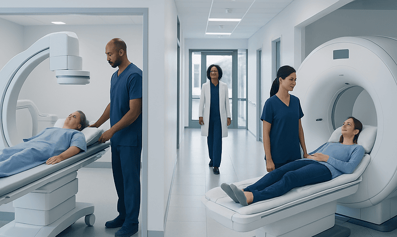
MRI or Ultrasound: How Doctors Choose the Right Test

MRI uses magnets and radio waves to create detailed images, especially of soft tissues, joints, the brain, and internal organs. Ultrasound uses sound waves and is often the first choice for pregnancy, abdominal organs, blood flow, and many children's and bedside examinations.
Key Takeaways
- MRI uses magnets and radio waves to create detailed images, especially of soft tissues, joints, the brain, and internal organs.
- Ultrasound uses sound waves and is often the first choice for pregnancy, abdominal organs, blood flow, and many children's and bedside examinations.
- The best test depends on the clinical question, not on which scan seems more advanced.
- Ultrasound is quick, portable, and does not use radiation; MRI also avoids radiation but usually takes longer and requires lying still.
- Doctors may start with ultrasound and then recommend MRI if more detail is needed.
MRI and ultrasound are both valuable imaging tests, but they answer different medical questions. Doctors choose between them based on the body area being examined, the type of tissue involved, the urgency of the situation, and what information is needed to guide care.
Overview: MRI and Ultrasound at a Glance
When a doctor orders imaging, the goal is to choose the test that will answer the medical question as clearly and safely as possible. MRI and ultrasound are both widely used, but they work in very different ways and are suited to different situations.
MRI, or magnetic resonance imaging, uses a strong magnetic field and radio waves to create detailed pictures of structures inside the body. It is especially helpful for soft tissues such as the brain, spinal cord, muscles, ligaments, cartilage, and many internal organs. Ultrasound uses high-frequency sound waves to produce real-time images. It is commonly used to look at the abdomen, pelvis, pregnancy, blood vessels, thyroid, breasts, and some muscles and tendons.
Neither test is universally “better.” In practice, doctors choose the one that is most likely to provide useful information with the least burden to the patient. In some cases, both tests may be used at different stages of diagnosis to build a more complete picture.
How the Tests Work and What They Show Best
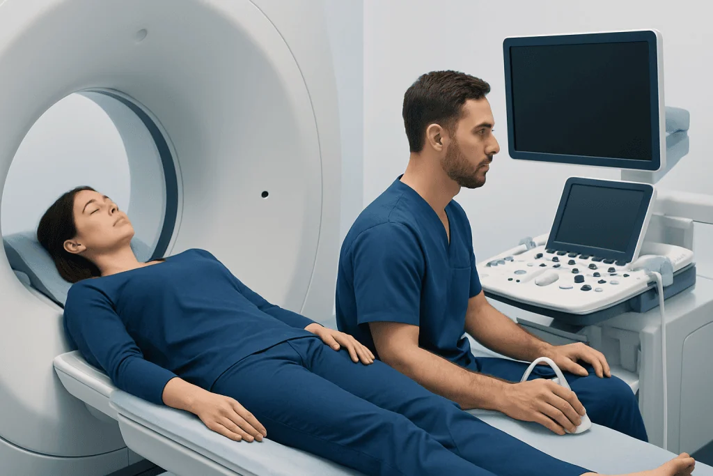
Ultrasound sends sound waves into the body through a handheld device called a transducer. The returning echoes are converted into images on a screen. Because it shows motion in real time, ultrasound is very useful for watching structures move, such as a fetus during pregnancy, blood flowing through vessels, or tendons gliding with movement. Doppler ultrasound can also help assess circulation.
MRI works differently. The patient lies inside a scanner while the machine aligns and detects signals from hydrogen atoms in the body’s tissues. A computer then creates highly detailed cross-sectional images. MRI can distinguish between many types of soft tissue and often reveals subtle changes that are not visible on ultrasound.
In general, ultrasound is especially helpful for superficial structures and fluid-filled organs, while MRI is often preferred when deeper anatomy or very fine tissue detail is needed. For example, MRI may be selected for complex joint injuries or neurological concerns, while ultrasound may be chosen first for gallbladder pain, pelvic assessment, or evaluation of a lump close to the skin.
- Ultrasound often shows best: gallbladder, liver, kidneys, uterus, ovaries, thyroid, blood vessels, pregnancy, and many soft tissue lumps.
- MRI often shows best: brain, spine, ligaments, cartilage, muscles, bone marrow, pelvic organs, and some tumors or inflammatory conditions.
What Doctors Consider When Choosing MRI or Ultrasound

The most important factor is the clinical question. Doctors ask what they are trying to confirm or rule out. If the concern is a tendon tear near the surface, fluid collection, or blood flow problem, ultrasound may give the answer quickly. If the concern involves the brain, spinal nerves, deep pelvic structures, or a complex joint, MRI may be more informative.
The body area also matters. Sound waves do not travel well through bone or air, so ultrasound has limitations when looking inside the skull, through the lungs, or in areas hidden behind bowel gas. MRI is better able to image many deep structures, although it may not be ideal for every emergency or every patient.
Doctors also consider timing, comfort, and practical issues. Ultrasound is usually faster, more portable, and can often be performed at the bedside. MRI takes longer, requires the patient to remain still, and may be difficult for people with severe claustrophobia or certain implanted devices. In addition, image quality in ultrasound depends more on the operator and on body habitus, while MRI tends to provide more standardized, detailed images.
Another common approach is to begin with ultrasound because it is accessible and efficient, then move to MRI if the ultrasound findings are uncertain or if more detail is needed for treatment planning. This stepwise process helps avoid unnecessary testing while still supporting accurate diagnosis.
Common Situations Where Ultrasound Is Preferred
Ultrasound is often the first imaging test when a doctor needs a quick, dynamic, and radiation-free examination. It is commonly used in pregnancy to monitor fetal growth and development. It is also a standard first-line test for many abdominal complaints, especially when gallstones, kidney swelling, or pelvic conditions are suspected.
Because ultrasound shows movement and flow, it is also very useful for examining blood vessels and guiding procedures. For example, it can help identify blood clots in the legs, evaluate circulation in arteries and veins, and guide needle placement during biopsies or fluid drainage. In children, ultrasound is particularly valuable because it avoids radiation and can often answer important questions without sedation.
Doctors may prefer ultrasound in situations such as:
- Suspected gallstones or gallbladder inflammation
- Pelvic pain, ovarian cysts, or uterine conditions
- Pregnancy assessment
- Evaluation of thyroid nodules or neck lumps
- Suspected deep vein thrombosis
- Assessment of soft tissue masses near the skin
In breast imaging, ultrasound is often used to evaluate a specific area of concern, especially in younger patients or to help distinguish a fluid-filled cyst from a solid lump. It may also complement other tests when further clarification is needed.
Common Situations Where MRI Is Preferred
MRI is often chosen when doctors need highly detailed images of soft tissues or when the anatomy is too deep or complex for ultrasound alone. It is a key tool in evaluating the brain and spinal cord, where ultrasound has very limited use in adults. MRI can also help assess joints, cartilage, ligaments, and bone marrow in a way that supports both diagnosis and treatment planning.
For musculoskeletal concerns, MRI is frequently used when symptoms suggest a meniscus tear, ligament injury, disc problem, or hidden stress injury. It may also be recommended when pain continues despite a normal X-ray or ultrasound. In these settings, MRI can show inflammation, small tears, and tissue changes in detail.
MRI is also used for many internal organ and pelvic questions, especially if ultrasound findings are incomplete. It can provide additional detail for the liver, pancreas, uterus, ovaries, prostate, and other structures. In some cases, contrast material may be used to better characterize lesions or assess inflammation.
Doctors may lean toward MRI for:
- Persistent neurological symptoms such as weakness, numbness, or severe headaches
- Spinal pain with suspected nerve compression
- Sports injuries involving ligaments, cartilage, or deep muscles
- Further evaluation of complex pelvic or abdominal findings
- Detailed assessment of some tumors or inflammatory disease
In broader imaging pathways, MRI may also complement tests used in breast cancer evaluation or support planning for neurosurgery and orthopedic care when anatomy needs to be mapped carefully.
Safety, Preparation, and Patient Experience
Both MRI and ultrasound avoid ionizing radiation, which is reassuring for many patients. Ultrasound is generally very well tolerated and usually requires little preparation, although fasting may be needed for some abdominal scans or a full bladder for certain pelvic examinations. The test is performed with gel on the skin and a moving transducer, and results can often be obtained quickly.
MRI is also considered safe for many people, but it has special considerations because of the strong magnet. Patients should tell their care team about any implanted devices, metal fragments, or prior surgeries. Some implants are MRI-compatible, while others may require special review. If contrast is planned, kidney function and allergy history may also be checked.
From a comfort perspective, MRI often takes longer than ultrasound and may involve lying inside a narrow scanner. This can be challenging for people who feel anxious in enclosed spaces or who have difficulty remaining still because of pain. In contrast, ultrasound is shorter and more flexible, making it easier in urgent care, bedside, and pediatric settings.
Preparation instructions vary by body area, so patients should follow the specific guidance given by their imaging center. Asking questions in advance can make the experience more comfortable and help ensure the best-quality images.
Why One Test May Lead to Another
Imaging is often a step-by-step process rather than a single decision. A doctor may start with ultrasound because it is fast, accessible, and effective for many common complaints. If the findings clearly explain the symptoms, no additional imaging may be needed. If the results are unclear or if there is concern about a deeper or more complex problem, MRI may be the next step.
This does not mean the first test was wrong or incomplete. Each imaging method has strengths and limitations, and many conditions are best assessed by combining information from the history, physical examination, laboratory tests, and more than one imaging study. Doctors choose tests to answer specific questions at each stage of care.
For example, a patient with abdominal or pelvic symptoms may have an initial ultrasound and later need advanced radiology evaluation with MRI for better tissue detail. In cancer care or difficult diagnostic cases, imaging may be reviewed by a multidisciplinary team that includes radiologists, surgeons, and other specialists. Acibadem International’s multidisciplinary specialists in JCI-accredited hospitals diagnose and treat complex conditions for international patients when advanced imaging and coordinated assessment are needed.
When to Speak With a Doctor About the Right Imaging Test
Patients do not need to decide alone whether MRI or ultrasound is the right test. A doctor considers symptoms, examination findings, age, medical history, and the most likely causes before recommending imaging. This helps avoid unnecessary scans and focuses on the test most likely to guide treatment.
It is reasonable for patients to ask why a particular test is being recommended, what information it is expected to provide, and whether another imaging option could also be appropriate. These questions can be especially helpful for people who are pregnant, have implanted devices, feel claustrophobic, or have concerns about contrast materials.
Medical review is especially important if symptoms are persistent, worsening, or associated with red flags such as significant weakness, severe pain, unexplained weight loss, or new neurological changes. In these situations, timely evaluation helps doctors choose the most appropriate next step, whether that is ultrasound, MRI, or another test altogether.
Frequently asked questions
Is MRI better than ultrasound?
Not always. MRI provides more detailed images for many deep soft tissues, the brain, spine, and joints, but ultrasound is often better for real-time imaging, blood flow, pregnancy, and many abdominal or pelvic questions. The better test is the one that best answers the specific medical question.
Why would a doctor order an ultrasound first and then an MRI?
Ultrasound is often a practical first step because it is quick, accessible, and effective for many common concerns. If it does not fully explain the symptoms or if more anatomical detail is needed, MRI may be used next. This stepwise approach helps keep testing focused and appropriate.
Do MRI and ultrasound use radiation?
No. Ultrasound uses sound waves, and MRI uses magnets and radio waves. Because neither test uses ionizing radiation, they are often preferred in situations where radiation avoidance matters.
Which test is safer during pregnancy?
Ultrasound is the standard imaging method used during pregnancy and is widely used to monitor fetal development. MRI may also be used in selected situations when more information is needed and a doctor believes it is appropriate. The choice depends on the stage of pregnancy and the medical reason for imaging.
Can ultrasound see the same things as MRI?
Sometimes, but not always. Ultrasound is excellent for many superficial structures, fluid-filled organs, and blood flow, but it has limits in deep tissues and areas blocked by bone or air. MRI usually provides more detail for complex soft tissue problems and many neurological conditions.
What if a patient is claustrophobic and needs MRI?
Patients should tell their doctor or imaging team in advance. The team may offer strategies such as reassurance, scheduling support, or discussing whether an alternative test could answer the question. In some cases, special arrangements may help the patient complete the scan more comfortably.
This article is for general information only and is not a substitute for professional medical advice. Please consult a qualified doctor about your individual situation.
Ultrasound in Turkey — costs, top hospitals & a free quote
JCI-accredited · board-certified surgeons · reply within 24h
Add us as a Preferred Source to see more of our trusted health content across Google Search, AI Overviews and Discover.

Check-up & Preventive Medicine
Comprehensive health screenings designed for early detection and prevention.
1 specialists in this unitMore from the Health Library

What Is Quest Diagnostics and How Does It Work?
Who Needs Contrast Imaging? Benefits, Risks, and Safety Considerations

Urine Test Results Explained: Infection, Protein, Blood, and Other Common Findings

Thyroid Ultrasound: Nodules, Goiter, and When Biopsy Is Needed

Neurophysiology for Seizures and Fainting: Which Tests Help Find the Cause?