Why the Azygos Vein May Look Enlarged on Chest Scans

The azygos vein runs along the right side of the spine and drains blood from the chest wall into the superior vena cava. It often appears enlarged on imaging when pressure rises in the venous system or a central vein is blocked, so an abnormal finding usually prompts evaluation of the heart and major veins rather than the vein itself.
Key Takeaways
- The azygos vein is part of the body’s venous drainage system in the chest.
- It helps return blood to the superior vena cava and can serve as an alternate pathway if other veins are blocked.
- Problems usually relate to enlargement, compression, clotting, or anatomical variation rather than disease of the vein itself.
- Many azygos vein findings are discovered on chest imaging done for another reason.
- Treatment depends on the underlying cause, such as heart failure, blockage, clot, or a nearby chest condition.
- New chest symptoms, unexplained swelling, or shortness of breath should be assessed by a doctor.
You went in for a chest scan — maybe for a cough, maybe before surgery — and the report mentions something called the azygos vein looking enlarged. It is a fair question to ask what that means.
The azygos vein carries blood from the chest wall and upper lumbar region back toward the heart. Most people never notice it. It becomes medically important when imaging shows enlargement, an anatomic variation, or changes related to heart, lung, or vein conditions.
Overview: what the azygos vein is
The azygos vein is a vein in the chest that helps carry blood back to the heart. It usually runs along the right side of the spine, collects blood from parts of the chest wall and nearby structures, and then drains into the superior vena cava, the large vein that returns blood from the upper body to the heart.
For most people it is just a normal part of the anatomy that never causes any trouble. It comes up when a chest X-ray, CT scan, or MRI shows that it is enlarged, takes an unusual course, or is caught up in another condition affecting the heart, lungs, or major veins.
Here is why doctors pay attention to it: this vein can act as a backup route for blood flow. If the superior vena cava or other central veins become narrowed or blocked, blood may be diverted through the azygos system. This makes the vein larger and more visible on imaging.
Where it is and what it does
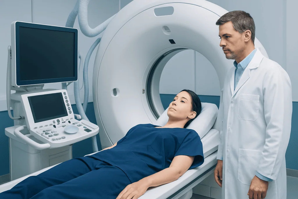
The azygos vein usually begins in the abdomen from connections with lumbar and subcostal veins, then enters the chest through the diaphragm or nearby openings. It travels upward in the back part of the chest and arches forward over the right lung root before joining the superior vena cava.
Its job is drainage. It collects blood from the posterior chest wall, some veins between the ribs, the esophagus, parts of the bronchi, and neighboring mediastinal structures. On the left side, related veins called the hemiazygos and accessory hemiazygos veins often cross the spine and drain into the azygos vein.
This system links the superior and inferior vena cava systems. Put simply, it gives blood another way home if the usual channels are partly blocked. That is why doctors pay attention to the azygos vein when evaluating central venous obstruction, pressure overload, or certain congenital variations.
Natural anatomical differences are common. Some people have an enlarged azygos arch, an azygos lobe of the lung, or other variations that are harmless but may be noticed on scans. Distinguishing a normal variant from a true abnormality is an important part of radiology and vascular assessment.
Common conditions and why the azygos vein may look abnormal
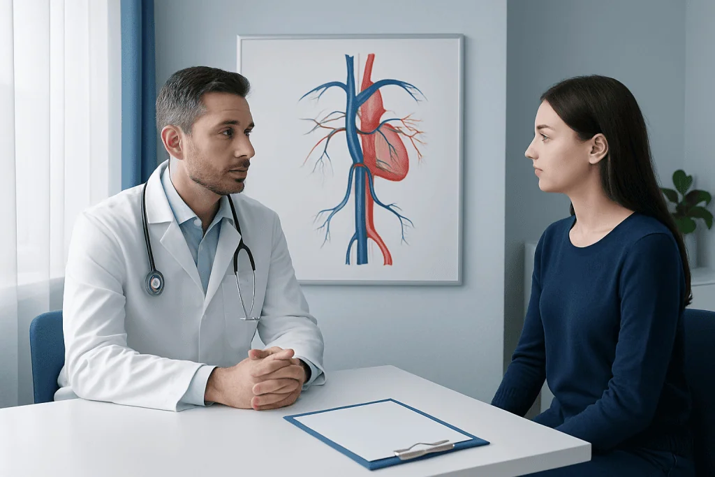
In most cases, the vein itself is not the problem. Instead, the vein may appear enlarged or prominent because of another issue elsewhere in the body. A common example is increased pressure in the venous system, such as from heart failure, fluid overload, or obstruction of the superior vena cava.
Another reason is blockage or narrowing in major veins. When this happens, the azygos vein may enlarge as it carries extra blood around the blockage. Doctors may evaluate this possibility if imaging suggests superior vena cava syndrome or another central venous problem.
Less commonly, the azygos vein can be affected by thrombosis, external compression from a tumor or enlarged lymph nodes, traumatic injury, or congenital anomalies. Nearby chest diseases can also alter its appearance on imaging. In some patients, the finding is incidental and ultimately benign.
Examples of situations that can involve the azygos vein include:
- Heart failure or increased right-sided heart pressures
- Superior vena cava obstruction or narrowing
- Portal hypertension in selected cases with collateral vein formation
- Congenital venous variants
- Chest masses, inflammation, or scar tissue causing compression
- Rare vein clotting or injury
Symptoms and possible signs
An azygos vein issue may cause no symptoms at all, especially when it is a normal anatomical variant or an incidental imaging finding. In those cases, the person often feels well and the vein is simply noted during testing for another reason.
When symptoms do occur, they usually come from the underlying cause rather than from the vein itself. For example, someone with central venous obstruction may develop swelling of the face, neck, or arms, visible chest wall veins, headache, or a sense of fullness in the head. A person with heart failure may have shortness of breath, fatigue, or leg swelling.
Compression or clotting related to the azygos system is uncommon, but chest discomfort, breathing difficulty, or signs of reduced venous return can prompt further assessment. If a chest mass or another lung condition is involved, symptoms may overlap with cough, weight loss, or recurrent infections depending on the cause.
Because these symptoms are so general, no one can pin down an azygos vein problem from symptoms alone. Imaging and an examination are usually needed to tell whether the finding is harmless or points to a more significant vascular or chest disorder.
How doctors diagnose azygos vein abnormalities
Diagnosis usually starts with the reason testing was ordered in the first place. A chest X-ray may show a widened azygos arch or another unexpected shadow, but CT scanning often gives a much clearer view of the vein and surrounding structures. CT with contrast is especially helpful when doctors need to assess obstruction, collateral circulation, masses, or clot.
MRI can also provide detailed information, particularly in selected patients who need further evaluation of blood vessels or soft tissues. Ultrasound has a more limited role because the azygos vein lies deep in the chest, though it may help assess related veins in the neck or upper limbs. When lung or chest symptoms are present, doctors may also investigate conditions such as pulmonary embolism or other cardiopulmonary causes.
Additional tests depend on the suspected underlying problem. These may include echocardiography to look at heart function, blood tests, or venography in selected cases. If a clot is suspected, doctors assess the broader risk of thrombosis and consider whether there is cancer, a catheter-related issue, or a clotting disorder.
Your doctor is not just trying to describe the vein. The questions are: is this normal for you? Is it enlarged because it is compensating for a blockage somewhere else? Is something nearby pressing on it? The treatment plan follows from those answers.
Treatment options and ongoing care
There is no one treatment for an azygos vein finding, because everything depends on the cause. A harmless anatomical variation usually needs nothing at all — reassurance, and follow-up only if symptoms change.
If the vein is enlarged because of heart or vascular disease, treatment focuses on the underlying condition. This may include medical management for heart failure, investigation of central venous obstruction, or specialist assessment for clotting. In some cases, patients may need advanced vascular imaging or procedures such as angiography to define the anatomy and plan treatment.
When there is significant narrowing or blockage in a major vein, interventional or surgical treatment may be considered. Depending on the location and cause, options can include catheter-based procedures, stenting, or management by cardiovascular surgery teams. If a chest mass is responsible, treatment is directed at that condition rather than the vein itself.
People with symptoms such as shortness of breath, facial swelling, arm swelling, or unexplained chest findings may be referred to specialists in vascular medicine, cardiology, thoracic imaging, or surgery. Near the end of the care pathway, coordinated centers such as Acibadem International can evaluate these cases through multidisciplinary specialists in JCI-accredited hospitals for international patients when complex diagnosis or treatment is needed.
Prevention, self-care, and when to seek medical care
You cannot prevent the anatomy you were born with, or most of its natural variations. But looking after your heart and blood vessels may lower the risk of some conditions that raise venous pressure or disturb blood flow. This includes following treatment plans for heart disease, staying active as medically appropriate, avoiding smoking, and attending follow-up appointments for chronic lung or vascular disorders.
People who have central lines, clotting risks, or a history of major vein obstruction should follow their doctor’s advice carefully. Staying well hydrated when appropriate, moving regularly during long periods of sitting, and reporting new swelling or breathing changes early can be helpful. Whatever self-care plan you follow should fit your overall health and medical history.
Medical care should be sought promptly for new or worsening shortness of breath, swelling of the face or arms, chest pain, coughing up blood, fainting, or visible enlarged veins across the upper chest. These symptoms do not always mean an azygos vein problem, but they can indicate an important heart, lung, or central vein condition that needs assessment.
If your imaging showed an unusual vein pattern or enlargement, ask what it means, whether you need follow-up, and whether any other tests are recommended. Those are fair questions. In some cases, related vascular assessment may involve specialized imaging such as CT angiography to better understand nearby chest anatomy and circulation.
Frequently asked questions
What is the azygos vein in simple terms?
The azygos vein is a vein in the chest that helps carry blood back to the heart. It drains blood from parts of the chest wall and nearby structures and connects to the superior vena cava.
Is an enlarged azygos vein dangerous?
Not always. An enlarged azygos vein can be a harmless anatomical variation, but it can also suggest increased venous pressure, a blockage in major veins, or another chest condition, so doctors interpret it in context.
Can a person feel an azygos vein problem?
Usually not directly. If symptoms are present, they are more often related to the underlying cause, such as shortness of breath, swelling of the face or arms, or chest symptoms from heart, lung, or vascular disease.
How is an azygos vein abnormality found?
It is often discovered on imaging such as a chest X-ray or CT scan done for another reason. CT with contrast is especially useful because it shows the vein, nearby structures, and possible blockages more clearly.
Does every azygos vein finding need treatment?
No. Many findings need no treatment if they represent normal anatomy or a benign variation. Treatment is considered when there is an underlying problem such as venous obstruction, clotting, heart disease, or a compressing mass.
When should someone talk to a doctor about the azygos vein?
A doctor should review the finding if imaging reports mention enlargement, obstruction, or an unclear abnormality. Immediate medical attention is important for symptoms such as sudden shortness of breath, chest pain, facial swelling, or coughing up blood.
This article is for general information only and is not a substitute for professional medical advice. Please consult a qualified doctor about your individual situation.
Joint and spine care in Turkey — expert assessment & treatment
7 JCI-accredited hospitals · board-certified surgeons · reply within 24h
Add us as a Preferred Source to see more of our trusted health content across Google Search, AI Overviews and Discover.

Medically reviewed by the Acıbadem International Medical Board — August 24, 2026
See our medical review board →
Update history
- PublishedAugust 4, 2026
- Medical review approvedAugust 24, 2026
- Last content updateAugust 24, 2026
References1
- Chest CT scan - MedlinePlus — medlineplus.gov
Radiology
Advanced imaging and image-guided interventions supporting diagnosis across every specialty.
124 specialists in this unitRadiology Specialists at Acibadem





More from the Health Library
SPECT Imaging Shows How Organs Function, Not Just Anatomy

Radioligand Therapy Delivers Radiation to Cancer Cells Directly

What an X Ray Shows and How the Test Is Done
Pelvic Ultrasound: How the Scan Works and Why It Is Ordered
When Doctors Order a CT Scan and How the Test Works