

Brain MRI vs Cerebral Angiography: Which Neuroradiology Test Is Used and Why
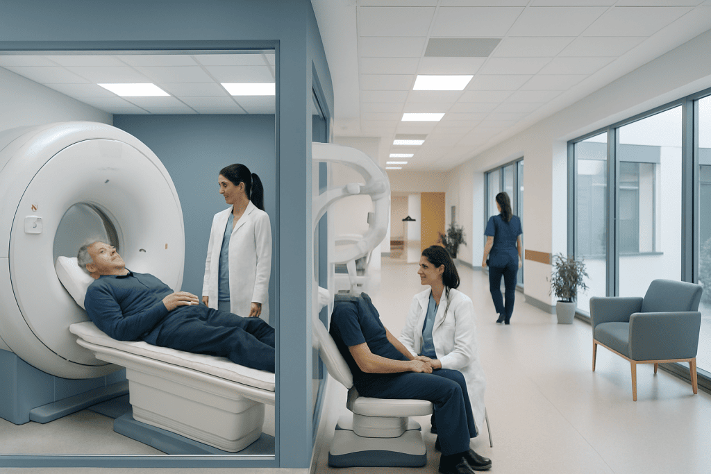
Brain MRI is best for detailed images of brain tissue, nerves, fluid spaces, and many structural abnormalities. Cerebral angiography focuses on blood vessels and is often used when a doctor needs precise vascular detail.
Key Takeaways
- Brain MRI is best for detailed images of brain tissue, nerves, fluid spaces, and many structural abnormalities.
- Cerebral angiography focuses on blood vessels and is often used when a doctor needs precise vascular detail.
- The choice depends on symptoms, suspected diagnosis, urgency, and whether treatment may be needed during the procedure.
- MRI is noninvasive, while catheter angiography is invasive and may carry a small risk of bleeding, stroke, or contrast-related problems.
- In many cases, these tests are complementary rather than interchangeable.
Brain MRI and cerebral angiography are different neuroradiology tests used for different clinical questions. MRI is usually chosen to examine brain tissue and surrounding structures, while cerebral angiography is used to look closely at blood vessels in the brain and neck.
Overview: How These Two Tests Differ
When comparing brain MRI vs cerebral angiography, the most important point is that these tests answer different questions. A brain MRI creates highly detailed images of the brain’s soft tissues, including the brain itself, nearby nerves, fluid-filled spaces, and sometimes the blood vessels depending on the technique used. It is commonly used to investigate headaches, seizures, weakness, balance problems, memory concerns, tumors, inflammation, infection, and signs of stroke.
Cerebral angiography, by contrast, is focused on the arteries and veins that supply and drain the brain. In everyday use, the term may refer to several vessel-imaging methods, but it most often means catheter angiography, also called digital subtraction angiography. In this procedure, a thin tube is guided through a blood vessel and contrast dye is injected so specialists can see the shape, size, and flow within cerebral vessels in great detail.
Because they look at different aspects of the brain, one test is not automatically “better” than the other. MRI is often the preferred first test when the main concern is brain tissue. Angiography is often chosen when a doctor suspects a blood vessel problem such as an aneurysm, arteriovenous malformation, narrowing, blockage, or certain causes of stroke and bleeding.
What a Brain MRI Is Used For
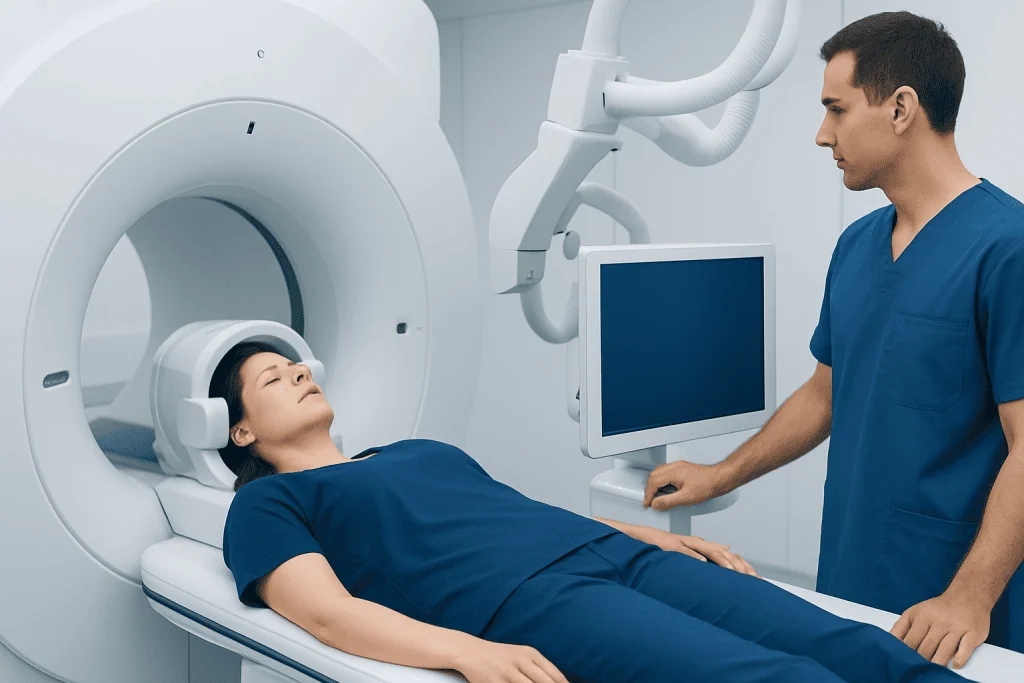
A brain MRI uses a strong magnetic field and radio waves to create detailed cross-sectional images. It does not use ionizing radiation. Depending on the reason for the test, it may be done with or without contrast material, and special MRI techniques can provide additional information about blood flow, acute stroke, inflammation, tumors, or small structural changes.
Doctors often request brain MRI when they need a close look at brain tissue. It can help identify conditions such as stroke, multiple sclerosis, tumors, infection, swelling, trauma-related changes, or causes of neurological symptoms that are not visible on simpler tests. It may also be used to assess disorders affecting the pituitary gland, cranial nerves, or inner structures involved in vision, hearing, or balance.
In some cases, MRI can also include magnetic resonance angiography, or MRA, which provides a noninvasive view of blood vessels. This can be helpful when vascular disease is suspected but a catheter-based study may not be necessary right away. For patients being evaluated for a brain tumor or for suspected stroke, MRI often plays a central role in diagnosis and treatment planning.
- Best for brain tissue and many neurological conditions
- Noninvasive and usually done as an outpatient test
- May use contrast to improve detail
- Can be combined with vessel imaging in selected cases
What Cerebral Angiography Is Used For

Cerebral angiography is a specialized test that shows the brain’s blood vessels in very fine detail. In catheter angiography, a doctor inserts a thin catheter, usually through an artery in the wrist or groin, and guides it toward the vessels supplying the brain. Contrast dye is then injected while rapid X-ray images are taken. This allows specialists to assess vessel shape, narrowing, blockage, abnormal connections, aneurysms, or active bleeding.
This test is especially useful when a highly accurate map of the blood vessels is needed. It may be recommended if noninvasive imaging suggests an aneurysm, arteriovenous malformation, vasculitis, vessel narrowing, or another vascular abnormality that needs confirmation or treatment planning. It can also help in the evaluation of some stroke cases, particularly when endovascular treatment is being considered.
One key advantage of catheter angiography is that diagnosis and treatment may sometimes happen in the same session. For example, if an abnormal blood vessel or aneurysm is identified, the interventional team may proceed with a planned endovascular procedure when appropriate, such as cerebral angiography as part of a broader diagnostic and treatment pathway or embolization for certain vascular lesions. This makes angiography especially valuable in interventional neuroradiology.
When Doctors Choose One Test Over the Other
The choice between brain MRI and cerebral angiography depends on the clinical question. If symptoms suggest a problem within the brain tissue itself, such as a tumor, inflammation, infection, demyelinating disease, or subtle stroke changes, MRI is often the first choice. It is also commonly preferred for planned, nonurgent investigations because it provides broad anatomical information without requiring an invasive catheter procedure.
If the main concern is a blood vessel abnormality, angiography may be more informative. This is especially true when doctors need to see very small vessel details, understand blood flow patterns, or plan an endovascular treatment. In emergencies, time-sensitive decisions matter. A patient with suspected bleeding from an aneurysm or a blocked artery causing stroke may undergo rapid vascular imaging, and catheter angiography may follow if treatment is needed.
In many real-world cases, doctors use several tests together. A person may first have a CT or MRI, followed by MRA, CTA, or catheter angiography depending on the findings. Rather than competing tests, they are often complementary tools. For patients who may need minimally invasive treatment, a specialist may also discuss procedures such as thrombectomy in selected stroke cases or aneurysm treatment when a vascular lesion is confirmed.
How the Tests Are Performed and What Patients Can Expect
During a brain MRI, the patient lies on a table that slides into the scanner. The test is painless, but it can be noisy and may take time depending on the number of image sequences needed. The patient must remain still for clear images. Some people receive contrast through a vein in the arm. Patients with claustrophobia, certain implants, or metal fragments in the body should tell the medical team in advance so the safest approach can be chosen.
During cerebral angiography, the skin is cleaned and numbed, and a catheter is inserted into an artery. The doctor guides the catheter under imaging and injects contrast dye while taking detailed pictures. The patient may feel brief warmth when contrast is injected. Afterward, the catheter is removed and pressure or a closure device is used to reduce bleeding. The patient is then monitored for a period of time, often for several hours.
Preparation differs slightly between the two tests. MRI preparation often focuses on metal safety, kidney function when contrast is planned, and managing anxiety if needed. Angiography usually requires more procedural preparation, including review of medications, bleeding risk, kidney function, allergy history, and fasting instructions when sedation is planned.
Benefits, Limitations, and Risks
Brain MRI offers excellent soft-tissue detail and can detect many conditions that angiography cannot. It is noninvasive and does not use radiation, which is an advantage for many patients. However, MRI may be less suitable for people with certain implanted devices, severe claustrophobia, or an inability to lie still. It can also be less ideal when the question is focused on tiny vessel anatomy or rapid endovascular planning.
Cerebral angiography provides extremely detailed vessel images and remains the reference standard for many complex vascular problems. It can reveal abnormalities that may not be fully characterized on MRI or CT-based vascular imaging. Its major limitation is that it is invasive. Although complications are uncommon in experienced hands, risks include bruising or bleeding at the puncture site, reaction to contrast, kidney stress in some patients, infection, vessel injury, and a small risk of stroke.
Contrast materials used in either test are also different. MRI contrast and angiographic contrast are not the same, and doctors consider kidney function, allergy history, and the clinical need before using them. The safest and most useful test is the one that best answers the medical question with the least risk for the individual patient.
Common Conditions These Tests Help Diagnose
Brain MRI and cerebral angiography are used across a wide range of neurological conditions. MRI is especially helpful for disorders involving brain tissue, while angiography is favored for vessel disorders. In some situations, the diagnosis becomes clearer only when both tissue imaging and vessel imaging are reviewed together by a multidisciplinary team.
Conditions that may be evaluated with brain MRI include tumors, inflammation, infection, demyelinating disease, hydrocephalus, structural causes of seizures, and many types of stroke. Conditions that may be assessed with cerebral angiography include aneurysms, arteriovenous malformations, dural fistulas, vasospasm, vessel narrowing, and selected causes of bleeding or recurrent stroke.
- Sudden neurological symptoms may prompt urgent vessel imaging to look for blockage or bleeding.
- Persistent unexplained neurological symptoms often lead to MRI for detailed tissue assessment.
- Known vascular abnormalities may require angiography for precise mapping before treatment.
- Follow-up imaging may use MRI, MRA, or angiography depending on the condition and prior treatment.
When to Seek Medical Advice and Questions to Ask
Anyone with sudden weakness, difficulty speaking, severe sudden headache, new seizures, loss of vision, confusion, or loss of consciousness should seek urgent medical attention. These symptoms may require rapid neurological assessment and imaging. For nonurgent symptoms such as chronic headaches, balance changes, memory concerns, numbness, or ongoing dizziness, a doctor can help determine whether imaging is needed and which test is most appropriate.
Patients often benefit from asking why a specific test is recommended, what it is expected to show, whether contrast will be used, and whether any alternative tests are reasonable. It is also helpful to mention kidney disease, pregnancy, implanted devices, past reactions to contrast, blood thinners, or concerns about claustrophobia or sedation.
Near the end of the decision process, care may involve neurologists, neuroradiologists, neurosurgeons, and interventional specialists working together. Acibadem International’s multidisciplinary specialists and JCI-accredited hospitals diagnose and treat complex neurological and vascular conditions for international patients when advanced assessment is needed.
Frequently asked questions
Is a brain MRI the same as cerebral angiography?
No. A brain MRI mainly shows brain tissue and surrounding structures, while cerebral angiography is designed to show the brain’s blood vessels in much finer detail. They may be used separately or together depending on what doctors need to learn.
Which test is better for stroke?
That depends on the type of stroke and the clinical situation. MRI is very useful for identifying brain tissue injury, while angiography is especially valuable when doctors need to assess blocked or abnormal blood vessels and consider an endovascular procedure.
Is cerebral angiography painful?
Patients usually receive local anesthesia at the insertion site, so the procedure is often more uncomfortable than painful. Some may feel pressure at the catheter site or brief warmth when contrast is injected. The care team monitors the patient closely throughout the procedure.
Does brain MRI use radiation?
No. MRI uses magnetic fields and radio waves rather than ionizing radiation. This is one reason it is commonly used for many neurological evaluations.
Can MRI replace cerebral angiography?
Sometimes MRI or MRA provides enough information, but not always. If doctors need the most precise view of brain blood vessels or are planning a procedure, catheter angiography may still be necessary.
How do doctors decide which test I need?
They consider symptoms, examination findings, urgency, medical history, and the suspected diagnosis. They also think about safety factors such as implanted devices, kidney function, bleeding risk, and whether treatment might be needed during the same session.
References
- World Health Organization
- American College of Radiology
- Radiological Society of North America
- National Institute of Neurological Disorders and Stroke
- Society of NeuroInterventional Surgery
This article is for general information only and is not a substitute for professional medical advice. Please consult a qualified doctor about your individual situation.
Neuroradiology in Turkey — costs, top hospitals & a free quote
JCI-accredited · board-certified surgeons · reply within 24h
Add us as a Preferred Source to see more of our trusted health content across Google Search, AI Overviews and Discover.

More from the Health Library



Related Specialists

Prof. Dr. Levent Eralp
Orthopedic Surgery & Traumatology
Assoc. Prof. Dr. Alper Karakuş
Cardiology
Prof. Dr. Yüksel Ersoy
Physical Medicine & Rehabilitation
Dt. Pelin Dülgeroğlu Karamık
Oral & Dental Health