Brain MRI vs CT Scan: Which Test Is Better for Stroke, Headache, or Injury?
CT scans are usually the first choice in emergencies such as suspected stroke or serious head injury because they are fast and widely available. MRI provides more detailed images of the brain and can detect many problems that may not appear clearly on CT.
Key Takeaways
- CT scans are usually the first choice in emergencies such as suspected stroke or serious head injury because they are fast and widely available.
- MRI provides more detailed images of the brain and can detect many problems that may not appear clearly on CT.
- The best test depends on the symptoms, timing, medical history, and whether urgent treatment decisions are needed.
- Neither test is universally “better”; each has strengths, limitations, and safety considerations.
- A doctor or radiologist chooses the most appropriate scan based on the clinical situation.
Brain MRI and CT scans are both important imaging tests, but they answer different clinical questions. In general, CT is often preferred when speed matters, while MRI is often better when more detailed brain tissue imaging is needed.
Overview: How Brain MRI and CT Scans Differ
When doctors need to look inside the brain, the two most common tests are computed tomography (CT) and magnetic resonance imaging (MRI). Both produce pictures of the brain, but they work in very different ways. A CT scan uses X-rays to create cross-sectional images, while an MRI uses a strong magnetic field and radio waves to show brain structures in greater soft-tissue detail.
In many situations, the question is not whether one test is always better than the other. The real question is which test is better for the person’s symptoms and how urgently answers are needed. For example, in an emergency room setting, speed can be critical. In a non-emergency outpatient setting, image detail may be more important.
CT scans are usually quicker, easier to access, and very good at detecting fresh bleeding, major fractures, and some urgent causes of neurological symptoms. MRI scans take longer but are often more sensitive for early tissue changes, small tumors, inflammation, infection, and many causes of persistent neurological symptoms.
Doctors may also use both tests at different times. A person with sudden stroke symptoms may have a CT first to guide immediate treatment, and later have an MRI for more detail. In this way, CT and MRI are often complementary rather than competing tests.
Which Test Is Better for Suspected Stroke?
For suspected stroke, a CT scan is often the first test because it is fast and can quickly show bleeding in or around the brain. This matters because early treatment depends on knowing whether symptoms are caused by a blocked blood vessel or a bleed. In emergency stroke care, rapid imaging can help doctors act without delay.
A non-contrast head CT is commonly used first to rule out hemorrhagic stroke. If doctors suspect a blocked artery, they may also use CT angiography or perfusion imaging to look at blood vessels and blood flow. This helps identify people who may benefit from urgent treatment, including stroke treatment and other time-sensitive interventions.
MRI can be more sensitive than CT for early ischemic stroke, especially small or deep brain infarcts and strokes in the posterior part of the brain. Diffusion-weighted MRI is particularly good at showing early injury to brain tissue. However, MRI is not always the best first test in an emergency because it usually takes longer, may be less available, and is not suitable for every patient.
In practical terms, CT is often better for the first emergency decision, while MRI is often better for confirming the extent and exact location of brain injury afterward. If a person has sudden weakness, speech difficulty, vision loss, severe dizziness, or facial drooping, emergency assessment matters more than waiting for a “perfect” test. Some patients may later be found to have conditions such as stroke or transient neurological events that require further imaging and specialist follow-up.
Which Test Is Better for Headache?
Most headaches do not require brain imaging. Doctors usually recommend a scan only when the headache pattern suggests a possible underlying problem, such as sudden severe onset, new neurological symptoms, fever, trauma, cancer history, immune suppression, or a major change from the person’s usual headaches.
When urgent causes need to be ruled out quickly, CT is often used first. A sudden “worst headache of life,” for example, may prompt an emergency CT scan to look for bleeding around the brain. CT is also commonly chosen in emergency departments because it is quick and effective for detecting many acute abnormalities.
MRI is often preferred for ongoing, unexplained, or recurrent headaches when doctors want a more detailed evaluation. It can better show small structural changes, tumors, inflammation, signs of prior injury, and some causes of secondary headache that may be subtle on CT. This is one reason MRI is frequently used in outpatient neurology or headache clinics.
If a person has a long-standing pattern of migraine without warning signs and a normal neurological exam, imaging may not be necessary at all. The decision depends on the person’s age, symptoms, exam findings, and medical history. A clinician will weigh whether imaging is likely to change treatment or provide important reassurance.
Which Test Is Better for Head Injury?
For a recent head injury, CT is usually the first and most useful test in the emergency setting. It can quickly identify skull fractures, bleeding inside the skull, brain swelling, and other urgent problems that may require observation, surgery, or immediate treatment. Because it is so fast, CT is especially valuable when a person is unconscious, confused, vomiting, or medically unstable.
Doctors do not perform CT for every bump to the head. They use clinical decision rules based on symptoms and risk factors, such as loss of consciousness, worsening headache, seizure, repeated vomiting, use of blood thinners, age, dangerous mechanism of injury, or signs of fracture. These rules help avoid unnecessary radiation while still identifying people who need urgent imaging.
MRI is not usually the first test for acute trauma, but it can be very helpful later. It may detect subtle brain injury, small areas of bruising, diffuse axonal injury, and lingering changes that are not easily seen on CT. If symptoms continue after a normal CT, a doctor may consider MRI to look more closely.
In moderate to severe trauma, CT and MRI may both have roles during recovery. CT answers the urgent question of whether there is dangerous bleeding or fracture now. MRI can later provide additional detail in people with persistent symptoms or suspected complications related to brain conditions found incidentally during imaging workup.
How Doctors Choose Between MRI and CT
The choice between MRI and CT depends on more than the symptom alone. Doctors consider how quickly the answer is needed, what diagnosis is suspected, how stable the patient is, and whether the person can safely undergo MRI. In an emergency, the fastest suitable test is often the best starting point.
CT may be preferred when:
- There is suspected bleeding, fracture, or severe trauma
- Stroke symptoms have just started and rapid treatment decisions are needed
- The patient cannot stay still for long
- MRI is not available immediately
MRI may be preferred when:
- Detailed brain tissue imaging is needed
- Symptoms are ongoing but not immediately life-threatening
- Doctors suspect small stroke, tumor, inflammation, infection, demyelination, or subtle injury
- A prior CT did not explain the symptoms
Contrast material may also affect the choice. Some CT scans and MRI scans use contrast dye to highlight blood vessels or abnormal tissue. Doctors check kidney function, allergy history, pregnancy status, and other safety factors before giving contrast. For some patients, advanced imaging such as MRI or CT imaging is part of a larger neurological assessment rather than a stand-alone answer.
Benefits, Limitations, and Safety Considerations
Each test has clear strengths. CT is fast, widely available, and highly useful for fresh bleeding, fractures, and many emergencies. MRI does not use ionizing radiation and generally provides better soft-tissue detail, making it especially helpful for evaluating many non-emergency neurological problems.
CT does expose the body to radiation, although the amount is controlled and the benefit often outweighs the risk when urgent diagnosis is needed. MRI avoids radiation, but the test can be noisy, longer, and uncomfortable for people with claustrophobia. Some people cannot have MRI because of certain implanted devices or metal fragments in the body, although many modern implants are MRI-compatible.
Both scans sometimes require contrast agents. CT contrast contains iodine, while MRI contrast commonly uses gadolinium-based agents. These are generally used carefully when needed, but doctors assess allergy history and kidney function beforehand. Not every scan needs contrast, and many important emergency brain scans are done without it.
Image quality also depends on patient movement. MRI is more sensitive to motion because it takes longer. For children, very anxious adults, or patients who cannot lie still, this may influence the decision. The imaging team works to make the process as safe and comfortable as possible.
What Patients Can Expect During the Scan
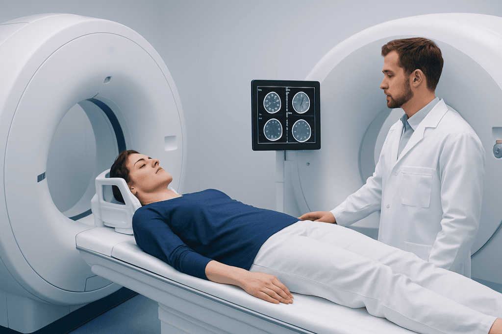
A brain CT scan is usually brief. The person lies on a table that moves through a donut-shaped scanner, and the test often takes only a few minutes. It is painless, and in emergency settings it can often be done almost immediately after medical evaluation.
An MRI takes longer, often 20 to 45 minutes or more depending on the images needed. The person lies on a table that slides into the scanner, and the machine makes loud tapping sounds during imaging. Ear protection is usually provided, and the staff can communicate throughout the test.
Before either scan, patients should tell the team about pregnancy, kidney disease, allergies, implanted devices, pacemakers, prior surgeries, or metal exposure. For MRI, this safety screening is especially important because the magnet can affect some metal objects and implants.
Results are interpreted by a radiologist and shared with the treating doctor. In emergencies, key findings may be reported very quickly. In planned outpatient imaging, final results are usually discussed during follow-up, when the doctor explains what the scan showed and whether further testing or treatment is needed.
When to Seek Medical Care and Final Thoughts
Urgent medical evaluation is important for sudden stroke-like symptoms, a severe sudden headache, new seizures, significant confusion, loss of consciousness, weakness, difficulty speaking, or concerning symptoms after head trauma. In these situations, the priority is prompt assessment rather than choosing a test independently. Doctors can decide whether CT, MRI, or both are appropriate.
For non-emergency symptoms such as persistent headaches, dizziness, memory changes, or ongoing neurological complaints, a scheduled consultation with a qualified doctor is the best first step. The clinician may decide that imaging is unnecessary, or may recommend one test based on the suspected cause and the person’s overall health.
In summary, CT is often better when speed is critical, especially for acute stroke assessment, sudden headache with concern for bleeding, and recent head injury. MRI is often better when more detailed brain imaging is needed, especially for subtle stroke, persistent headaches with warning signs, and ongoing symptoms not explained by CT.
For patients who need further evaluation, Acibadem International’s multidisciplinary specialists and JCI-accredited hospitals diagnose and treat neurological conditions for international patients, including imaging-guided assessment and, when needed, neurosurgical care. The most appropriate scan is always the one that best answers the clinical question safely and without unnecessary delay.
Frequently asked questions
Is an MRI better than a CT scan for the brain?
Not always. MRI usually gives more detailed images of brain tissue, but CT is often better when a very fast answer is needed, especially in emergencies. The best test depends on the symptom, timing, and clinical question.
Why is CT often used first for stroke?
CT is fast and can quickly show bleeding in the brain, which is a key first step in stroke treatment decisions. It is also widely available in emergency departments. MRI may be added later for more detail.
Which scan is better for headaches?
For sudden severe headaches or emergency situations, CT may be used first to look for bleeding. For ongoing or unexplained headaches, MRI is often preferred because it shows more detail. Many routine headaches do not need imaging at all.
Is MRI safer than CT?
MRI does not use ionizing radiation, which is an advantage for some patients. However, it is not suitable for everyone, especially certain people with metal implants or other contraindications. CT does use radiation, but it is often the safest and most useful option when urgent diagnosis is needed.
Can a CT scan miss something that an MRI can find?
Yes. MRI can detect some small strokes, subtle injuries, inflammation, tumors, and other changes that may not be obvious on CT. That is why doctors sometimes order MRI after a normal CT if symptoms continue.
How long do brain MRI and CT scans take?
A brain CT scan usually takes only a few minutes. A brain MRI usually takes longer, often 20 to 45 minutes or more depending on the study. Preparation and safety screening can add extra time, especially for MRI.
References
- American College of Radiology
- National Institute of Neurological Disorders and Stroke
- Radiological Society of North America
- World Stroke Organization
- National Health Service
This article is for general information only and is not a substitute for professional medical advice. Please consult a qualified doctor about your individual situation.
Explore treatments in Turkey — costs, top hospitals & a free quote
JCI-accredited · board-certified surgeons · reply within 24h
Add us as a Preferred Source to see more of our trusted health content across Google Search, AI Overviews and Discover.