CT Urography vs MRI for Urologic Problems: How Doctors Choose

CT urography is often preferred when doctors need a fast, detailed look at the kidneys, ureters, and bladder, especially for stones or blood in the urine. MRI is especially useful when radiation should be avoided or when soft-tissue detail is important, such as in some prostate, bladder, or kidney evaluations.
Key Takeaways
- CT urography is often preferred when doctors need a fast, detailed look at the kidneys, ureters, and bladder, especially for stones or blood in the urine.
- MRI is especially useful when radiation should be avoided or when soft-tissue detail is important, such as in some prostate, bladder, or kidney evaluations.
- The best test depends on the reason for imaging, kidney function, pregnancy status, implanted devices, and prior test results.
- Neither scan is universally better; each has strengths and limits that help guide diagnosis and treatment.
- Doctors may start with ultrasound and then use CT or MRI if more information is needed.
CT urography and MRI are both valuable imaging tests for urinary tract and male urologic problems, but they answer different clinical questions. Doctors choose between them based on the person’s symptoms, suspected condition, need for speed, radiation exposure, and whether contrast agents are safe to use.
Overview: Why CT Urography and MRI Are Used in Urology
Urologic problems can affect the kidneys, ureters, bladder, prostate, urethra, and male reproductive organs. When symptoms such as blood in the urine, flank pain, repeated urinary infections, difficulty urinating, or a suspected mass need closer evaluation, imaging helps doctors understand what is happening inside the body. Two of the most important imaging tools are CT urography and MRI.
CT urography is a specialized computed tomography study that focuses on the urinary tract. It commonly uses contrast dye to show how the kidneys filter and how urine moves through the ureters into the bladder. MRI uses magnetic fields and radio waves rather than radiation. It can provide excellent soft-tissue detail and may be used with or without contrast, depending on the clinical question.
Doctors do not usually choose between these tests at random. They match the imaging method to the suspected problem. In many cases, one test is clearly more practical or informative than the other. In others, a person may have both studies at different times because each provides a different type of information.
What CT Urography Shows Best
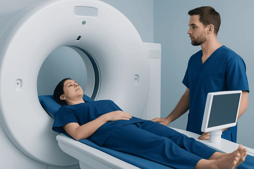
CT urography is especially strong at showing the anatomy of the urinary tract in fine detail. It can detect kidney stones, blockage of urine flow, structural abnormalities, bleeding sources, and many masses involving the kidneys or urinary collecting system. It is also commonly used when a person has visible blood in the urine or unexplained microscopic blood in the urine and doctors need to look carefully for stones, tumors, or other causes.
Because CT is fast and widely available, it is often chosen in urgent situations. A person with sudden severe side pain, suspected obstruction, trauma, or a possible complicated infection may benefit from rapid CT imaging. In many hospitals, CT can be completed in a matter of minutes, which is important when quick decision-making is needed.
CT urography does have limits. It exposes the body to ionizing radiation, and contrast dye may not be suitable for everyone, especially some people with reduced kidney function or a history of significant contrast reactions. Even so, when the goal is to evaluate stones, upper urinary tract anatomy, or possible urinary tract tumors, CT urography is frequently the first-choice test. Broader imaging strategies for CT and MRI in urology are often built around these strengths.
What MRI Shows Best

MRI is often selected when doctors need more detailed soft-tissue information. It can be particularly useful for evaluating the kidneys, bladder wall, prostate, and surrounding tissues. MRI may help distinguish one type of tissue from another more clearly than CT in certain situations, which can support the assessment of complex cysts, masses, inflammation, or possible spread of disease.
Another important advantage is that MRI does not use ionizing radiation. That makes it a valuable option for younger patients, people who may need repeated imaging, and some patients in whom radiation exposure is a particular concern. MRI may also be considered when a CT result is unclear and more characterization is needed.
MRI is not always the best first test. It generally takes longer than CT, can be more sensitive to movement, and may be difficult for people with claustrophobia unless support is arranged. Some implanted devices may also limit MRI use, although many modern implants are MRI-compatible under specific conditions. In kidney and urinary imaging, MRI can complement CT and ultrasound rather than fully replace them.
How Doctors Choose Between CT Urography and MRI
The choice depends first on the clinical question. If doctors suspect a kidney stone, acute obstruction, or need a rapid survey of the urinary tract, CT is often preferred. If they need better soft-tissue contrast, wish to avoid radiation, or want a more detailed look at structures such as the prostate or certain kidney lesions, MRI may be favored.
The person’s medical history matters too. Kidney function is important because iodinated contrast used for CT and gadolinium-based contrast used for MRI are not suitable in every situation. A doctor will review blood test results, especially kidney function, as well as past contrast reactions, allergies, pregnancy status, and any metal implants or devices. These practical details can change which test is safest and most useful.
Doctors also consider what previous tests have shown. Often, ultrasound is the first imaging step for urinary symptoms because it is simple and radiation-free. If the ultrasound leaves unanswered questions, CT or MRI may follow. For example, a patient being evaluated for recurrent infections may first have renal ultrasonography and then move on to cross-sectional imaging if obstruction, stones, or an anatomic problem is suspected.
Finally, the urgency of the situation matters. CT is usually easier to obtain quickly in emergency settings. MRI may be planned when the situation is stable and more specific tissue characterization is needed. In this way, choosing imaging is less about which test is “better” overall and more about which test is better for that person, at that moment, for that suspected problem.
Common Urologic Problems and the Imaging Doctors Often Prefer
For suspected kidney stones, non-contrast CT is commonly the most informative test because it is highly effective at showing stone location and size. If there is concern about repeated stone episodes, anatomy, or complications, CT may guide next steps in care. This is especially relevant in people being assessed for kidney stone disease.
For blood in the urine, CT urography is often a key study because it can evaluate the kidneys, ureters, and bladder in a comprehensive way. However, MRI may be used if CT contrast is unsuitable or if doctors need additional detail about a suspicious finding. Cystoscopy, a camera examination of the bladder, may still be needed because imaging and direct visualization answer different questions.
For recurrent or complicated urinary infections, imaging aims to look for blockage, stones, abscesses, reflux, or structural abnormalities. CT may be preferred in an acute or severe presentation, while MRI may be used in selected cases when deeper soft-tissue assessment is helpful. In some people with complicated infection patterns, doctors may evaluate concerns related to urinary infections alongside imaging findings.
For masses in the kidney, bladder, or nearby tissues, both CT and MRI may play a role. CT often detects the lesion, while MRI may better characterize it or help define local extension. In bladder dysfunction, such as suspected neurologic causes of urinary retention or incontinence, imaging may be combined with functional testing, particularly if there is concern for conditions such as neurogenic bladder.
Preparing for the Scan and Understanding Risks
Preparation depends on the test. Before CT urography or contrast-enhanced MRI, patients may be asked about kidney disease, diabetes medications, prior reactions to contrast, pregnancy, and hydration. Blood tests may be checked to confirm kidney function. In some cases, fasting is recommended for a few hours before the exam, although instructions vary by center.
CT uses radiation, so doctors avoid unnecessary scans and choose CT when the expected benefit outweighs the exposure. MRI avoids radiation, but it has its own considerations. Some people cannot have MRI because of certain devices or metal fragments, and others may need help if they feel anxious in enclosed spaces. Hearing protection is typically provided because MRI can be noisy.
Contrast agents for CT and MRI differ, and each has potential risks. Most people tolerate them well, but doctors remain careful in those with reduced kidney function or a previous reaction. Patients should always tell the imaging team about all medical conditions, implants, and past experiences with contrast. This information helps make the scan safer and more comfortable.
What Results Mean and How Imaging Guides Treatment
Imaging findings help doctors decide what happens next, but the scan itself is only one part of the diagnosis. Results are interpreted together with symptoms, examination findings, urine tests, blood tests, and sometimes endoscopy or biopsy. A stone seen on CT, for example, may explain pain and guide treatment, while a complex kidney lesion on MRI may need further specialist review.
Sometimes imaging provides reassurance by ruling out serious causes of symptoms. At other times, it identifies a problem early, when treatment may be simpler. Follow-up imaging may also be used to monitor a known condition, check whether an obstruction has resolved, or evaluate response to treatment.
In more complex cases, care may involve radiologists, urologists, nephrologists, and other specialists working together. Acibadem International’s multidisciplinary specialists and JCI-accredited hospitals diagnose and treat urologic conditions for international patients, including assessments that may involve renal radiology and coordinated kidney-urology care.
When to See a Doctor for Urologic Symptoms
A person should seek medical advice for blood in the urine, repeated urinary infections, pain in the side or lower back, painful urination, difficulty emptying the bladder, new urinary leakage, or swelling in the scrotum or lower abdomen. These symptoms can have many causes, and imaging may or may not be needed depending on the situation. Prompt evaluation helps doctors decide the safest and most useful next step.
Urgent care is especially important for severe pain, fever with urinary symptoms, inability to pass urine, vomiting with suspected kidney stones, or symptoms after trauma. These situations can signal blockage, infection, or another condition that needs timely attention. Fast imaging, often with CT, may be appropriate in emergency settings.
People should also follow up if symptoms continue despite treatment or if a previous scan found something that needs monitoring. Asking why a specific test has been chosen can be helpful. Understanding the reason for CT or MRI often makes the process less stressful and helps patients take an active role in their care.
Frequently asked questions
Is CT urography better than MRI for all urinary problems?
No. CT urography is often better for stones, urgent evaluation, and a detailed overview of the urinary tract, while MRI is often better for selected soft-tissue questions and when avoiding radiation is important. The best test depends on the symptom, the suspected diagnosis, and the patient’s medical history.
Why would a doctor choose MRI instead of CT?
A doctor may choose MRI when more soft-tissue detail is needed or when radiation should be avoided. MRI may also be helpful if a CT result is inconclusive or if a patient should not receive iodinated CT contrast.
Can CT urography detect kidney stones better than MRI?
In many cases, yes. CT is generally the preferred imaging test for suspected kidney stones because it shows stone size and location clearly and can be performed quickly. MRI is not usually the first-line test for stone detection.
Is contrast always needed for CT urography or MRI?
Not always. Some CT scans for stones are done without contrast, while CT urography for blood in the urine often uses contrast to evaluate the urinary tract lining and urine flow. MRI may be done with or without contrast depending on what doctors need to assess.
Which scan is safer for people with kidney problems?
Safety depends on the type and severity of kidney impairment and whether contrast is needed. Doctors review kidney function before contrast-enhanced scans and may choose a different test, use a non-contrast study, or delay imaging if necessary. The safest approach is individualized.
Can someone have both CT and MRI for the same urologic issue?
Yes. Sometimes CT finds a problem and MRI is then used to characterize it in more detail, or the tests are used at different stages of care. This does not usually mean something is wrong; it often reflects the fact that the tests provide different kinds of information.
References
- American College of Radiology
- National Institute of Diabetes and Digestive and Kidney Diseases
- European Association of Urology
- Radiological Society of North America
- National Kidney Foundation
This article is for general information only and is not a substitute for professional medical advice. Please consult a qualified doctor about your individual situation.
Explore treatments in Turkey — costs, top hospitals & a free quote
JCI-accredited · board-certified surgeons · reply within 24h
Add us as a Preferred Source to see more of our trusted health content across Google Search, AI Overviews and Discover.