

Diagnostic Ultrasound: What It Shows and How It Works

Diagnostic ultrasound uses high-frequency sound waves, not radiation, to produce images. It is commonly used to assess organs, soft tissues, blood vessels, and pregnancy.
Key Takeaways
- Diagnostic ultrasound uses high-frequency sound waves, not radiation, to produce images.
- It is commonly used to assess organs, soft tissues, blood vessels, and pregnancy.
- The test is usually painless, quick, and performed without sedation.
- Preparation depends on the body area being examined, such as fasting or drinking water.
- Results are interpreted by a radiologist or another trained specialist in the context of symptoms and other tests.
Diagnostic ultrasound is a safe, widely used imaging test that uses sound waves to create pictures of structures inside the body. It can help doctors examine organs, soft tissues, blood flow, and pregnancy in real time without using ionizing radiation.
Overview: What Is Diagnostic Ultrasound?
Diagnostic ultrasound is an imaging method that creates pictures of the inside of the body using high-frequency sound waves. A handheld device called a transducer sends sound waves into the body and receives the returning echoes. A computer then converts those echoes into images that can be viewed in real time.
Because it does not use ionizing radiation, diagnostic ultrasound is often chosen when doctors want a safe and practical way to examine soft tissues, organs, or blood flow. It is commonly used in the abdomen, pelvis, thyroid, breasts, muscles, joints, and blood vessels, and it is also well known for monitoring pregnancy.
One of the main advantages of ultrasound is that it shows movement as it happens. This allows clinicians to assess structures such as a beating heart, a developing fetus, or blood moving through arteries and veins. In many cases, it is used as a first-line imaging test because it is accessible, well tolerated, and informative.
How Ultrasound Works
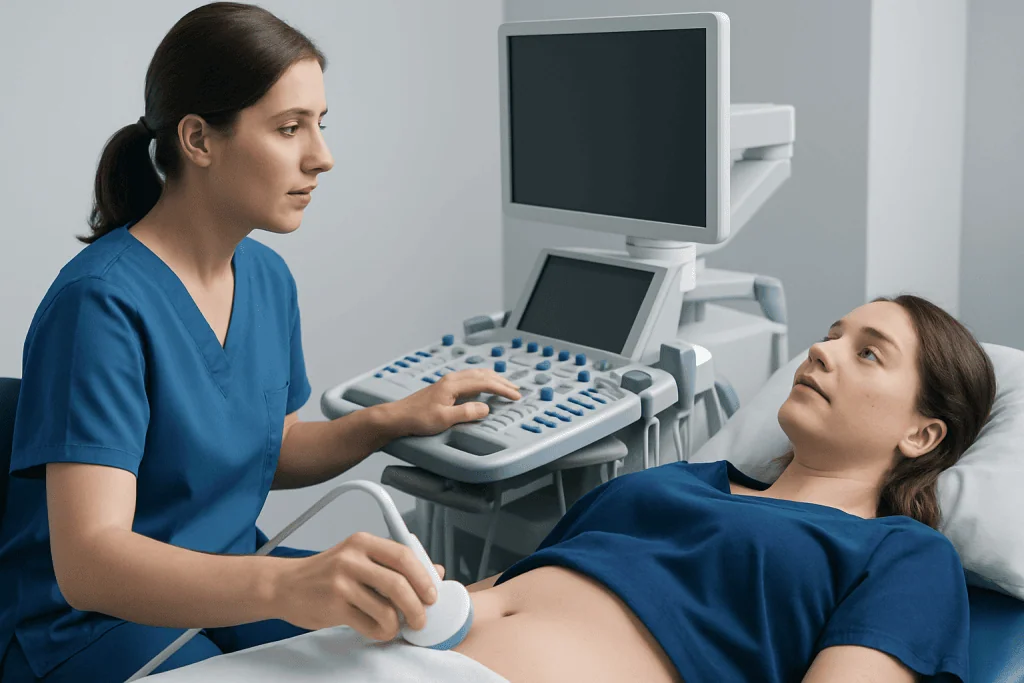
Ultrasound works by using sound waves above the range of human hearing. When these waves pass into the body, different tissues reflect them in different ways. Fluid, soft tissue, muscle, and dense structures each produce a distinctive pattern of echoes, helping form an image.
The transducer is placed on the skin after a clear gel is applied. The gel helps the sound waves travel efficiently by removing tiny air pockets between the transducer and the skin. During some examinations, a special internal transducer may be used, such as in transvaginal or transrectal ultrasound, to obtain clearer images of nearby structures.
There are several ultrasound techniques. Standard ultrasound creates still and moving images of anatomy. Doppler ultrasound evaluates blood flow and can help identify narrowing, blockage, or abnormal circulation. In some settings, ultrasound may also guide procedures, such as fluid drainage, biopsy, or needle placement, improving precision and safety.
What Diagnostic Ultrasound Can Show

Diagnostic ultrasound can reveal the size, shape, position, and texture of many organs and soft tissues. In the abdomen, it may be used to examine the liver, gallbladder, pancreas, spleen, kidneys, and bladder. In the pelvis, it can help assess the uterus, ovaries, and prostate. It is also frequently used to evaluate the thyroid, breasts, testicles, and some musculoskeletal structures.
Ultrasound is especially useful for identifying fluid-filled and soft tissue abnormalities. It may help detect cysts, gallstones, kidney stones in some situations, enlarged organs, inflamed tissues, or masses that need further assessment. It can also show whether a lump appears fluid-filled or solid, which can guide the next steps in care.
Doppler ultrasound adds information about circulation. This can help doctors look for blood clots, reduced blood supply, varicose vein problems, or abnormal blood flow in arteries and veins. During pregnancy, ultrasound can assess fetal growth, heartbeat, position, the placenta, and amniotic fluid levels.
- Abdominal pain or bloating
- Pelvic pain or abnormal bleeding
- Neck swelling or thyroid nodules
- Breast or testicular lumps
- Swelling in the legs or suspected blood clots
- Monitoring pregnancy and fetal development
When Doctors Recommend an Ultrasound
Doctors recommend diagnostic ultrasound when they need more information about symptoms, physical examination findings, or laboratory results. For example, it may be ordered for abdominal pain, suspected gallbladder disease, urinary symptoms, swelling, or a newly discovered lump. It is often one of the first tests used because it is quick and does not expose the patient to radiation.
Ultrasound is also commonly used for follow-up. A clinician may repeat it over time to monitor a known cyst, thyroid nodule, pregnancy, or blood vessel condition. In people with chronic diseases, ultrasound can help track changes and guide treatment planning without repeated radiation exposure.
Sometimes, ultrasound is used alongside other imaging tests. If the findings are unclear or a deeper area of the body needs more detailed assessment, a doctor may suggest additional imaging such as MRI scanning or CT scanning. In breast care, it may complement mammography when evaluating certain lumps or dense breast tissue.
How to Prepare and What Happens During the Test
Preparation depends on the area being examined. For some abdominal ultrasounds, a person may be asked not to eat for several hours beforehand, which can reduce gas in the intestines and improve visibility. For a pelvic ultrasound, the bladder may need to be full, so drinking water before the appointment may be recommended. In many other cases, no special preparation is needed.
During the examination, the patient usually lies on an exam table while the sonographer or doctor moves the transducer over the skin. The gel may feel cool, and mild pressure may be applied, especially if a tender area needs to be examined. Most ultrasound scans are painless, although there may be slight discomfort if the area is sore or if an internal transducer is used.
The test typically takes a short time, though the exact length depends on the purpose of the exam and the body part being studied. After the scan, the gel is wiped away and normal activities can usually be resumed immediately. There is generally no recovery period, and complications from routine diagnostic ultrasound are very uncommon.
Understanding the Results and Limitations
Ultrasound images are interpreted by a radiologist or another trained specialist, who considers the findings together with symptoms, medical history, and other test results. A normal scan can provide reassurance, while an abnormal scan may help explain symptoms or show that more evaluation is needed. Results may be available quickly in urgent situations, but routine reports can take longer depending on the facility.
Although ultrasound is very useful, it has limitations. It does not show all parts of the body equally well. Sound waves do not travel effectively through air or dense bone, so the lungs, bowel gas, and some deep or hidden structures can be more difficult to assess. Body shape and the location of the problem may also affect image quality.
An ultrasound finding does not always provide a complete diagnosis by itself. Sometimes it shows a change that is nonspecific, meaning it could have more than one cause. In those cases, doctors may recommend follow-up imaging, blood tests, or a tissue sample. For example, if a suspicious area is seen, a person may need evaluation for a related condition such as thyroid cancer or breast cancer, depending on the organ involved.
Safety, Benefits, and When to Seek Medical Advice
Diagnostic ultrasound has an excellent safety profile when performed appropriately by trained professionals. It does not use X-rays, which is why it is often preferred in pregnancy and in situations where repeat imaging may be needed. For many patients, the main benefits are convenience, real-time imaging, and the ability to assess both anatomy and blood flow.
Even though ultrasound is safe, it should still be used for a clear medical reason and interpreted by qualified clinicians. A normal ultrasound does not always rule out every problem, and persistent symptoms should not be ignored. If pain, swelling, bleeding, fever, or a new lump continues despite a normal scan, it is sensible to return to a doctor for further advice.
People should seek prompt medical care if they have severe abdominal pain, signs of a possible blood clot, heavy bleeding, sudden swelling, or pregnancy-related symptoms that concern them. In experienced centers, ultrasound is often part of a broader diagnostic pathway. Acibadem International’s multidisciplinary specialists and JCI-accredited hospitals diagnose and treat conditions investigated by ultrasound for international patients.
Frequently asked questions
Is diagnostic ultrasound safe?
Yes. Diagnostic ultrasound is generally considered very safe when used appropriately by trained professionals. It uses sound waves rather than ionizing radiation, which is one reason it is widely used in pregnancy and for repeat follow-up imaging.
Does an ultrasound hurt?
Most ultrasound scans are painless. A person may feel cool gel on the skin and light pressure from the transducer, and there can be mild discomfort if the area is tender or if an internal ultrasound is needed.
How long does an ultrasound take?
Many ultrasound exams take between about 15 and 45 minutes, depending on the body part and the reason for the scan. More complex studies or Doppler assessments can sometimes take longer.
Do people need to prepare for an ultrasound?
Sometimes. Preparation depends on the type of exam, and it may involve fasting or drinking water to fill the bladder. The imaging center or doctor usually provides clear instructions before the appointment.
Can ultrasound detect cancer?
Ultrasound can identify unusual masses or tissue changes, but it usually cannot confirm cancer on its own. If a suspicious area is found, doctors may recommend more imaging, follow-up scans, or a biopsy to make a diagnosis.
What is the difference between ultrasound, CT, and MRI?
Ultrasound uses sound waves and gives real-time images, especially of soft tissues and blood flow. CT uses X-rays and is often helpful in emergencies or for a broad view of internal structures, while MRI uses magnetic fields to provide detailed images of soft tissues and certain organs.
References
- World Health Organization
- Radiological Society of North America
- American College of Radiology
- National Institute of Biomedical Imaging and Bioengineering
- National Health Service
This article is for general information only and is not a substitute for professional medical advice. Please consult a qualified doctor about your individual situation.
Ultrasound in Turkey — costs, top hospitals & a free quote
JCI-accredited · board-certified surgeons · reply within 24h
Add us as a Preferred Source to see more of our trusted health content across Google Search, AI Overviews and Discover.

Check-up & Preventive Medicine
Comprehensive health screenings designed for early detection and prevention.
2 specialists in this unitMore from the Health Library



Related Specialists

Prof. Dr. Ercan Karaarslan
Radiology
Prof. Dr. Bora Peynircioğlu
Interventional Radiology
Prof. Dr. Koray Güven
Interventional Radiology
Assoc. Prof. Dr. Deniz Can Aliş
Radiology