Echocardiogram: What It Shows About the Heart
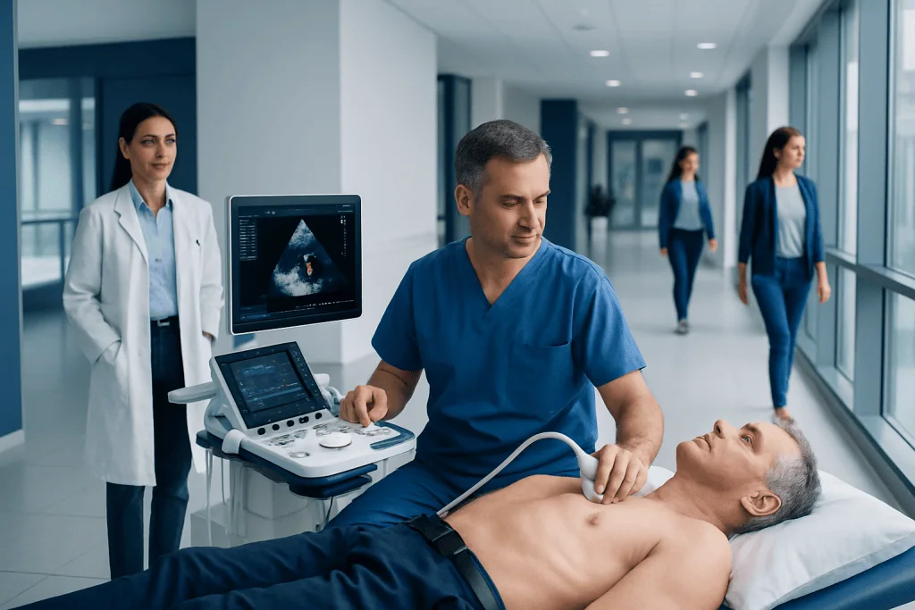
An echocardiogram uses sound waves, not radiation, to show the heart in motion. It can assess heart pumping strength, valve function, chamber size, wall motion, and fluid around the heart.
Key Takeaways
- An echocardiogram uses sound waves, not radiation, to show the heart in motion.
- It can assess heart pumping strength, valve function, chamber size, wall motion, and fluid around the heart.
- The most common type is a transthoracic echocardiogram, performed with a probe on the chest.
- Some people may need a transesophageal, stress, Doppler, or contrast echocardiogram for more detailed information.
- Results should be interpreted by a qualified clinician together with symptoms, examination findings, and other tests.
An echocardiogram is a safe ultrasound test that creates moving images of the heart. It helps doctors see how the heart chambers, valves, muscle, and blood flow are working.
Overview
An echocardiogram, often called an echo test or heart ultrasound, is a diagnostic examination that uses high-frequency sound waves to create images of the heart. Unlike an X-ray or CT scan, it does not use ionizing radiation. The test allows clinicians to see the heart beating in real time and to evaluate how blood moves through the heart.
During the test, a trained specialist places a small handheld device called a transducer on the chest or, in certain cases, uses a probe placed in the esophagus. The transducer sends sound waves toward the heart and receives the returning echoes. A computer turns these echoes into moving pictures on a screen.
An echocardiogram is commonly used when a person has chest discomfort, shortness of breath, a heart murmur, fainting, palpitations, leg swelling, abnormal ECG findings, or known heart disease. It is also used to monitor conditions over time, such as heart valve disease, heart failure, cardiomyopathy, congenital heart problems, and the effects of some treatments on the heart.
What an Echocardiogram Shows
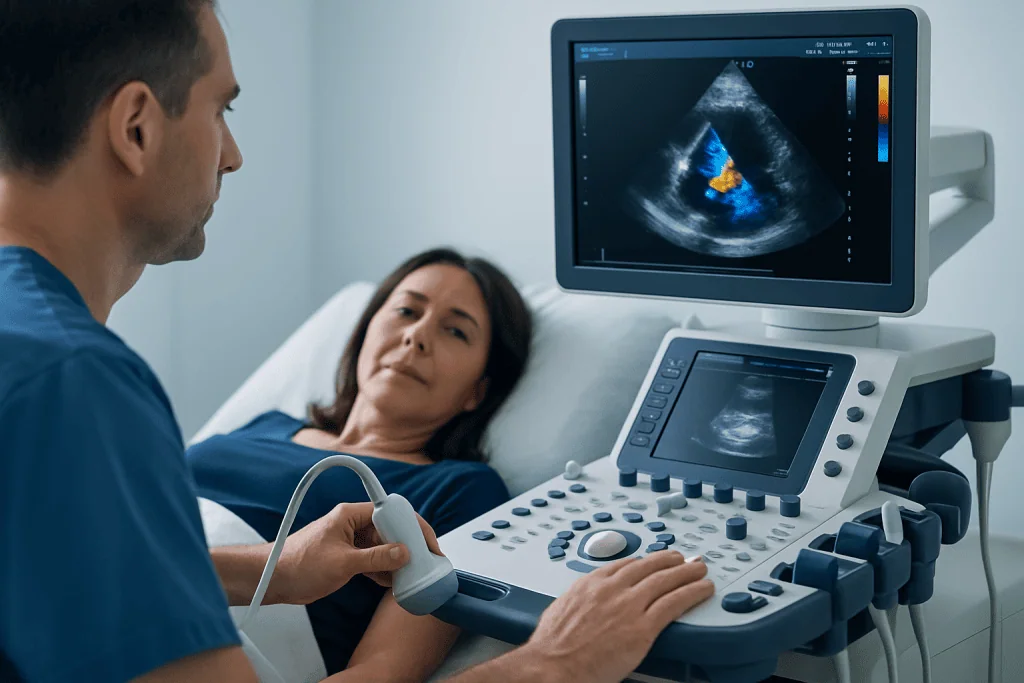
An echocardiogram provides detailed information about the heart’s structure and function. It can measure the size of the heart chambers, the thickness and movement of the heart walls, and the strength of the heart’s pumping action. One of the most familiar measurements is the ejection fraction, which estimates how much blood the left ventricle pumps out with each heartbeat.
The test is especially useful for assessing the heart valves. It can show whether a valve is narrowed, known as stenosis, or leaking, known as regurgitation. Doppler echocardiography adds information about the speed and direction of blood flow, helping doctors understand how significant a valve problem may be.
An echo can also identify fluid around the heart, called pericardial effusion, and may suggest increased pressures in the lungs or right side of the heart. It can detect abnormal heart wall motion after a heart attack, signs of cardiomyopathy, and some congenital heart defects. In selected situations, it may help identify blood clots, tumors, or infections affecting the heart valves.
Although an echocardiogram is highly informative, it is only one part of a complete heart evaluation. The images are interpreted alongside a person’s symptoms, physical examination, medical history, blood tests, ECG, and sometimes additional imaging or catheter-based tests.
Types of Echocardiogram
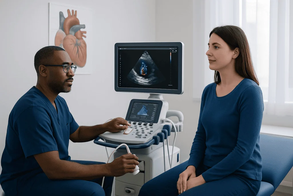
The most common type is a transthoracic echocardiogram. In this test, the transducer is moved over the chest with ultrasound gel to obtain different views of the heart. It is noninvasive, painless for most people, and usually performed without sedation.
A transesophageal echocardiogram provides closer images of certain heart structures by placing a thin probe into the esophagus, which lies behind the heart. This type may be recommended when standard chest images are not clear enough or when doctors need a detailed view of the valves, the left atrium, the aorta, or possible clots or infection. It is usually performed with throat numbing and sedation or anesthesia support, depending on the patient and facility.
A stress echocardiogram evaluates how the heart works during physical exertion or medication-induced stress. Images are taken at rest and again when the heart is working harder. This can help assess reduced blood flow to the heart muscle, exercise capacity, valve function during stress, or symptoms that appear mainly with activity.
Other specialized forms include Doppler echocardiography, which measures blood flow; contrast echocardiography, which uses an injected ultrasound contrast agent to improve image quality; and fetal echocardiography, which evaluates the heart of an unborn baby when there is a specific medical reason.
Preparing for the Test and What to Expect
Preparation depends on the type of echocardiogram. For a standard transthoracic echocardiogram, little or no special preparation is usually needed. Patients can typically eat, drink, and take usual medicines unless their doctor gives different instructions. Comfortable clothing is helpful, and jewelry or clothing around the chest may need to be removed.
During a transthoracic echo, the patient lies on an examination table, often on the left side. Small ECG stickers may be placed on the chest to record the heart rhythm during imaging. The sonographer applies gel and moves the transducer across different areas of the chest. Mild pressure may be felt, especially when the probe is positioned between the ribs, but the test should not be painful.
For a transesophageal echocardiogram, patients are usually asked not to eat or drink for several hours before the test. Because sedation may be used, someone else may need to accompany the patient and drive them home afterward. The throat can feel mildly sore for a short time after the procedure.
For a stress echocardiogram, patients may be asked to avoid certain foods, caffeine, or medications before the test, depending on the clinical plan. They should tell the medical team about chest pain, breathing problems, dizziness, asthma, pregnancy, allergies, or implanted devices before the examination begins.
Understanding Echocardiogram Results
Echocardiogram results are usually reported in a structured way. The report may describe image quality, heart chamber sizes, wall thickness, pumping function, valve appearance, blood flow patterns, estimated pressures, and whether there is fluid around the heart. It may also compare findings with previous echocardiograms if available.
A common term in the report is left ventricular ejection fraction. This reflects the pumping function of the main chamber that sends blood to the body. A low ejection fraction may be seen in heart failure, cardiomyopathy, prior heart attack, or other conditions, while some people can have symptoms even when ejection fraction is preserved. For this reason, the number must be interpreted in context.
Valve findings may be described as mild, moderate, or severe. Mild valve leakage is common and may not require treatment, while more significant valve disease may need monitoring, medication, or a procedure depending on symptoms and heart changes. The report may also mention wall motion abnormalities, which can suggest reduced blood supply or previous injury to the heart muscle.
Patients should review their results with the doctor who ordered the test or a cardiologist. A report can contain technical language, and not every abnormal finding is urgent or dangerous. The most important question is how the findings relate to the person’s symptoms, risks, and overall treatment plan.
Benefits, Risks, and Limitations
A major benefit of echocardiography is that it provides real-time information about the heart without radiation. It is widely used because it is noninvasive in its standard form, repeatable, and helpful for diagnosis as well as follow-up. It can guide treatment decisions for many heart conditions, including valve disease, heart failure, congenital heart problems, and suspected complications after a heart attack.
For a standard transthoracic echocardiogram, risks are very low. Some people may feel brief discomfort from the transducer pressure or from removing ECG stickers. Ultrasound gel may feel cool on the skin but is harmless.
Transesophageal and stress echocardiograms involve additional considerations. A transesophageal echo may cause temporary throat discomfort, and sedation requires monitoring. Rare complications can occur, especially in people with esophageal problems. A stress echo can bring on symptoms such as breathlessness, palpitations, dizziness, or chest discomfort, which is why it is performed under medical supervision.
Like all tests, an echocardiogram has limitations. Image quality can be affected by body shape, lung disease, chest anatomy, or the ability to lie in certain positions. Some heart structures may not be fully visible, and additional tests such as cardiac MRI, CT, nuclear imaging, coronary angiography, or blood tests may be recommended when more information is needed.
When to See a Doctor
A person should seek medical advice if they have symptoms that could be related to the heart, such as unexplained shortness of breath, chest pressure, fainting, persistent palpitations, swelling of the legs, unusual fatigue, or reduced ability to exercise. These symptoms do not always mean a serious heart problem, but they deserve appropriate assessment.
An echocardiogram may also be recommended for people with a newly detected heart murmur, abnormal ECG, high blood pressure affecting the heart, a family history of certain inherited heart conditions, previous heart attack, suspected heart failure, or known valve disease. It can also be used to monitor heart function during or after treatments that may affect the heart.
Urgent medical care is needed for severe or persistent chest pain, sudden shortness of breath, fainting with injury, stroke-like symptoms, or rapid heartbeat with weakness or chest discomfort. In less urgent situations, a primary care doctor or cardiologist can determine whether an echocardiogram is the right next step.
For international patients, Acibadem International offers assessment by multidisciplinary specialists in JCI-accredited hospitals, including cardiology teams who diagnose and treat heart conditions using echocardiography and other evidence-based tests. The right evaluation should always be individualized by a qualified clinician.
Frequently asked questions
Is an echocardiogram the same as an ECG?
No. An ECG records the electrical activity of the heart through electrodes on the skin, while an echocardiogram uses ultrasound to create images of the heart's structure and motion. Doctors often use both tests because they provide different, complementary information.
Does an echocardiogram hurt?
A standard transthoracic echocardiogram is usually painless. Some people feel mild pressure when the transducer is pressed against the chest to improve the image. A transesophageal echocardiogram may cause temporary throat discomfort, but numbing medicine and sedation are commonly used.
How long does an echocardiogram take?
A standard transthoracic echocardiogram often takes about 30 to 60 minutes, depending on the number of images needed and how easy it is to see the heart. Stress or transesophageal echocardiograms may take longer because they include preparation, monitoring, and recovery time.
Can an echocardiogram detect blocked arteries?
A resting echocardiogram does not directly show the coronary arteries in most adults. However, it can show areas of the heart muscle that move abnormally, which may suggest reduced blood flow or previous injury. A stress echocardiogram can help evaluate whether the heart muscle is getting enough blood during exertion or medication-induced stress.
Is an echocardiogram safe during pregnancy?
A standard echocardiogram uses ultrasound and does not involve ionizing radiation, so it is generally considered safe when medically indicated. Pregnant patients should still tell the medical team they are pregnant so the test can be planned appropriately. Fetal echocardiography may also be used to assess a baby's heart when recommended by a specialist.
What happens if the echocardiogram is abnormal?
An abnormal result does not automatically mean an emergency or that treatment is needed right away. The doctor will consider the type and severity of the finding, symptoms, medical history, and other test results. Follow-up may include monitoring, lifestyle advice, medication, additional imaging, or referral to a cardiologist or heart valve team.
This article is for general information only and is not a substitute for professional medical advice. Please consult a qualified doctor about your individual situation.
Explore treatments in Turkey — costs, top hospitals & a free quote
JCI-accredited · board-certified surgeons · reply within 24h
Add us as a Preferred Source to see more of our trusted health content across Google Search, AI Overviews and Discover.

Cardiology Department
Diagnosis and treatment of heart and vascular conditions, from prevention to advanced interventional procedures.
93 specialists in this unitRelated Treatments
Related Conditions
More from the Health Library

Ablation for PVCs: When Extra Heartbeats Need Specialist Treatment

Coronary Artery Bypass Surgery Cost: What Is Included and What Changes the Price?

What Does a Cardiologist Do at Your First Visit?

Coronary Bypass Graft Types: Artery vs Vein Grafts Explained

Dental Clearance Before Heart Bypass Surgery: Infection Risk and Preparation